

Sudden Blur or a Shower of Floaters With Diabetes: When to Seek Same-Day Retinal Care

Key Takeaways
- A sudden shower of floaters in a person with diabetes most often signals bleeding from fragile new vessels, but it can also be a retinal tear, and the two cannot be told apart without a dilated exam.
- Blur from high blood sugar typically affects both eyes and rises and falls with glucose, whereas retinal blur is usually one-sided and does not track the meter.
- Stage 1 (background) retinopathy causes no symptoms and needs no eye treatment, so sudden symptoms in someone with a stage 1 letter mean progression or a different cause.
- Diabetic macular edema is the most common cause of vision loss in retinopathy and the form with the most treatment options, according to the National Eye Institute.
- Laser for proliferative retinopathy protects remaining vision rather than restoring what is lost, and can narrow peripheral or night vision, per NHS guidance.
- A gas bubble after vitrectomy makes flying and high altitude unsafe until it has been absorbed, a period Mayo Clinic puts at roughly two to eight weeks depending on the gas.
If you have diabetes and notice a sudden shower of new floaters, flashes of light, a dark curtain or shadow, or blur in one eye that does not follow your blood sugar, guidance from NHS and Mayo Clinic is to seek same-day eye assessment. These signs can mean bleeding inside the eye, macular swelling or a retinal detachment, and delay can cost sight that treatment cannot bring back.
It usually starts on an ordinary morning. Someone reaches for the kettle and notices the kitchen window has grown a swarm of tiny dark specks that drift when the eyes move. They blink. The specks stay. A few hours later the words on a phone screen look as if they are underwater, and the first instinct is to check the glucose meter, because blur and diabetes have been companions for years.
That instinct is understandable, and sometimes correct. But with diabetic retinopathy, sudden vision loss or a burst of floaters is one of the few eye symptoms that should not be given a day to settle. The retina, the paper-thin layer at the back of the eye that turns light into nerve signals, does not tolerate waiting well.
This explainer walks through what those symptoms usually mean, who is seen the same day, what actually happens at a retinal appointment, and what the weeks afterward tend to look like. The decisions belong to your eye team; the point here is to help you recognize when to make the call.
Why diabetic retinopathy sudden vision loss is a same-day problem, not a wait-and-see one
Most diabetic eye disease is quiet. The National Eye Institute describes retinopathy that can progress for years without symptoms, which is exactly why annual dilated exams exist. When a symptom finally announces itself abruptly, it usually means the disease has crossed a threshold: a fragile new blood vessel has bled, fluid has pooled under the center of the retina, or scar tissue has begun to lift the retina off the wall of the eye.
Each of those events has a clock. Retinal cells depend on a blood supply from the layer beneath them, and Mayo Clinic notes that the longer a retina stays detached, the greater the risk of permanent loss. Bleeding into the eye is less immediately destructive, but it hides the retina from view, and what it hides may be a tear or a detachment that needs prompt repair. Swelling at the center, left for months, can leave lasting damage even after the fluid is drained.
The stakes are not abstract. The CDC lists diabetic retinopathy among the leading causes of blindness in working-age adults in the United States, and the NEI estimates that roughly one in three adults with diabetes over age 40 already has some degree of retinopathy. Most will never lose useful sight, particularly those who attend screening and keep glucose and blood pressure in range. The people who do lose sight are disproportionately those who waited.
So the opinion this article holds, grounded in NHS and Mayo Clinic guidance, is simple: a sudden change in one eye, a shower of floaters, flashes or a shadow is a same-day call to an eye clinic or emergency service. Not tomorrow, and not after seeing whether a good night’s sleep clears it.
What actually happens inside the eye when vision changes suddenly
Picture the eye as a small globe filled with clear gel, called the vitreous, with the retina lining the inside of the back wall like wallpaper. Years of raised glucose stiffen and weaken the tiny vessels that feed that wallpaper. Some leak. Some close off entirely, leaving patches of retina short of oxygen. Starved tissue sends out a chemical distress signal, vascular endothelial growth factor, or VEGF, which is a protein that tells the body to grow new vessels.
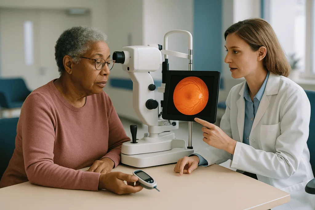
The new vessels are the problem. They sprout in the wrong place, on the retinal surface and into the gel, and they are built badly: thin walled, leaky, and prone to rupture. This is proliferative diabetic retinopathy, the advanced stage the NHS calls stage 3. When one of those vessels tears, blood spills into the vitreous. Red blood cells floating in the gel cast shadows on the retina, and the person sees dozens of dark specks, a reddish haze, or a smear that moves with the eye. That is a vitreous hemorrhage.
New vessels also drag scar tissue behind them. As that tissue contracts, it can pull the retina away from the wall, a tractional retinal detachment. The person may notice a grey curtain creeping in from one side, or straight door frames bowing. Light flashes, described as sparks or lightning at the edge of vision, often mean the retina is being tugged.
A third mechanism does not involve new vessels at all. Leaky older vessels near the macula, the small central area responsible for reading and recognizing faces, allow fluid to seep into the tissue. The macula swells, and the center of vision blurs or warps. This is diabetic macular edema, and the NEI identifies it as the most common reason people with retinopathy lose vision.
Floaters and diabetes: what a sudden shower usually means
Almost everyone over 50 has a floater or two. The vitreous gel liquefies with age, strands clump, and a single grey thread drifts lazily across a bright wall. That is ordinary, and the NHS describes it as generally harmless. The picture that should change your plans is different in three ways: number, speed and company.
Number first. A shower is not one new floater; it is a sudden cloud, often described as pepper, soot, cobwebs or a swarm of gnats. Speed second. It arrives over minutes or hours, not months. Company third. It is frequently joined by flashes of light, a red or brown tint to vision, or a shadow in one corner that was not there at breakfast.
In a person with diabetes, the most likely explanation for that combination is blood from a fragile new vessel. But it is not the only one, and this matters because you cannot tell the causes apart from the inside. The same shower can come from a posterior vitreous detachment, the age-related peeling of the gel away from the retina, which Mayo Clinic notes can tear the retina as it separates. A tear that is found early can often be sealed with laser in the clinic; a tear that progresses to detachment needs surgery.
There is one comforting truth here that also becomes a trap. A vitreous hemorrhage sometimes settles under gravity within a day or two, and vision partly clears. People read that as recovery. It is not. The vessel that bled is still there, and whatever the blood was covering has not been examined. Clearing symptoms do not downgrade the urgency; the NHS still advises urgent assessment for any sudden floaters with diabetes.
Sudden blur: telling a glucose swing from a retinal event
Blurred vision is on every list of high blood sugar symptoms, and for a good reason that has nothing to do with the retina. When glucose runs high, the lens at the front of the eye absorbs extra fluid and changes shape, shifting the eye’s focus. Mayo Clinic lists blurred vision among the signs of hyperglycemia, and the pattern is fairly recognizable once you know it: both eyes, a general softness rather than a missing patch, drifting in step with your readings, and easing over days to a few weeks once glucose steadies. Low glucose can blur vision too, along with sweating and shakiness, and it lifts once sugar is treated.

Retinal blur behaves differently. It tends to affect one eye more than the other. Cover each eye in turn and the difference is often obvious. It may sit in the center of what you see, so that a face is clear at the edges but smudged in the middle, or straight lines take on a wave. It does not follow your meter. You can have a perfect fasting reading and still not read the label on a jar.
Two further clues push toward the retina. Distortion, where a tiled floor or a page of text looks bent, points to macular swelling. A dark patch, shadow or curtain that stays put when you move your eye points to bleeding or detachment.
The honest guidance is that people should not be asked to make this diagnosis at home. If blur is sudden, one-sided, or arrives with floaters or a shadow, the NHS and Mayo Clinic both place it in the seek-help-now category. If it is mild, in both eyes and tracking your glucose, tell your diabetes team, and still keep the annual dilated exam that would have caught quieter changes.
Who is usually seen the same day, and who is usually asked to wait
Eye clinics triage on the assumption that time matters most for a retina that is detaching or bleeding. Retinal detachment is treated as a medical emergency by Mayo Clinic, with repair typically arranged within days. New hemorrhage in someone with known proliferative disease is also generally assessed the same day, because the view of the retina is what determines whether anything more urgent is hiding behind the blood.
At the other end are people whose annual screening shows early background changes and who have no symptoms. The NHS pathway for that group is a yearly review with attention to glucose, blood pressure and cholesterol, not urgent referral. Between those poles sit people with gradual central blur, who are typically seen within days to a few weeks so that macular swelling can be measured and treated before it becomes entrenched.
| What you notice | What it can mean | Usual urgency (NHS, Mayo Clinic) |
|---|---|---|
| Sudden shower of floaters, flashes | Vitreous hemorrhage, retinal tear or posterior vitreous detachment | Same day |
| Dark curtain or shadow spreading across one eye | Retinal detachment | Emergency, same day |
| Sudden blur or wavy lines in the center of one eye | Macular edema or bleeding near the macula | Same day or within 24 hours |
| Severe eye pain, redness, nausea with blur | Raised eye pressure, including neovascular glaucoma | Emergency, same day |
| Mild blur in both eyes that rises and falls with glucose | Lens swelling from high sugar | Tell your diabetes team; routine exam |
| One or two long-standing floaters, unchanged for years | Age-related vitreous change | Routine annual screening |
Neovascular glaucoma deserves a word: it is a rise in eye pressure caused by new vessels blocking the eye’s drainage channel, and it can present as a painful, red, blurry eye. It is uncommon but serious, and pain with vision change is always an emergency sign.
What is stage 1 retinopathy, and can it cause sudden symptoms?
The NHS describes three stages plus maculopathy. Stage 1, also called background or mild non-proliferative retinopathy, is the earliest: tiny bulges in the retinal vessel walls, known as microaneurysms, and perhaps a few pinpoint hemorrhages visible on a screening photograph. The vessels leak a little. The retina itself still works normally, and the person sees normally.
The answer to the second half of the question is therefore no. Stage 1 does not produce a shower of floaters or a sudden dark patch. If someone whose last screening reported background changes develops those symptoms, one of two things has happened: the disease has advanced faster than the annual cycle caught, or the cause lies elsewhere, such as a posterior vitreous detachment or a sugar-related lens shift. Either way the response is the same, an urgent examination rather than reassurance based on an old letter.
What does stage 1 mean for the years ahead? The NHS guidance is that no eye treatment is needed at this point. What is needed is attention to the three levers that slow progression: glucose, blood pressure and blood lipids. The CDC frames this plainly, noting that managing diabetes and keeping blood pressure and cholesterol in range lowers the risk of vision loss. People with stage 1 stay on yearly screening; the NHS may shorten the interval if changes are progressing.
Stage 2, pre-proliferative retinopathy, shows more widespread damage: blocked vessels, cotton-wool spots where nerve fibers have swollen, and beaded veins. Symptoms are still usually absent, but the risk of moving to stage 3 rises, and screening intervals tighten. Stage 3 is the proliferative disease with new vessels that produces most of the sudden events this article concerns. Maculopathy can occur alongside any stage.
What is diabetic maculopathy, and what are its symptoms?
The macula is a patch of retina a few millimeters across that handles everything you look at directly: print, faces, the road ahead. Diabetic maculopathy means the vessels serving that patch have become leaky or blocked. When they leak, fluid collects within the tissue and the macula thickens; this is macular edema. When they block, the center simply goes short of blood, a form called ischemic maculopathy that is harder to treat.
The symptoms are central and specific. Words blur while the edges of the page stay sharp. A door frame bends. Colors look washed out. Faces are recognizable by hair and outline but not by features. Peripheral vision, the ability to navigate a room, is usually preserved, which is why people with maculopathy often say they can see fine until they try to read.
Maculopathy tends to build gradually, but it frequently feels sudden. The stronger eye compensates for months, and the problem is discovered only when that eye is covered, or when the second eye begins to fail as well. This is one reason clinicians ask patients to check each eye separately from time to time; the NEI lists central blur and wavy lines among the signs that should prompt an eye exam.
Diagnosis rests on optical coherence tomography, or OCT, a scan that uses light to map the retina in cross-section and measures thickness to the micron. It is painless, takes minutes and is repeated at follow-up to judge response. Treatment, discussed later, includes anti-VEGF injections, steroid implants and focal laser. The NEI’s point that macular edema is the leading cause of vision loss in diabetic retinopathy carries a hopeful corollary: it is also the form of the disease with the most treatment options, provided it is found while the tissue is still healthy enough to recover.
What happens at a same-day retinal appointment
Plan for two things: several hours, and a lift home. The exam almost always involves dilating drops, which widen the pupil so the clinician can see the whole retina; Mayo Clinic notes they blur near vision and cause light sensitivity for a few hours afterward, so driving is unsafe and sunglasses help.
The sequence is fairly standard. Vision in each eye is measured on a letter chart. Eye pressure is checked, because bleeding and new vessels can push it up. Once the pupils are wide, the ophthalmologist uses a slit lamp, a microscope with a bright beam, and a handheld lens to inspect the retina and vitreous directly. If blood is blocking the view, an ultrasound probe placed on the closed eyelid can show whether the retina beneath is flat or lifted.
Imaging follows. OCT maps the macula. A wide-field photograph records the periphery. Fluorescein angiography, in which a yellow dye is injected into an arm vein and photographed as it travels through the retinal vessels, shows where vessels are leaking or closed and whether new vessels have formed. The dye can turn skin and urine yellow for a day; the NHS describes this as expected.
Bring a list of your medicines, your recent glucose readings and the date of your last screening. Mention blood thinners, since they affect how a hemorrhage is managed. The visit ends with one of a few pathways: observation with a date to return, laser performed the same day or booked shortly, a course of injections scheduled, or referral for surgery if the retina is detached. The clinician will explain which, and why; it is a reasonable moment to ask what would change the plan.
Is laser treatment for diabetic retinopathy effective?
Yes, with an important qualification about what it is effective at. Laser treatment has been the backbone of proliferative retinopathy care for decades, and the NHS summary is that it can stabilize the disease and reduce the risk of further vision loss. What it does not usually do is restore vision that has already gone. People who expect a laser to sharpen their sight are often disappointed; people who understand it as a way to keep the sight they have tend to feel differently about the same result.
There are two main forms. Panretinal photocoagulation places hundreds of small burns across the peripheral retina, away from the macula. It sounds destructive, and in a sense it is: the aim is to reduce the amount of oxygen-hungry tissue so that the distress signal driving new vessel growth quietens and the vessels shrink back. Focal or grid laser targets leaking spots near the macula to reduce swelling; since anti-VEGF injections arrived, it is used less often for edema on its own but still has a role.
The treatment is done in the clinic with numbing drops and sometimes a contact lens held against the eye. Most people describe flashes and a dull ache rather than pain. The NHS notes that more than one session is often needed for panretinal treatment.
Trade-offs are real and worth hearing before you consent. Peripheral vision may narrow, night vision can worsen, and some people notice glare or a small blind spot. Vision may be blurry for a day or so afterward. The NHS lists these effects, and your specialist will weigh them against the risk of untreated new vessels, which is generally considerably higher. The decision is theirs to recommend and yours to accept, ideally after that conversation.
Injections and vitrectomy: how they work and what the typical timeline looks like
Anti-VEGF injections block the protein described earlier, the distress signal that makes vessels leak and sprout. The medicine is injected into the vitreous through a numbed eye wall, a procedure that takes seconds and that most people describe as pressure rather than pain. Because the effect wears off, treatment is a course rather than a single event: the NHS describes a series of injections over many months, with the interval set and adjusted by the specialist according to how the retina responds on OCT. Macular thickness often falls within weeks, and the NHS notes that for macular edema these injections can improve as well as stabilize vision, which is why they have become first-line for that problem.
Risks are uncommon but specific: a rise in eye pressure, bleeding on the eye surface, and rarely an infection inside the eye called endophthalmitis, which presents as increasing pain, redness and worsening vision in the days after an injection and needs emergency treatment. Steroid implants, which release medicine slowly inside the eye, are an alternative for some people, with their own trade-offs including cataract and pressure rise. Which option, if any, suits you is a decision for your prescribing clinician.
Vitrectomy is surgery. Through tiny openings in the eye wall, the surgeon removes the vitreous gel, and with it any blood, then peels away scar tissue and, if the retina is detached, flattens it and seals tears with laser. The gel is replaced with fluid, or with a gas bubble or silicone oil to hold the retina in place while it heals. The NHS describes vitrectomy as the usual route when a hemorrhage does not clear or when scar tissue is pulling on the retina.
Complications include cataract, further bleeding, raised pressure and re-detachment. Recovery is measured in weeks, and the surgeon’s estimate depends heavily on what was found inside.
What the following days and weeks usually look like
Timelines here are typical ranges drawn from NHS and Mayo Clinic patient information, not promises, and your own team’s instructions override anything general.
After a vitreous hemorrhage managed by observation, the blood often settles and clears on its own over weeks to months. People are commonly asked to sleep with the head raised and avoid heavy lifting or bending, so that blood pools low in the eye and the macula stays visible. A follow-up exam checks that nothing is developing behind the haze.
After laser, expect a few hours of dilation blur, a dull ache that evening, and sometimes headache. Night vision or peripheral awareness may feel different for weeks, and for some people permanently. Routine activity usually resumes the next day.
After an injection, the eye may feel gritty for a day. A small dark circle drifting in vision is often an air bubble and clears within a day or so. The warning pattern is different: pain that builds rather than fades, increasing redness, or vision that worsens over the following two to three days should be reported immediately.
After vitrectomy with a gas bubble, the view is dim and wobbly at first, like looking through water, and improves as the bubble is absorbed over roughly two to eight weeks depending on the gas used, per Mayo Clinic. You may be asked to hold a specific head position for several days. Flying, travelling to high altitude and certain anesthetics are unsafe while gas remains, because the bubble expands and raises pressure dangerously; the NHS and Mayo Clinic both flag this. Visual recovery continues for weeks, and the final result depends on how healthy the retina was before surgery. Follow-up visits are frequent early and then space out.
What people often get wrong about diabetic retinopathy sudden vision loss
My sugars are good, so my eyes are fine. Good control lowers risk substantially and is the single most useful thing you can do, but it does not erase damage from earlier years, and retinopathy can still progress. Screening is for everyone with diabetes, as the CDC’s yearly dilated exam recommendation makes clear.
No symptoms means no disease. The reverse is closer to the truth. Stages 1 and 2, and much of stage 3, are symptom-free. By the time a floater shower arrives, the disease has usually been present for years.
The floaters are fading, so it has sorted itself out. Blood settling is not the vessel healing, and it says nothing about what lies beneath. Urgent review still applies.
Laser will fix my vision. Laser protects remaining sight. Improvement, when it happens, is more often seen with injections for macular edema, and even then the NHS describes it as a possibility, not an expectation.
One injection and I am done. Anti-VEGF treatment is a course over months, sometimes years, with intervals adjusted to response.
Tightening glucose fast is always good for the eyes. Rapid improvement in control can, for some people with established retinopathy, temporarily worsen retinal changes before the long-term benefit arrives. This is not a reason to avoid better control, and never a reason to stop or change a prescribed medicine on your own. It is a reason to tell your eye team when your diabetes treatment changes so they can watch more closely.
Screening photos are the same as a full eye exam. Retinal photography is excellent at catching retinopathy, which is its purpose, but it does not replace an ophthalmologist’s dilated examination when you have new symptoms.
Supplements or special glasses will protect me. No supplement has mainstream evidence for preventing diabetic retinopathy. Glucose, blood pressure, lipids and attendance at screening are what the evidence supports.
Questions to ask your care team
A same-day appointment moves quickly, and the questions you wanted to ask surface in the car park afterward. Writing a few down beforehand changes that. The list below is a starting point rather than a script; pick the ones that matter to you.
- What stage of retinopathy do I have in each eye, and is there maculopathy?
- What caused the floaters or blur I noticed, and has the cause been fully seen, or is some of the retina still hidden by blood?
- Is there a retinal tear or detachment, and if so, how soon does it need repair?
- What are you recommending, what are the alternatives, and what would happen if we waited?
- If laser is proposed, is the aim to protect the vision I have, or is improvement realistic? What side effects should I expect on night and side vision?
- If injections are proposed, roughly how long is a course likely to run, how will you decide when to space them out, and what signs of infection should I watch for afterward?
- If surgery is proposed, will a gas bubble be used, how long should I avoid flying or altitude, and is there a head position I need to hold?
- Which symptoms should bring me back the same day rather than at my next scheduled visit, and what number do I call out of hours?
- Should I keep driving? Is there a legal reporting requirement where I live?
- Does anything about my diabetes treatment, blood pressure or blood thinners need to change, and who coordinates that between my eye team and my diabetes team?
- How often will I be seen, and who do I contact if my vision changes between visits?
You are entitled to clear answers in plain language, and to a copy of the plan. If the answer to any of these is uncertain, a good clinician will say so and explain what would make it clearer.
When to call your doctor
Some of what follows repeats earlier sections deliberately, because this is the part people come back to in a hurry. If you have diabetes and any of these begins suddenly, contact an eye clinic, your ophthalmologist or emergency services the same day, following NHS and Mayo Clinic guidance on urgent eye symptoms:
- A sudden shower of new floaters, particularly with flashes of light
- A dark curtain, shadow or veil moving across the vision of one eye
- Sudden blur, a missing patch or distortion in one eye that does not track your blood sugar
- A red or brown haze over vision
- Severe eye pain, a red eye, headache or nausea accompanying any vision change
- Increasing pain, redness or worsening vision in the days after an eye injection or surgery
- Sudden loss of vision of any degree, even if it partly recovers
Do not drive yourself; the dilating drops alone will make that unsafe. Do not wait to see whether it clears overnight, and do not assume an earlier screening letter reporting mild changes rules anything out.
Contact your diabetes team, rather than emergency services, for blur in both eyes that comes and goes with glucose readings, for questions about whether a new medicine could be affecting your vision, and to arrange your annual dilated exam if it has lapsed. Never stop or change a prescribed medicine because of a vision symptom without speaking to the prescriber.
Every judgment about urgency, testing and treatment sits with the clinicians who examine your eye. This article can tell you when to pick up the phone. What happens next is theirs to decide with you.
Frequently asked questions
What is stage 1 retinopathy?
Stage 1, or background retinopathy, is the earliest form of diabetic eye disease, with tiny bulges and pinpoint leaks in retinal vessels but normal vision. The NHS describes no eye treatment at this stage, only attention to glucose, blood pressure and cholesterol and continued yearly screening. It does not cause sudden floaters or blur; if those occur, something else is happening and needs urgent assessment.
How do you live with diabetic retinopathy day to day?
Most people live well with it by attending every screening or clinic visit, keeping glucose, blood pressure and lipids in the ranges their diabetes team sets, not smoking, and knowing the red-flag symptoms that mean same-day care. Checking each eye separately now and then catches central changes the stronger eye hides. Low-vision services, good lighting and larger print help if sight is already affected.
What is diabetic maculopathy?
Diabetic maculopathy is damage to the vessels serving the macula, the small central part of the retina used for reading and recognizing faces. Leaking vessels cause swelling called macular edema; blocked vessels starve the center of blood. Symptoms are central blur, wavy lines and faded colors with side vision preserved. It is diagnosed with an OCT scan and commonly treated with injections or focal laser.
Is laser treatment for diabetic retinopathy effective?
For proliferative retinopathy, laser is effective at stabilizing the disease and lowering the risk of further vision loss, according to NHS guidance, and remains a standard treatment. It does not usually restore vision already lost, and it can reduce night and peripheral vision. Whether laser, injections or a combination suits you is a decision for your ophthalmologist based on your retina’s findings.
Can high blood sugar cause sudden blurry vision?
Yes. High glucose draws fluid into the lens and shifts the eye’s focus, producing blur that usually affects both eyes and eases over days to weeks once glucose steadies, as Mayo Clinic notes. Low glucose blurs vision too. Blur in one eye, a dark patch, distortion or floaters is a different pattern and should be assessed the same day rather than attributed to sugar.
Are floaters a serious sign with diabetes?
A single long-standing floater is usually the ordinary aging of the eye’s gel. A sudden shower of many new floaters, especially with flashes or a shadow, is serious in someone with diabetes because it often means bleeding from new vessels or a retinal tear. The NHS advises urgent same-day assessment for that pattern, even if the floaters seem to be fading.
How quickly can diabetic retinopathy cause vision loss?
The disease itself usually develops over years without symptoms, which is why yearly dilated exams are recommended by the CDC. The events that cause abrupt loss, a vitreous hemorrhage or retinal detachment, can happen within hours once proliferative disease is present. A detached retina loses function progressively, so Mayo Clinic treats it as an emergency needing repair within days.
Does vision come back after a vitreous hemorrhage?
Often it improves as blood is absorbed, which can take weeks to months, and vitrectomy can clear blood that does not settle on its own. How much vision returns depends on the health of the retina underneath, not on the blood itself. Your ophthalmologist will examine or ultrasound the retina to check for tears or detachment and will discuss realistic expectations for your eye.
Can I drive with sudden vision changes or after retinal treatment?
Not on the day of any dilated exam, laser or injection; the drops blur vision for hours. Beyond that, driving depends on your vision meeting the legal standard in your state or country, which your eye team can assess. Many places require drivers to report conditions affecting sight. Ask your clinician directly and follow their advice.
Do I still need a yearly dilated eye exam if my vision seems fine?
Yes. Most diabetic retinopathy, including stages that put you at risk of sudden bleeding, produces no symptoms until late. The CDC recommends a dilated eye exam at least once a year for people with diabetes, and the NHS offers annual screening. Finding changes early, when treatment protects sight, is the point of the exam.
References
- NHS: Diabetic retinopathy
- NHS: Diabetic retinopathy, stages
- National Eye Institute (NIH): Diabetic retinopathy
- CDC: Diabetes and vision loss
This article is for general information only and is not a substitute for professional medical advice. Please consult a qualified doctor about your individual situation.

More from the Blog

What Smart Lens Surgery Can and Cannot Do for Near, Middle and Distance Vision
Smart lens surgery is a marketing name for replacing the eye's natural lens with a multifocal, trifocal or extended-depth-of-focus intraocular lens. It can restore…

Who Is Smart Lens Surgery For? Presbyopia, Early Cataract and Lens Choices Explained
Smart lens surgery, the marketing name for refractive lens exchange, is usually considered for adults over roughly 45 who have presbyopia, an early cataract,…

Why Your Surgeon Sends You Home With Eye Shields After ReLEx SMILE
Surgeons give eye shields after ReLEx SMILE to protect the cornea while it seals, mainly during sleep, when people rub their eyes or press…

ReLEx SMILE vs LASIK: What the Missing Flap Changes for Dry Eye and Recovery
The core smile vs lasik difference is the corneal flap. LASIK lifts a hinged flap of the cornea's surface and reshapes the tissue underneath;…

Who Is a Poor Match for Multifocal Lenses? Retina, Cornea and Personality Factors
People are usually a poor match for multifocal lenses when the eye has a condition that limits contrast or sharp focus, such as macular…

Scleral Lenses vs Intracorneal Rings for Keratoconus: What Each Does for Vision and Comfort
Scleral lenses and intracorneal ring segments treat keratoconus differently. Scleral lenses are large, removable contact lenses that vault over the irregular cornea and rest…