

The CT Scan Before ESWL: How Stone Density and Position Predict Whether Shock Waves Will Work
Key Takeaways
- Non-contrast CT measures four things that shape the ESWL decision: stone size, density in Hounsfield units, exact location, and skin-to-stone distance.
- Research series and major urology guideline groups commonly treat a density above roughly 1,000 Hounsfield units as a marker that shock waves fragment the stone less reliably.
- A skin-to-stone distance beyond about 10 centimetres weakens the energy reaching the stone and may exceed the fixed focal depth of the lithotripter.
- Lower-pole stones can fragment well yet clear poorly, because the pieces have to drain uphill out of a dependent pocket of the kidney.
- Dual-energy CT can identify uric acid stones, which may sometimes be dissolved with supervised urine alkalinisation rather than treated with any procedure.
- ESWL fractures the stone but does not remove it; your own ureter clears the gravel over days to weeks, and blood in the urine for a few days afterwards is expected.
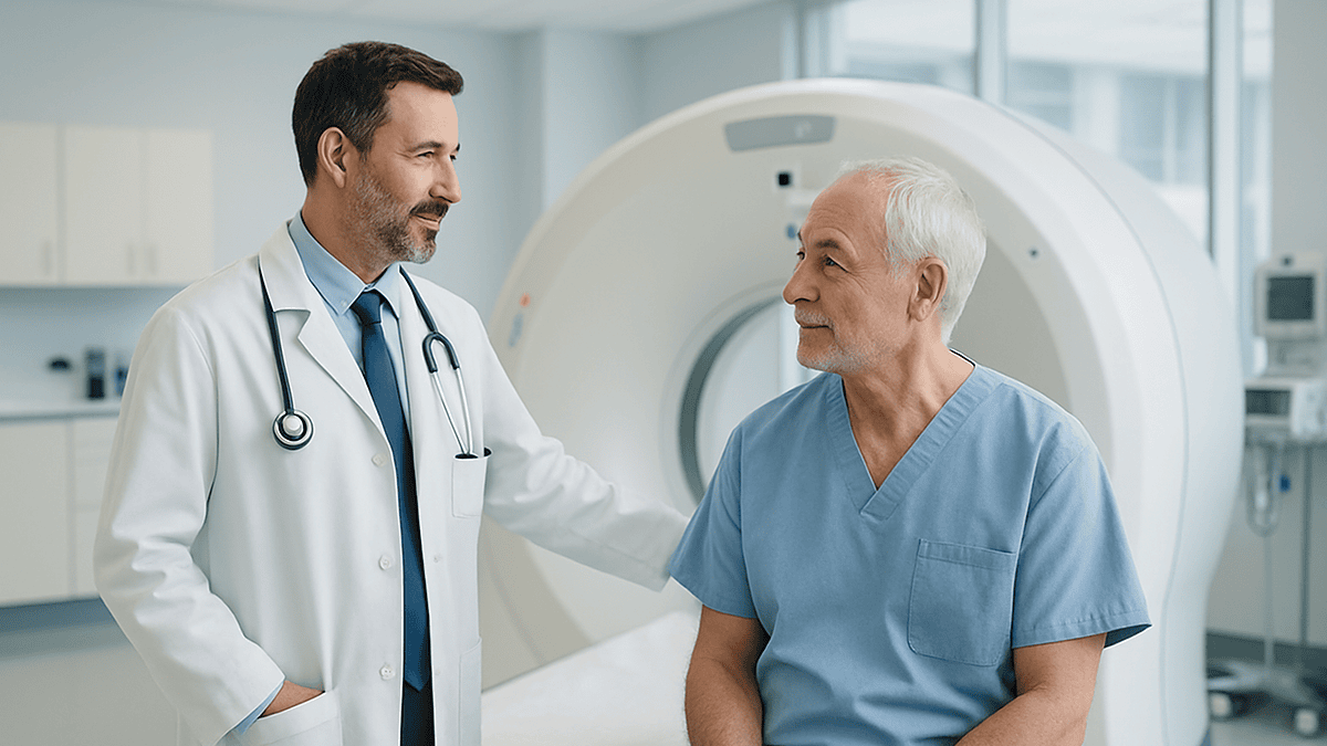
A CT scan before ESWL (extracorporeal shock wave lithotripsy) lets the urologist measure a kidney stone's size, its density in Hounsfield units, its exact position, and the distance from skin to stone. Very dense stones (commonly above roughly 1,000 Hounsfield units), stones far from the skin, and stones in the lower pole of the kidney tend to fragment less reliably, so the scan helps the treating team judge whether shock waves or another option is the better fit.
The urologist turns the monitor so you can see it. A grey slice of your own abdomen, and in the middle of the left kidney, a white fleck no bigger than a lentil. “That’s it,” she says. Then she clicks on it, and a number appears: 1,180. You came in to ask whether shock waves could break the stone without surgery. You did not expect the answer to hinge on a figure you have never heard of.
That number is why the CT scan before ESWL matters so much. Shock wave lithotripsy works from outside the body, which means the surgeon never sees or touches the stone directly. Everything depends on what the imaging reveals about the target: how hard it is, how deep it sits, and where exactly in the urinary tract it has lodged.
This article explains what radiologists and urologists read from that scan, which measurements genuinely predict whether a stone will fragment, and what the evidence says about the questions people ask most, from pain to misdiagnosis.
Why do urologists order a CT scan before ESWL?
Shock wave lithotripsy is the one stone treatment where nobody touches the stone. Energy is focused from outside the body at a target seen only on imaging, so the quality of the picture becomes the quality of the plan. The CT scan before ESWL is usually a non-contrast study: a scan of the abdomen and pelvis without injected dye, which shows calcium-containing and most other stones as bright white specks against soft tissue.
It answers four questions at once. How big is the stone? How dense is it? Where precisely is it sitting? And how much tissue lies between the skin and the stone? Each answer shifts the odds. A small, moderately dense stone close to the skin behaves very differently under shock waves from a large, rock-hard stone buried in a deep pocket of the lower kidney.
Ultrasound and plain X-ray can confirm that a stone exists, but neither gives a reliable density reading, and ultrasound tends to overestimate size. There is a safety dimension too. CT shows whether urine is backing up above the stone, whether there is more than one stone, and whether the drainage anatomy would let fragments pass. The Mayo Clinic and the NIH’s kidney disease institute both describe CT as the most detailed imaging test for suspected stones, which is why most people meet it either in the emergency department or in clinic before any treatment is chosen.
The decision still rests with the treating urologist. The scan simply gives that conversation measurements instead of guesses.
How shock wave lithotripsy actually breaks a stone
Picture a pulse of pressure, generated by a device pressed against your flank through a water-filled cushion, travelling through skin, muscle and kidney tissue to converge on a point a few millimetres wide. Soft tissue transmits that pressure almost like water, so it passes through largely unharmed. A stone does not. Stone is rigid and brittle, so the pulse creates compressive stress on the front face and tensile stress on the back face, and tiny cracks begin to form.
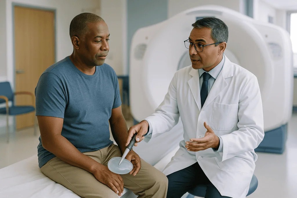
Two mechanisms do the work. The first is direct stress from the wave itself, repeated hundreds or thousands of times across a session. The second is cavitation: microscopic bubbles form in urine around the stone as the pressure drops behind each wave, then collapse violently against the surface, chipping it away. Together these turn one stone into gravel that is small enough to pass through the ureter, the narrow tube connecting kidney to bladder.
Targeting relies on live imaging during the procedure, usually X-ray fluoroscopy or ultrasound, and the operator adjusts the focal point as you breathe. According to the Mayo Clinic, a session typically lasts about 45 to 60 minutes and can cause moderate discomfort, so sedation or light anesthesia is common.
This mechanism explains why the pre-treatment CT matters. A very dense stone resists cracking. A stone far from the skin receives less energy because the wave spreads and weakens with distance. And a stone sitting in a dependent part of the kidney may fracture nicely yet leave fragments that have no easy route out. ESWL does not remove anything; it only makes pieces smaller, and your own urinary tract has to do the rest.
What a CT scan for kidney stones shows that an X-ray cannot
A plain abdominal X-ray is fast and uses little radiation, but it has blind spots. Stones made of uric acid, a waste product that crystallises in acidic urine, are often invisible on X-ray. Bowel gas, bone and overlapping shadows hide small stones, and X-ray gives no sense of depth. Ultrasound avoids radiation entirely and is the first choice in pregnancy, yet it struggles to see stones in the middle of the ureter and cannot measure density at all.
Non-contrast CT fills those gaps. According to the NIH’s kidney disease institute, it can detect stones of almost every composition and pinpoints their location without dye. Modern scanners use low-dose protocols for stone follow-up, and the NHS notes that a CT appointment usually takes around 10 to 20 minutes, with the actual scanning portion lasting well under a minute.
From the images, the reporting radiologist records several things the urologist will use:
- The longest diameter of the stone, and often its volume.
- Its attenuation in Hounsfield units, a measure of how strongly it blocks X-rays.
- Its exact position: which part of the kidney, or which third of the ureter.
- Skin-to-stone distance, measured along the path a shock wave would take.
- Signs of obstruction, such as a swollen kidney (hydronephrosis) or fluid around it.
- Other stones, kidney anatomy variants, and incidental findings elsewhere.
Some centres also use dual-energy CT, which scans at two X-ray energies and can distinguish uric acid stones from calcium stones. That distinction is useful because uric acid stones can sometimes be dissolved with urine-alkalinising treatment under medical supervision, a path that may avoid any procedure. The Mayo Clinic lists dual-energy CT among the imaging options that reveal even tiny stones.
Hounsfield units kidney stone density: the number that predicts fragmentation
The Hounsfield unit (HU) is the CT scanner’s brightness scale. Water sits at zero, air at roughly minus 1,000, and dense bone above 1,000. A radiologist places a small measurement circle over the stone and the software reports the average value inside it. That single figure is the closest thing imaging offers to tapping the stone with a hammer.

Why does it predict ESWL results? Attenuation tracks mineral composition and how tightly the crystals are packed. Calcium oxalate monohydrate and cystine stones are hard, tightly bound and typically read high. Uric acid and calcium oxalate dihydrate stones are softer and read lower. Harder stones need more shock waves to crack, often fragment into larger pieces, and are more likely to need a second session or a different procedure.
Research series and the major urology guideline groups in Europe and the US commonly use a cutoff around 1,000 HU, with some studies drawing the line nearer 900. Above that range, fragmentation becomes noticeably less reliable; below it, shock waves usually perform well, provided size and position are also favourable. These thresholds are guides rather than walls. A stone at 1,050 HU is not untreatable, and a stone at 700 HU is not guaranteed to shatter.
Two practical cautions. First, the measurement depends on technique: the slice thickness, the size of the measuring circle, and whether the radiologist used a bone or soft-tissue window all shift the number. Second, a stone with a dense core and softer shell may fracture differently from one with uniform density, which is one reason some reports describe the stone’s internal structure as well as its average value. Ask your urologist what your reading was and how it factored into the recommendation.
Skin to stone distance ESWL: why where the stone sits in your body matters
Shock waves lose energy the further they travel through tissue, in the same way a shout fades across a field. Skin-to-stone distance (SSD) is the straight-line measurement from the skin surface to the centre of the stone along the path the shock wave would take, usually averaged across three angles on the CT images. Longer distances mean a weaker pulse arriving at the target and a wider, less precise focal zone.
Studies in urology journals and the guideline statements that draw on them repeatedly identify an SSD above roughly 10 centimetres as a marker of lower fragmentation rates. The number is a convenient landmark rather than a precise cliff. Body habitus is the biggest driver: more abdominal fat or thick back musculature lengthens the path. So does a kidney that sits deep in the retroperitoneum, which is normal anatomical variation rather than anything you did.
SSD also interacts with the machine. Most lithotripters have a fixed focal depth, so if the stone lies beyond that range the operator simply cannot bring the energy to bear where it is needed. Positioning you prone (face down) rather than supine can shorten the path for some stones, and the treating team will factor that in.
Why mention this at all if you cannot change it on the day? Because it shapes the honest conversation. When a stone is small and soft but the SSD is long, the urologist may still offer ESWL with a clear statement that a second session or a switch to ureteroscopy is possible. When a stone is both dense and deep, the same surgeon may suggest going straight to a telescopic approach to avoid an unproductive first treatment. Neither path is a failure; both are the scan doing its job.
Does stone location in the kidney or ureter change the odds?
Yes, and in a way that has less to do with breaking the stone than with clearing the pieces. The kidney’s collecting system is shaped a little like a hand: a central palm (the renal pelvis) and finger-like pockets (calyces) in the upper, middle and lower poles. Stones in the lower pole sit in a pocket that drains uphill against gravity. Shock waves can fragment them well, yet the gravel often settles back into the same pocket instead of washing out.
Guideline groups therefore treat lower-pole stones with more caution, particularly larger ones, and may favour ureteroscopy, in which a thin telescope passes up through the bladder and ureter and a laser breaks the stone under direct vision. CT helps here beyond simply naming the pole. It shows the angle at which the lower calyx joins the pelvis, how long and narrow its neck is, and whether the anatomy would let fragments escape. A steeply angled, narrow-necked lower calyx is a well-recognised reason to think twice about shock waves.
Ureteral stones follow their own logic. Stones in the upper ureter, near the kidney, are usually easy to target and respond reasonably to ESWL. Stones in the lower ureter, deep in the pelvis, can be shielded by pelvic bone and harder to image during treatment, which is one reason many teams prefer ureteroscopy for that segment. The NHS describes both approaches as standard options for stones that will not pass on their own, chosen according to size and position.
Position also flags urgency. A stone that is completely blocking the ureter above an infected kidney is an emergency requiring drainage first, not lithotripsy, and CT is often what reveals it.
ESWL success factors at a glance: size, density, distance and position
No single measurement decides the question. Urologists weigh the CT findings together, often alongside your general health and preferences. The table summarises how each factor tends to tilt the balance, using the ranges that appear most often in guideline documents and patient information from major centres.
| CT finding | Tends to favour ESWL | Tends to favour another option | Why it matters |
|---|---|---|---|
| Stone size | Smaller stones, generally under about 2 cm (Cleveland Clinic patient information) | Larger stones, especially in the lower pole | Bigger stones produce more fragments and often need repeat sessions |
| Density (Hounsfield units) | Below roughly 900–1,000 HU | Above roughly 1,000 HU | Dense, hard stones resist cracking |
| Skin-to-stone distance | Under roughly 10 cm | Over roughly 10 cm | Energy weakens with distance; focal depth is limited |
| Location | Renal pelvis, upper or middle pole, upper ureter | Lower pole with narrow calyceal neck; lower ureter | Fragments must drain by gravity and anatomy |
| Obstruction or infection | No swelling, no signs of infection | Blocked, infected kidney | Drainage comes first; shock waves can spread infection |
Two caveats keep this table honest. The thresholds are drawn from research populations and will not predict any individual outcome, which is why no responsible clinician quotes a personal success percentage from a scan alone. And the factors stack: a stone that is slightly over one line but comfortably within the others may still be a good candidate, while a stone that is borderline on all four is a different proposition.
Other pieces of context sit outside the scan but matter just as much: whether you take blood-thinning medicines, whether you are pregnant, whether your kidney function is normal, and what you would prefer if a second procedure became necessary. The treating team puts all of this together before making a recommendation.
Who is usually offered ESWL, and who is usually asked to wait
The typical candidate has a single stone, modest in size, of moderate density, sitting in an accessible part of the kidney or upper ureter, with no obstruction or infection and normal blood clotting. For that person, shock waves offer a treatment with no incision and no telescope, usually done as a day case. The NHS and the NIH both list it as a first-line option for stones too large to pass unaided.
Several situations usually mean waiting or choosing something else. MedlinePlus lists pregnancy as a reason not to proceed, because the shock waves and the X-ray guidance pose a risk to the fetus. People taking anticoagulant medicines, which reduce the blood’s ability to clot, face a higher risk of bleeding around the kidney; the prescribing clinician decides whether and how to adjust these, never the patient alone. An active urinary infection has to be treated first. An aortic or renal artery aneurysm, a bulge in a major blood vessel near the kidney, is another reason to avoid shock waves in that area.
Anatomical factors can also shift the decision. A blockage below the stone would trap fragments. Very low-lying or unusually positioned kidneys may be hard to bring into focus. Severe skeletal deformity can block the shock wave path. Children can have ESWL, with preparation and anesthesia adapted to age, and the pathway is discussed in detail with the family.
Finally, there are people for whom ESWL is possible but perhaps not the best fit: someone with a dense, deep lower-pole stone, or someone who travels for work and cannot tolerate the uncertainty of fragments passing over weeks. For them a single definitive ureteroscopy may be preferable. None of these judgements are made from a checklist; they come from a conversation with the treating team that starts with the CT.
Can a kidney stone be misdiagnosed on a CT scan?
Non-contrast CT is the most accurate routine test for stones, but accuracy is not perfection. Mistakes fall into two groups: seeing a stone that is not there, and missing one that is.
The classic false positive is the phlebolith, a small calcified knot inside a pelvic vein. On a single slice it can look exactly like a stone in the lower ureter. Radiologists look for clues such as a soft-tissue rim around a true stone, a tapering vein leading to a phlebolith, and whether the ureter above is swollen. Calcified lymph nodes, arterial plaque and even swallowed material in the bowel can occasionally confuse the picture.
False negatives are rarer but real. Very small stones can be missed if slices are thick or the scan is motion-blurred. Stones formed from certain antiviral protease inhibitors, a medicine class used in HIV treatment, are famously invisible on CT. And a stone that has already passed into the bladder may be overlooked when attention is fixed on the kidney.
Measurement errors matter too. Stone size can be overestimated when the window settings are adjusted a certain way, and Hounsfield readings vary with technique, which is why a report that says 950 HU on one scan and 1,050 HU on another may reflect method rather than a change in the stone.
What protects you is context. The Mayo Clinic describes diagnosis as combining imaging with symptoms, urine tests and blood tests. If the scan and your symptoms disagree, or if a planned ESWL finds nothing to target on the day, the team re-examines rather than proceeds. Asking “how confident is the radiologist that this is a stone?” is a reasonable question.
How long does the CT scan take, and how painful is shockwave lithotripsy?
These are two of the most-searched questions, and both have reassuring, specific answers.
The CT comes first. The NHS states that a CT appointment usually takes around 10 to 20 minutes from arrival in the room to leaving it. The actual acquisition, when the table slides through the ring and the scanner spins, lasts seconds. For a stone study no contrast dye is needed, so there is no cannula and no need to fast, although departments vary on preparation, and you may be asked to arrive with a comfortably full bladder or to remove metal objects. You lie still, hold your breath briefly once or twice, and that is all. Radiation dose for a low-dose stone protocol is a fraction of a standard abdominal CT, and your referrer weighs it against the information gained.
ESWL is a different kind of experience. Most people describe the sensation of each shock wave as a firm tap or flick against the flank, repeated steadily, rather than sharp pain. The Mayo Clinic notes that the procedure can cause moderate discomfort, which is why sedation or light general anesthesia is typically offered; some centres use local anesthesia with intravenous pain relief instead. You lie on a padded table, the water cushion is pressed to your side, and the machine is positioned using live imaging before the waves begin.
Afterwards, the aching tends to come from two sources: bruising of the skin and muscle over the treatment area, and the kidney itself reacting to the energy and to fragments beginning to move. Many people feel pain closer to a renal colic episode as pieces travel down the ureter over the following days. Your team will discuss pain management with you before you go home.
What the days and weeks after ESWL usually look like
Most people go home the same day, once they are alert, have passed urine and feel comfortable enough to move. The pattern after that is fairly predictable, though the timeline varies from person to person.
In the first few days, blood in the urine is expected; MedlinePlus describes it as a common after-effect that usually settles within a few days. The urine may look pink or tea-coloured and then clear. A bruise over the flank is common and fades like any other bruise. Dull back ache is normal. Fragments can begin passing immediately or take days to weeks, and each small piece may announce itself with a wave of cramping pain as it moves down the ureter. Drinking fluids helps keep urine flowing, and many teams ask you to strain your urine through a fine sieve so that fragments can be sent for composition analysis, which guides long-term prevention.
Over the following weeks, you will usually be asked to return for imaging, often an X-ray or ultrasound, sometimes a repeat low-dose CT, to confirm whether fragments remain. This is the moment when the pre-treatment scan proves its worth a second time: the team compares before and after, and if a dense stone has only partly broken, they can plan a second session or a different approach without starting from scratch.
Some centres prescribe a medicine from the alpha-blocker class, which relaxes the muscle of the ureter and may help fragments pass; whether that is appropriate is a decision for the prescribing clinician. Return to desk work is often possible within a day or two, with heavier activity resuming as soreness allows. Any instructions you were given on the day take precedence over general ranges.
What people often get wrong about the CT scan before ESWL
Myth: “If the stone shows up on CT, shock waves can break it.” Visibility and fragility are different properties. A cystine stone is beautifully visible and notoriously resistant. The Hounsfield value, not the presence of a white dot, is what speaks to hardness.
Myth: “A high density number means I need surgery.” It means shock waves are less likely to be the single, final treatment. Some people choose ESWL anyway, accepting a higher chance of a second session; others prefer ureteroscopy from the outset. The number informs a choice rather than dictating one.
Myth: “The CT already told them everything, so I don’t need imaging on the day.” Stones move. A stone in the kidney at the time of the scan may have dropped into the ureter by the treatment date, and a ureteral stone may have passed entirely. Live imaging on the lithotripsy table is routine, and finding nothing to treat is a good outcome, not a wasted trip.
Myth: “ESWL removes the stone.” It only fractures it. Your own ureter clears the pieces, which is why location and drainage anatomy matter as much as density and why some discomfort after treatment is expected rather than a sign of failure.
Myth: “An old scan from months ago is fine.” Stones grow, shift and multiply, and swelling in the kidney can change. Most teams want recent imaging; how recent is their call.
Myth: “More shock waves equal better results.” Beyond a certain number, additional waves add bruising to the kidney without adding fragmentation, which is why sessions are capped and repeated rather than extended. The evidence on precise limits varies by machine, so the operating team sets the protocol.
Questions to ask your care team
Walking into a consultation with a short list turns a monologue into a conversation. These questions map directly onto what the CT scan before ESWL can and cannot tell you.
- What size is my stone, and where exactly is it sitting?
- What was the Hounsfield reading, and how did it influence your recommendation?
- What is my skin-to-stone distance, and does it fall within the range your lithotripter can reach?
- Is my stone in the lower pole, and if so, how is the calyx shaped for drainage?
- Did the scan show any swelling of the kidney, infection, or a second stone?
- If you had to guess from the scan, is this likely to need more than one session? What would we do next if it does?
- How do my other options, such as ureteroscopy or watchful waiting with fluids, compare for a stone like mine?
- Do any of my regular medicines, especially blood thinners, need review before the procedure, and who will manage that?
- What kind of anesthesia or sedation do you use, and what should I expect to feel during and afterwards?
- What follow-up imaging will I have, and when?
- Should I strain my urine to collect fragments for analysis?
- Which symptoms afterwards are normal, and which should bring me straight back?
Bring the printed radiology report or ask for a copy; it typically lists the measurements discussed in this article. If a figure is missing, for instance the Hounsfield value, it is fair to ask whether it can be measured from the existing images rather than repeating the scan. And if the recommendation surprises you, asking “what on the scan made you lean this way?” is one of the most useful sentences in the room. The urologist’s answer should come back to specific findings, and every final decision stays with you and the treating team together.
When to call your doctor
Before treatment, while you are waiting with a known stone, certain symptoms mean the plan changes from scheduled to urgent. A fever or shaking chills alongside flank pain can signal infection trapped behind an obstructing stone, a situation that needs drainage quickly. The NHS and the Mayo Clinic both advise seeking immediate care for pain so severe you cannot sit still or find a comfortable position, pain with nausea and vomiting that stops you keeping fluids down, difficulty passing urine, or visible blood in the urine with any of these.
After ESWL, some symptoms are expected: pink or light red urine for a few days, flank bruising, dull ache, and episodes of cramping as fragments pass. Call the team, or go to emergency care, if you notice any of the following:
- Fever above the threshold your team gave you, chills, or feeling suddenly unwell.
- Pain that is escalating rather than easing, or that is not controlled by the plan you were sent home with.
- Heavy bleeding, large clots, or urine that stays dark red rather than clearing.
- Inability to pass urine, or passing only tiny amounts over several hours.
- Persistent vomiting that prevents you drinking.
- Dizziness, fainting, or a racing heartbeat, which can accompany significant bleeding or infection.
- Severe pain on the opposite side from the treated kidney, or new swelling of the abdomen.
Trust the pattern, not a single reading. A fragment can lodge in the ureter and block it, sometimes days after you felt fine; that is manageable when caught early. If you have one working kidney, a kidney transplant, diabetes, or a weakened immune system, your team will probably ask you to call with a lower threshold, and that advice overrides any general list. When in doubt, the call is always worth making.
Frequently asked questions
Do I really need a CT scan for kidney stones before ESWL?
In most cases, yes, because CT is the only routine test that measures stone density and skin-to-stone distance, the two findings that best predict whether shock waves will fragment the stone. Ultrasound confirms a stone exists and is preferred in pregnancy, but it cannot judge hardness. If you already have a recent CT, your urologist may use it rather than repeat the scan.
How painful is shockwave lithotripsy?
Most people describe each shock wave as a firm tap against the flank rather than sharp pain. The Mayo Clinic notes the procedure can cause moderate discomfort, which is why sedation or light anesthesia is usually offered. Afterwards, flank bruising and aching are common, and cramping pain can occur as fragments move down the ureter over the following days. Your team will plan pain relief with you.
Can a kidney stone be misdiagnosed on a CT scan?
Occasionally. The most common confusion is a phlebolith, a small calcified vein in the pelvis that can mimic a lower ureteral stone. Very small stones can be missed, and stones formed from certain antiviral medicines are invisible on CT. Radiologists use features such as a soft-tissue rim and swelling above the stone to tell them apart, and clinicians combine the scan with symptoms and urine tests.
How long does a CT scan for kidney stones take?
According to the NHS, a CT appointment usually takes around 10 to 20 minutes, and the scanning itself lasts only seconds. A kidney stone study normally uses no contrast dye, so no cannula is needed. You lie still, hold your breath briefly, and the table moves through the scanner ring. Low-dose protocols are widely used for stone imaging to limit radiation.
What Hounsfield units kidney stone reading is too hard for ESWL?
There is no absolute limit, but research series and major guideline groups commonly use a threshold of roughly 1,000 Hounsfield units, with some studies closer to 900. Above that range, fragmentation becomes less reliable and a second session or ureteroscopy is more likely. Below it, shock waves usually perform well if size and position are also favourable. The reading can vary slightly with scanning technique.
Why does skin to stone distance ESWL planning matter if I can't change it?
Because it predicts how much energy reaches the stone and whether the lithotripter can focus at that depth. A distance beyond roughly 10 centimetres is linked with lower fragmentation rates in published series. Knowing it lets the urologist set realistic expectations, choose prone or supine positioning, or suggest ureteroscopy from the start rather than an unproductive first session.
Is a lower pole kidney stone a bad candidate for shock waves?
Not automatically, but it is treated with more caution. Lower-pole stones often fragment well yet clear poorly, because the pieces must drain upward out of a dependent pocket. CT shows how steep and narrow that pocket is. For smaller lower-pole stones with favourable anatomy, ESWL remains an option; for larger ones, guidelines tend to favour ureteroscopy.
Which ESWL success factors matter most on the scan?
Four findings carry most weight: stone size, density in Hounsfield units, skin-to-stone distance, and location within the kidney or ureter. Signs of obstruction or infection matter too, because an infected blocked kidney needs drainage before any lithotripsy. No single factor decides the question; the treating team weighs them together with your health, medicines and preferences.
Will I need another CT scan after ESWL?
Usually some follow-up imaging is arranged a few weeks after treatment to check whether fragments remain, but it is often an X-ray or ultrasound rather than CT. A repeat low-dose CT is used when the stone was not visible on X-ray or when results are unclear. Your urologist decides the type and timing based on your original scan and how you recover.
Can an old CT scan be used to plan ESWL?
Often not, if it is several months old. Stones can grow, move from the kidney into the ureter, or pass entirely, and kidney swelling can change. Most teams want recent imaging that reflects the stone’s current size and position, and live imaging is used again on the treatment table. How recent is recent enough is a judgement for the treating urologist.
References
- MedlinePlus: Lithotripsy
- NHS: Kidney stones, treatment
- NHS: CT scan
- NIH NIDDK: Diagnosis of kidney stones
This article is for general information only and is not a substitute for professional medical advice. Please consult a qualified doctor about your individual situation.

More from the Blog

HUS in Children: How the Hospital Pathway, Comfort and Recovery Are Planned for Families
Treatment for HUS (hemolytic uremic syndrome) in children is mainly supportive hospital care: careful fluid and salt balance, close monitoring of blood counts and…

Why the Bladder Stops Emptying: Urinary Retention Causes in Men and Women and Their Care
Urinary retention happens when the bladder cannot empty because something blocks the outlet, the bladder muscle is too weak to push, or the nerves…

What to Expect From Prostatitis Treatment: How Long Relief Takes and Why Follow-Up Matters
Prostatitis treatment results depend on the type. Acute bacterial prostatitis usually eases within days of antibiotics, though the full course runs weeks. Chronic bacterial…

Inside Varicocele Surgery: Anesthesia, the Small Groin Incision and How Long It Takes
Varicocele surgery, or varicocelectomy, is usually a same-day outpatient procedure done under general or regional anesthesia with local numbing. Through a small incision low…

How Hydronephrosis Is Treated: Relieving the Blockage and Protecting Kidney Function
Hydronephrosis is treated by relieving whatever is blocking urine flow and protecting the kidney while that happens. If pressure or infection threatens the kidney,…

How Kidney Amyloidosis Is Treated: Controlling Amyloid Production and Protecting the Filters
Kidney amyloidosis treatment works on two fronts: shutting down the source of the misfolded protein and protecting the kidneys from further damage. For AL…