

The DEXA Scan: How Bone Density Is Measured and Scores Are Read
Key Takeaways
- A T-score of −2.5 or lower at the hip or spine meets the WHO definition of osteoporosis, and each one-point drop in T-score roughly doubles fracture risk.
- Skip calcium supplements for 24 hours before the scan: an undissolved tablet can overlap the spine on the image and falsely inflate your reading.
- A central DEXA delivers about one-tenth the radiation of a chest X-ray, comparable to a few hours to a day of natural background exposure.
- Medicare Part B covers a screening bone density test every 24 months for qualifying adults, and cash prices for bone DEXA commonly run about $40 to $300.
- Z-scores, not T-scores, are the appropriate yardstick for premenopausal women, men under 50, and children: a Z-score of −2.0 or lower means density below the expected range for age.
- CDC data show 12.6 percent of US adults aged 50 and older have osteoporosis at the hip or spine, and most don't know it until a bone breaks.
Quick Answer
A DEXA scan is a quick, low-dose X-ray test that measures bone mineral density, usually at the hip and spine, to assess fracture risk and screen for osteoporosis. Results are reported as a T-score: −1.0 or higher is considered normal, between −1.0 and −2.5 indicates low bone mass, and −2.5 or lower meets the definition of osteoporosis. The painless test typically takes 10 to 20 minutes.
The first sign is rarely dramatic. A wrist that breaks catching a fall off a curb. A back that aches after lifting a grandchild. A favorite pair of pants that suddenly drags on the floor because you’ve quietly lost an inch of height. Bone loss doesn’t announce itself: it whispers, and by the time it shouts, something has usually fractured.
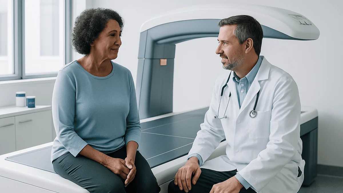
That’s exactly why the DEXA scan exists. It’s one of the least imposing tests in medicine: you lie on a padded table, fully clothed, while a scanner arm glides over you like a slow photocopier. No tunnel, no injection, no discomfort. Fifteen minutes later, you have hard numbers describing something you cannot feel.
Yet those numbers, T-scores, Z-scores, standard deviations, read like statistics homework. Here’s what the test actually measures, how to prepare so your results aren’t skewed, and how to translate the printout into plain English.
What does a DEXA scan check for?
A DEXA scan (dual-energy X-ray absorptiometry, often written DXA) measures bone mineral density, essentially, how much calcium and other minerals are packed into a defined segment of bone. Denser bone is generally stronger bone, and less likely to break under everyday stress.
Clinicians order the test for three main reasons. First, to screen for osteoporosis before a fracture happens, since the disease is silent until then. Second, to diagnose low bone mass in someone who has already broken a bone from a minor fall, what doctors call a fragility fracture. Third, to track whether bone density is stable, improving, or declining over time, especially in people taking prescription bone medications.
The scale of the problem justifies the screening. According to CDC survey data, 12.6 percent of US adults aged 50 and older, roughly one in eight, have osteoporosis at the hip or lumbar spine, and women are affected about four times as often as men. Millions more have low bone mass, the intermediate zone sometimes called osteopenia.
One clarification worth making early: a DEXA scan is not the same as a nuclear medicine “bone scan,” which uses an injected tracer to look for tumors, infections, or hidden fractures. Same word, entirely different test. DEXA answers one question, how dense is this bone, and answers it very well.
How does DEXA measure bone with two X-ray beams?
The clever part is hiding in the name: dual-energy. The machine sends two X-ray beams of different energy levels through your body. Soft tissue, muscle, fat, skin, absorbs the two beams differently than mineralized bone does. By comparing how much of each beam makes it through to the detector, software can mathematically subtract the soft tissue and calculate how much mineral sits in the bone alone.
The output is a number in grams per square centimeter: the mineral content of the scanned bone divided by its area. That raw figure means little on its own, so the machine compares it against reference databases and converts it into the standardized scores your report shows.
Why does density predict fractures? Bone is living tissue, constantly being broken down and rebuilt. When breakdown outpaces rebuilding, which accelerates after menopause and with age in everyone, the internal honeycomb structure of bone thins. Struts get sparser and more fragile, the way a bridge weakens when rivets are removed one by one. Mineral density is a measurable proxy for that structural change.
It’s a proxy, not a crystal ball. Two people with identical T-scores can have different real-world risk depending on age, fall history, and bone geometry. That’s why good clinicians read a DEXA result alongside the whole person, not instead of them.
What actually happens during the scan?
Expect one of the gentler appointments on your medical calendar. You lie on your back on an open, padded table, no enclosed tube, a genuine relief for anyone who finds MRI machines claustrophobic. For the spine portion, the technologist may rest your lower legs on a foam block to flatten your lumbar curve against the table. For the hip, your foot is positioned in a brace that rotates the leg slightly inward.
Then the scanner arm passes slowly overhead. You’ll be asked to hold still and occasionally to hold your breath for a few seconds, the same way you would for a photograph. There is no noise to speak of, no sensation at all, and nothing touches you except the positioning aids.
The whole visit typically runs 10 to 30 minutes, and the scanning itself is often shorter. You can usually keep your clothes on, provided they’re free of metal, more on that below. Afterward there are no restrictions: you can drive, eat, exercise, and return to work immediately.
Results are not usually delivered on the spot. A radiologist or your ordering clinician interprets the images and scores, and you’ll typically hear back within a few days to a week. When the report arrives, the numbers that matter most are the T-scores for your hip and spine, which deserve their own section.
What should you never do before getting a DEXA bone scan?
Preparation is minimal, but a few missteps can genuinely distort your results, and one matters more than all the others.
- Don’t take calcium supplements for at least 24 hours beforehand. An undissolved calcium tablet sitting in your gut can overlap the spine on the image and artificially inflate your density reading. Calcium-rich foods are fine; it’s the concentrated pills and chews that cause trouble.
- Don’t schedule DEXA right after imaging that uses contrast material. If you’ve recently had a barium swallow, a CT with contrast, or a nuclear medicine study, residual contrast or tracer can skew the measurement. Most centers ask you to wait roughly one to two weeks; tell the scheduler about any recent imaging so they can advise.
- Don’t wear metal. Zippers, underwire, belt buckles, rivets on jeans, and body jewelry all show up on the image. Soft clothing without metal fasteners means you likely won’t need to change into a gown.
- Don’t stay silent about a possible pregnancy. The radiation dose is very small, but X-ray tests are generally avoided during pregnancy unless clearly necessary. Tell the technologist if there’s any chance.
Everything else about your day can be completely normal. Eat breakfast, take your usual medications unless your clinician says otherwise, and drink what you like. Unlike cholesterol or glucose testing, DEXA requires no fasting, one of the few tests where showing up well-fed is perfectly acceptable.
How do you read a T-score?
Your T-score compares your bone density to the average peak density of a healthy young adult of the same sex, roughly, bone at its lifetime best, around age 30. The score is expressed in standard deviations, a statistical unit of distance from that average. Zero means you match the young-adult average; negative numbers mean you fall below it.
| T-score | Category (WHO criteria) | What it means |
|---|---|---|
| −1.0 and above | Normal | Bone density within the expected healthy range |
| Between −1.0 and −2.5 | Low bone mass (osteopenia) | Below normal but not osteoporosis; a signal to protect bone now |
| −2.5 and below | Osteoporosis | Meets the diagnostic threshold; fracture risk is substantially elevated |
| −2.5 and below, plus a fragility fracture | Severe (established) osteoporosis | The threshold plus a broken bone from minor trauma |
The scale is steeper than it looks. As a rule of thumb supported by decades of research, each one-point drop in T-score roughly doubles fracture risk. The gap between −1.0 and −2.5 is not cosmetic: it’s the difference between watchful maintenance and a formal diagnosis.
Your report will list separate T-scores for the lumbar spine, the total hip, and the femoral neck (the narrow top of the thigh bone). Clinicians generally act on the lowest of these. And remember the cutoffs are thresholds on a continuum, not cliffs: a −2.4 and a −2.6 describe very similar bone, even though only one carries the diagnostic label.
What’s a Z-score, and when does it matter more?
While the T-score compares you to a healthy 30-year-old, the Z-score compares you to people your own age, sex, and body size. For most postmenopausal women and men over 50, the T-score drives the diagnosis. But for younger adults and children, the T-score is the wrong yardstick, of course a 35-year-old shouldn’t be graded against her own peak, because she’s still near it.
For premenopausal women, men under 50, and children, guidelines from bone health organizations favor the Z-score. A Z-score of −2.0 or lower is described as “below the expected range for age,” a deliberately careful phrase. In a younger person, that finding doesn’t automatically mean osteoporosis; it means something may be interfering with bone that shouldn’t be at that stage of life.
Even in older adults, a strikingly low Z-score carries a message. If your bones are notably thinner than those of your same-age peers, who have all weathered the same decades, a clinician will often look for a secondary cause. Possibilities include an overactive parathyroid or thyroid gland, celiac disease and other absorption problems, kidney disease, very low vitamin D levels, or long-term use of certain prescription medicines known to thin bone.
In other words: the T-score tells you where you stand; the Z-score hints at why. A thoughtful report reads them together.
Who should get a DEXA scan, and at what age?
Screening recommendations from major medical bodies converge on a few groups.
- All women 65 and older. This is the most consistent recommendation across US guidelines, regardless of risk factors.
- Postmenopausal women under 65 with risk factorslow body weight, a parent who fractured a hip, current smoking, heavy alcohol use, early menopause, or a prior fracture as an adult.
- Men, generally starting around 70, or earlier with risk factors. Evidence in men is thinner and guidelines vary more, but men account for a meaningful share of hip fractures and are frequently underdiagnosed.
- Adults of any age who break a bone from a fall at standing height or less. A fragility fracture is itself strong evidence of weakened bone and should trigger evaluation, not just a cast.
- People with conditions or long-term medications known to accelerate bone loss, including certain endocrine, rheumatologic, and gastrointestinal diseases. Your clinician can tell you whether yours qualifies.
Height loss is an underappreciated flag. Losing more than about an inch and a half from your young-adult height can indicate silent compression fractures in the spine, and it warrants a conversation even if you feel fine.
If none of these apply, say, you’re a healthy 45-year-old without risk factors, routine screening isn’t recommended. The test is safe, but a result you don’t need can create worry without changing anything you’d actually do.
Why do they scan the hip and spine instead of the wrist?
Central DEXA, the standard test, measures the lumbar spine and hip for two practical reasons. These are the sites where fractures do the most damage, and they’re where measurements best predict future breaks. A hip fracture in an older adult often means surgery, months of rehabilitation, and a lasting loss of independence; spinal compression fractures cause chronic pain, height loss, and the stooped posture once called a dowager’s hump. Measuring risk where the stakes are highest simply makes sense.
You may also encounter peripheral devices, portable machines at health fairs or pharmacies that scan the heel, finger, or forearm. These have real uses: they’re inexpensive, mobile, and can flag people who need a full evaluation. But their results don’t map neatly onto the standard T-score categories, and a normal heel reading doesn’t rule out low density at the hip or spine. Treat a peripheral screening as a prompt, not a verdict.
Occasionally the forearm is deliberately included in a central DEXA, for instance, when the hip or spine can’t be measured accurately because of prior surgery, hardware, or severe arthritis, or when a clinician suspects an overactive parathyroid gland, which preferentially thins forearm bone.
One quirk worth knowing: arthritis and calcium deposits in an aging spine can falsely raise the spine T-score. When spine and hip numbers disagree sharply, the hip is often the more trustworthy witness.
How much radiation does a DEXA scan involve?
Remarkably little: this is one of the lowest-dose X-ray tests in all of medicine. A central hip-and-spine DEXA delivers on the order of one-tenth the radiation of a standard chest X-ray, and far less than a CT scan. Put in everyday terms, it’s comparable to the natural background radiation you absorb from soil, air, and cosmic rays over a few hours to a day of ordinary living. A cross-country flight exposes you to more.
That’s why the technologist typically stays in the room during the scan rather than retreating behind a lead barrier, and why repeat scans every couple of years raise no meaningful cumulative concern. MedlinePlus and Mayo Clinic both characterize the dose as very low, and it’s a large part of why DEXA is considered suitable for routine screening of healthy people: a bar many imaging tests can’t clear.
The standard cautions still apply. Radiation of any dose is generally avoided in pregnancy unless the test is clearly necessary, so mention any possibility of pregnancy before you’re positioned on the table. And low risk is not a reason to scan without purpose; a test is worthwhile when its result could change what you and your clinician do next.
For everyone else weighing the decision, radiation should sit near the bottom of the list of concerns. Cost and usefulness are the more legitimate questions, so let’s take those next.
How much does a DEXA scan typically cost in the US?
For a screening test, DEXA is refreshingly affordable, though, as with most American healthcare, the price depends heavily on where you go and how you pay.
Paying cash, a central bone density scan commonly runs between roughly $40 and $300. Freestanding imaging centers tend to sit at the lower end; hospital outpatient departments at the higher end, sometimes with a separate radiologist’s reading fee added. It’s fair, and increasingly normal, to call ahead and ask for the all-in self-pay price.
With insurance, screening is often covered when you meet guideline criteria. Medicare Part B covers a bone mass measurement every 24 months for qualifying beneficiaries, including women whose clinicians determine they’re at risk for osteoporosis, and more often when medically necessary, such as monitoring treatment. Commercial plans generally follow similar logic, though prior authorization rules vary, so a quick call to your insurer before scheduling can spare you a surprise bill.
Body composition DEXA scans, the fitness-oriented version measuring fat and muscle, are a different story. They’re typically considered elective, rarely covered by insurance, and usually priced somewhere between $50 and $150 per scan out of pocket.
Context helps here: a hip fracture routinely generates tens of thousands of dollars in surgical, hospital, and rehabilitation costs, quite apart from its human toll. Against that backdrop, a periodic double-digit screening test for the people who need it is one of medicine’s better bargains.
Is a DEXA scan worth it?
For the right person, yes, and the case rests on a simple chain of logic. Osteoporosis is common, silent, and consequential. DEXA detects it reliably before the first fracture. And once detected, bone loss is genuinely actionable: weight-bearing and resistance exercise, adequate calcium and vitamin D intake, fall-proofing the home, quitting smoking, moderating alcohol, and, when risk is high enough, prescription treatments your clinician can discuss. Screening only earns its keep when a positive result changes what happens next, and here it clearly can.
The evidence is strongest for women 65 and older and for younger postmenopausal women with risk factors, which is why the major US guidelines recommend screening in those groups. For men, the evidence base is smaller and recommendations vary, but a man over 70, or younger with fractures or risk factors, has a reasonable claim to the test.
Where the value proposition weakens is at the healthy end of the spectrum. A low-risk 40-year-old getting a DEXA “just to know” will most likely receive a normal result that confirms what age alone predicted, or a borderline number that generates anxiety without changing management. The test is safe and cheap, but not every safe, cheap test is useful.
An honest summary: DEXA is worth it when your age, history, or risk factors put low bone density plausibly on the table. It is a screening tool, not a wellness souvenir.
What about DEXA body composition scans?
The same machine that measures bone can, with different software, divide your entire body into three compartments: bone mineral, lean mass (mostly muscle), and fat mass. Some systems also estimate visceral fat: the metabolically active fat packed around abdominal organs, which correlates more strongly with cardiovascular and metabolic risk than the fat you can pinch.
This is a legitimately good measurement. Sports medicine programs and research labs use DEXA body composition because it’s more precise than bathroom-scale bioimpedance devices and far more informative than BMI, which can’t distinguish a muscular athlete from someone carrying excess fat at the same height and weight. For an athlete tracking whether a training block added muscle or a person confirming that weight loss is coming from fat rather than lean tissue, the regional breakdown, arms, legs, trunk, offers detail few other accessible tools match.
Some honest caveats belong alongside the enthusiasm. A body composition DEXA is not a medical screening and doesn’t diagnose anything; it’s a measurement, and its value depends entirely on what you do with it. Day-to-day hydration shifts can nudge lean-mass readings, so single scans matter less than trends measured on the same machine under similar conditions. Insurance rarely covers it, and no major guideline recommends it for the general public.
If the numbers would genuinely guide your training or motivate durable habits, it can be a reasonable elective purchase. Just know you’re buying information, not health.
How often should a bone density scan be repeated?
There is no single correct interval: the honest answer is “it depends on your first result,” and the logic is intuitive: the closer your bones are to a threshold that would change management, the sooner it’s worth looking again.
The most common rhythm in practice is every one to two years for people diagnosed with osteoporosis or undergoing treatment, which aligns with Medicare’s coverage of a scan every 24 months. Bone changes slowly, and scanning more often than the disease can move mostly measures machine noise rather than biology.
At the other end, reassuring results buy long stretches of peace. A well-known study of older women found that those with normal bone density or mild osteopenia at their first scan could reasonably wait many years, potentially up to 15, before fewer than 10 percent progressed to osteoporosis. Women with T-scores approaching −2.5, by contrast, warranted rechecks within about a year. Your clinician will tailor the interval to your starting point, age, and risk factors.
Two practical notes improve the value of any repeat scan. First, use the same facility and ideally the same machine when possible; different manufacturers calibrate differently, and cross-machine comparisons introduce error. Second, don’t over-read small changes: a shift of a percent or two may fall within the measurement’s margin of error. Trends across scans matter more than any single delta.
When should you see a doctor about your bones?
Because bone loss itself causes no symptoms, the signals that warrant a medical conversation are mostly indirect, and easy to dismiss if you’re not looking for them.
Make an appointment if you’ve broken a bone from a fall at standing height or less, or from an activity that shouldn’t crack a healthy skeleton, like a firm cough or lifting a laundry basket. See someone if you’ve lost more than about an inch and a half of height since young adulthood, if your upper back has become noticeably rounded, or if you have new, persistent mid-back pain, spinal compression fractures often masquerade as ordinary backache. And if you’re a woman turning 65, a man around 70, or a younger adult with risk factors such as early menopause, long-term use of bone-thinning medications, heavy smoking or drinking, or a parent with a hip fracture, raise screening at your next routine visit even if you feel perfectly well.
Seek prompt medical care, don’t wait for an appointment, after any fall that leaves you unable to bear weight on a leg, with severe hip or groin pain, or with sudden intense back pain, especially if it comes with numbness, weakness, or changes in bladder or bowel control.
The larger point deserves repeating: the best time to discuss bone density is before the first fracture, not after. A fifteen-minute scan is a small ask; a hip fracture is not.
Frequently asked questions
What does a DEXA scan check for?
A DEXA scan measures bone mineral density, most often at the hip and lumbar spine, to screen for osteoporosis and estimate fracture risk before a break occurs. It’s also used to confirm low bone mass after a fragility fracture and to track whether density is stable or changing over time. With different software, the same machine can measure body composition, fat, muscle, and bone, though that’s a separate, usually elective, test.
What should you never do before getting a DEXA bone scan?
Don’t take calcium supplements within 24 hours of the scan, undissolved tablets can appear on the image and artificially raise your spine reading. Also avoid scheduling DEXA within about one to two weeks of a barium study, contrast CT, or nuclear medicine test, since residual contrast skews results. Wear clothing without zippers, buckles, or underwire, and tell the technologist if there’s any chance you’re pregnant. No fasting is required.
How much does a DEXA scan typically cost in the US?
Cash prices for a central bone density DEXA commonly range from roughly $40 to $300, with freestanding imaging centers usually cheaper than hospital outpatient departments. Medicare Part B covers a bone mass measurement every 24 months for qualifying beneficiaries, and commercial insurers generally cover guideline-based screening. Elective body composition DEXA scans typically cost about $50 to $150 out of pocket and are rarely covered. Calling ahead for the all-in price is reasonable and routine.
Is a DEXA scan worth it?
For people who meet screening criteria, yes. Osteoporosis is common, silent, and treatable, and DEXA detects it before the first fracture, when exercise, nutrition, fall prevention, and prescription options can still change the trajectory. The evidence is strongest for women 65 and older, younger postmenopausal women with risk factors, and adults with fragility fractures. For a healthy, low-risk 40-year-old, routine screening isn’t recommended and is unlikely to change anything you’d do.
What is a bad T-score?
A T-score of −2.5 or below meets the World Health Organization’s definition of osteoporosis, while scores between −1.0 and −2.5 indicate low bone mass, sometimes called osteopenia. Anything at −1.0 or above is considered normal. The thresholds sit on a continuum, so a −2.4 and a −2.6 describe very similar bone. Clinicians typically act on the lowest score among your spine, total hip, and femoral neck measurements, weighed alongside age and other risk factors.
How long does a DEXA scan take?
The appointment usually takes 10 to 30 minutes, and the actual scanning is often shorter. You lie on an open padded table while a scanner arm passes slowly overhead: there’s no tunnel, no injection, and no discomfort. You can typically keep your clothes on if they’re free of metal, and there are no restrictions afterward. Results are read by a radiologist or your ordering clinician, usually within a few days to a week.
Is a DEXA scan the same as a bone scan?
No. A DEXA scan is a low-dose X-ray test that measures bone density to assess osteoporosis and fracture risk. A nuclear medicine bone scan involves injecting a radioactive tracer to look for tumors, infections, or hidden fractures: a different test answering different questions. The similar names cause frequent confusion at scheduling desks, so it’s worth confirming which test your clinician ordered before you book.
How often should you get a DEXA scan?
It depends on your first result. People with osteoporosis or on treatment are often rescanned every one to two years, matching Medicare’s 24-month coverage cycle. Research in older women suggests those with normal density or mild osteopenia may safely wait many years before rescreening, while T-scores near −2.5 warrant rechecks within about a year. Using the same facility and machine for repeat scans makes comparisons more reliable, since manufacturers calibrate differently.
Can you improve a low DEXA score?
Bone density can be stabilized and sometimes modestly improved, though results vary by person and cause. Evidence supports weight-bearing and resistance exercise, adequate calcium and vitamin D intake, not smoking, and limiting alcohol. For higher-risk individuals, prescription treatments exist that your clinician can discuss based on your scores and history. Equally important is fall prevention, clearing trip hazards, checking vision, and building balance, since a fracture requires both fragile bone and a fall.
What is the difference between a T-score and a Z-score?
A T-score compares your bone density to the average peak density of a healthy young adult of your sex; a Z-score compares you to people of your own age, sex, and body size. T-scores drive diagnosis in postmenopausal women and men over 50. Z-scores are the preferred measure for younger adults and children, where −2.0 or lower means density below the expected range for age: a finding that often prompts a search for an underlying cause.
References
- Bone Density Scan: MedlinePlus Medical Test
- DEXA (DXA) Scan: Bone Density Test: Cleveland Clinic
- Bone Mineral Density Tests: What the Numbers Mean: NIH NIAMS
- Osteoporosis or Low Bone Mass in Older Adults: United States, 2017–2018: CDC/NCHS
This article is for general information only and is not a substitute for professional medical advice. Please consult a qualified doctor about your individual situation.

More from the Blog

Recovery After a Stereotactic Biopsy: Headache, Pin-Site Soreness, Driving and Results
Stereotactic biopsy recovery is usually short. After a breast biopsy, most people go home the same day and have bruising or soreness for a…
After a PET-CT Scan: Clearing the Tracer, Contact Precautions and When Results Arrive
After a PET-CT scan, most people go home the same day, drink extra fluids and empty their bladder often to help clear the radioactive…
Preparing for an MRI: Eating, Metal, What to Wear
For most MRI scans you can eat and drink normally beforehand, because the magnet images tissue, not stomach contents. The exceptions are scans of…

What Does a Comprehensive Check-Up Include? Blood Work, Imaging and the Follow-Up Plan
A comprehensive check-up usually includes a medical and family history, a physical examination with blood pressure and body measurements, and blood and urine tests…

What a CT Scan Shows, and Why Doctors Order One
A CT scan shows detailed cross-sectional images of bones, organs, blood vessels and soft tissue by combining many X-ray views into slices. Doctors most…

Can an Ultrasound Detect Cancer? What It Can Show, Its Limits and the Next Steps
An ultrasound can detect abnormal lumps, masses and changes in organs that may be cancer, and it is especially good at telling a fluid-filled…