

The Digital Dentistry Workflow Step by Step: Scan, Design, Milling and Fitting in Practice
Key Takeaways
- An intraoral scanner records only the surfaces of teeth and gum; it cannot see decay under fillings, bone levels or nerve canals, which is why implant planning still adds a 3D X-ray.
- In implant design the software starts from the ideal final tooth and works backward to where the post must sit, a method called prosthetically driven planning that exposes bone shortfalls before surgery.
- A surgical guide is a printed resin template with metal sleeves that steer the drill to the planned angle and depth, but it cannot feel bone density or replace surgical judgment.
- Zirconia crowns are milled oversized in a soft state and then shrunk and hardened in a furnace, one reason a zirconia restoration is not always ready the same afternoon.
- Bone fuses with an implant at the same pace whatever the workflow; Mayo Clinic describes implant treatment as a staged process lasting many months.
- The fitting appointment is the true test of every upstream step, and small bite adjustments after crown placement are common rather than a sign of failure.
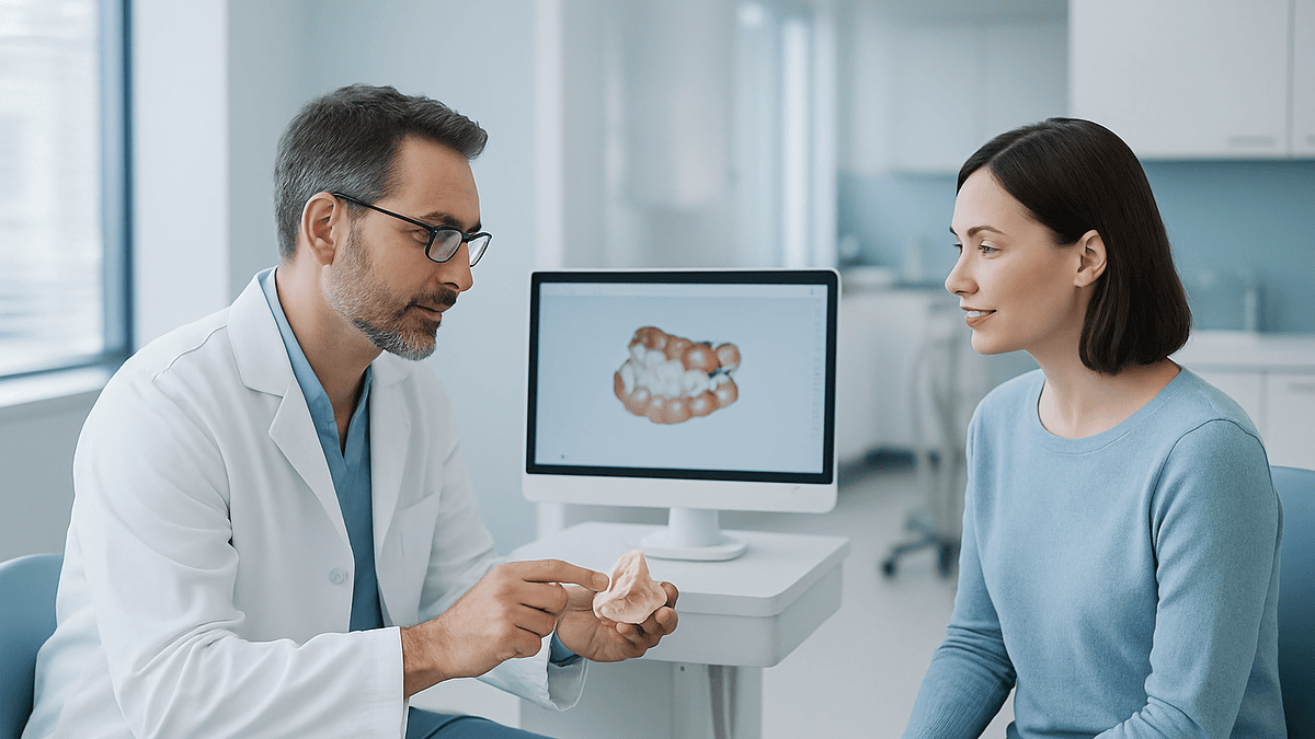
A digital dentistry workflow replaces putty impressions and hand-poured models with data: an intraoral scanner captures the teeth, 3D X-ray imaging maps the jaw when implants are planned, software designs the crown or surgical guide, a milling machine or 3D printer manufactures it, and the dentist fits and adjusts it in the chair. Healing biology and clinical judgment stay unchanged; what changes is how information moves between steps.
You have braced yourself for the tray. Anyone who has had a crown before remembers it: a mouthful of cold putty, the instruction to breathe through your nose, the long minute that feels like five. Instead, the dentist picks up something that looks like an oversized electric toothbrush, asks you to open, and a picture of your teeth begins to assemble itself on a screen beside you, tooth by tooth, in color.
That small surprise is the front door of the digital dentistry workflow. Behind it sits a chain of steps most patients never see: a scan becomes a file, the file becomes a design, the design becomes a block of ceramic carved by a machine, and the ceramic comes back to your mouth for the part no machine can do, the fitting.
Here is what actually happens at each stage, where the evidence is solid, where it is still thin, and what to ask before you say yes.
What is a digital dentistry workflow, in plain language?
Strip away the jargon and a digital dentistry workflow is a hand-off system. The shape of your mouth is captured as data rather than in a rubbery mold, the restoration is designed on a screen rather than in wax, and it is manufactured by a machine reading that design rather than by a technician layering porcelain by eye. The mouth still heals at the same speed. The dentist still decides. What changes is how information travels from your teeth to the laboratory and back.
Two acronyms come up constantly. CAD stands for computer-aided design, the software step where a crown or bridge is drawn in three dimensions. CAM stands for computer-aided manufacturing, the step where a milling machine or printer turns that drawing into a physical object. Together they are usually written CAD/CAM.
In the implant setting, three more terms matter. A dental implant is a screw-shaped post, usually titanium, placed in the jawbone to act as an artificial tooth root, as MedlinePlus describes. An abutment is the connector that sits on top of the implant and pokes through the gum. The crown is the visible tooth that attaches to the abutment. A digital workflow can touch every one of these: planning where the post goes, designing the abutment to fit the gum, and shaping the crown to meet the opposing teeth.
It helps to think of the workflow as five stations: scan, image, design, make, fit. Some practices run all five in one building. Many scan in the clinic and send the file to a laboratory that does the design and manufacturing. Either way, the order rarely changes, and the biology at the end of it, a titanium post fusing with living bone, is identical to what it was before any of this existed.
Intraoral scanner vs impression: what actually happens during the scan
An intraoral scanner is a handheld wand with a small camera and a light source at its tip. It projects a pattern of light onto your teeth, records how that pattern bends over every surface, and stitches thousands of overlapping snapshots into a single three-dimensional model in real time. A conventional impression, by contrast, is a tray of soft material pressed over the teeth and left to set, then poured in stone at the laboratory to make a physical model.
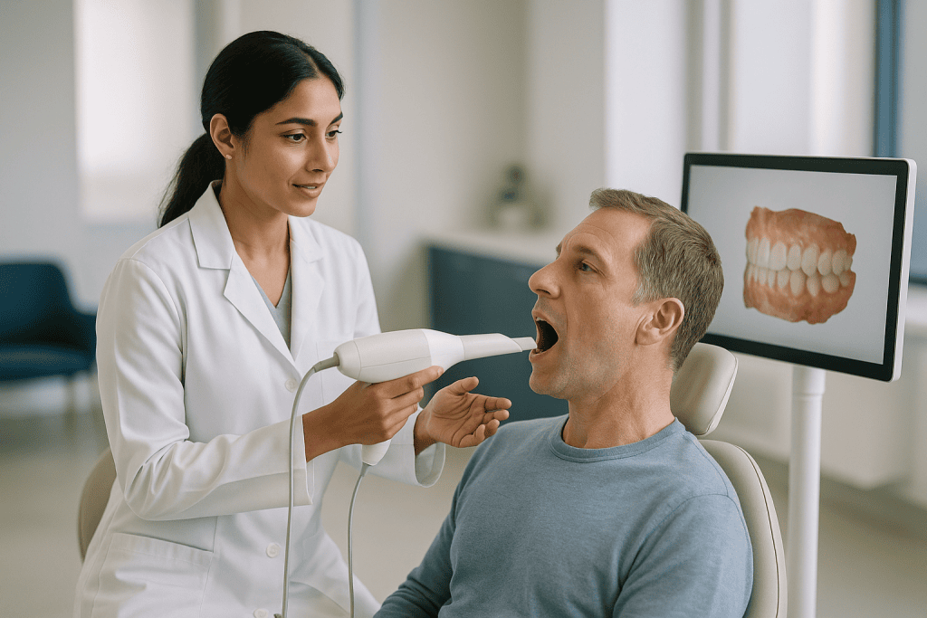
From the chair, the scan feels undramatic. Your dentist or assistant dries the teeth with air, holds your cheek or tongue aside, and glides the wand slowly along the outside, the inside and the biting surfaces of each arch. If a region turns out blurry, they go back over it; the software highlights gaps in a different color. You will be asked to bite together at the end so the two arches can be related to each other, a step called a bite registration.
If you already have an implant in place, a small metal or plastic cap called a scan body is screwed onto it first. The scanner recognizes the cap’s shape and, through it, works out exactly where the implant sits and at what angle. That geometry is what the laboratory uses to design an abutment that emerges through the gum in the right place.
Scanners have limits worth knowing. They record surfaces only, so they cannot see under the gum line, and a margin hidden by bleeding or saliva may be captured poorly. In those cases many dentists still take a conventional impression of that tooth, and that is a judgment call rather than a failure. People with a strong gag reflex often prefer the wand because nothing sits at the back of the throat, though the mouth must stay open for several short passes.
Why implant planning needs 3D imaging, not just a surface scan
A surface scan shows the dentist the shape of what is visible. An implant lives in what is not: bone, nerve canals, the floor of the sinus. For that, the workflow adds a cone beam computed tomography scan, usually shortened to CBCT. It is a dental X-ray machine that rotates once around your head and reconstructs a three-dimensional image of the jaw, rather than the flat picture a conventional dental film gives.
Sitting or standing still with your chin on a rest, you will hear the arm sweep around you. Nothing touches the inside of your mouth. The image lets the team measure how much bone height and width is available, where the nerve that supplies feeling to the lower lip runs, and how close the upper back teeth sit to the sinus. Mayo Clinic notes that this planning stage, including imaging and models, is part of the standard preparation before implant surgery.
The digital step that makes the rest possible is called registration or merging: software overlays the surface scan onto the CBCT so that the visible teeth and gum line up precisely with the bone beneath them. The planned implant can then be positioned on screen with both the soft tissue and the hard tissue in view, and a surgical guide can be designed from the same combined file.
Radiation is the question people ask most. MedlinePlus describes dental X-rays as low-dose, and a CBCT delivers more than a single small film but is chosen when the extra information changes the plan. Tell your dentist if you are or might be pregnant, and ask whether an existing scan can be reused rather than repeated. The decision to image, and how much of the jaw to include, belongs to the treating clinician weighing benefit against exposure.
The design stage: how CAD software plans your crown or implant
Once the files exist, someone has to make decisions with them. A dentist or dental technician opens the merged scan in design software and, for a crown, marks the edge where the restoration will meet the prepared tooth. The program proposes a tooth shape from a library of anatomical forms and adapts it to the neighbors and the opposing bite. The designer then refines it, adjusting how tightly it contacts the adjacent teeth, where the biting cusps land, and how thick the material needs to be to resist fracture.
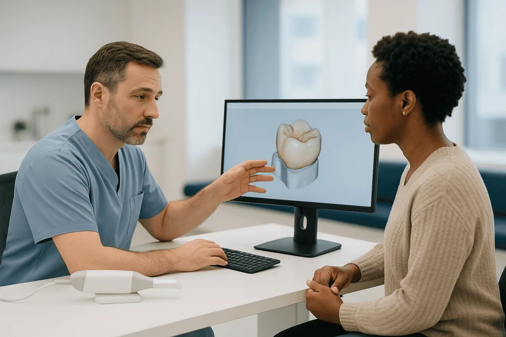
Implant design runs in the opposite direction from the way surgery used to be planned. Rather than placing the post wherever bone happens to be thickest and building a tooth on top afterward, the software starts with the ideal final tooth and asks where an implant would need to sit to support it. Clinicians call this prosthetically driven planning. If the bone in that ideal position is thin, the plan makes the shortfall visible early, and the team can discuss a bone graft, a different angle or a different position before anything is drilled.
Color is the one thing the scanner does not reliably capture. Shade is usually chosen separately with a shade guide held beside the tooth, sometimes with a photograph sent to the laboratory, and it is worth asking to be involved in that choice under daylight rather than under the operatory lamp.
None of this is automatic. The library tooth is a starting point, not an answer. A design finished in a few clicks can still be wrong in a way that only shows when it meets a real bite, which is why the fitting appointment remains the true test of everything upstream.
Guided implant surgery: turning the plan into a printed surgical guide
Guided implant surgery means the implant is placed through a template made from the digital plan rather than positioned freehand. The template, called a surgical guide, is a thin resin plate 3D printed to fit over your remaining teeth, or over the gum if no teeth remain. Metal sleeves set into it direct the drill at the depth, angle and position chosen on screen.
Guides come in degrees. A pilot guide directs only the first, narrow drill and the surgeon completes the site by hand. A fully guided kit directs every drill and sometimes the implant itself. Your team chooses based on how much room there is to work, how many implants are planned, and whether teeth are available to anchor the plate securely.
Surgery day looks much like any implant appointment. Mayo Clinic describes the procedure as typically done in stages under local anesthetic, with sedation or general anesthesia offered in some situations. The surgeon seats the guide, confirms it does not rock, opens the gum or uses a small punch, drills through the sleeves, places the implant and closes. A cover screw or a small healing cap sits on top while the bone heals.
What a guide does not do is remove the need for surgical skill. It cannot feel bone density, it cannot see if it has shifted on a slippery gum, and it cannot decide what to do when the bone under the sleeve turns out softer than the scan suggested. Studies in the dental literature generally find guided placement reproduces the planned position more consistently than freehand placement, but a good plan on a poorly seated guide is still a poor result. Ask how the fit of the guide will be checked before the first drill turns.
Milling, 3D printing and the same day dental crown
Manufacturing splits into two families. Milling is subtractive: a machine with fine rotating burs carves the crown out of a solid block of ceramic, zirconia or resin, following the design file. Printing is additive: a machine builds an object layer by layer from liquid resin cured by light. Milling dominates for the final crown because the materials strong enough to bite on are not yet printable to the same standard; printing dominates for models, surgical guides, temporary teeth and, increasingly, denture bases.
Materials behave differently once they leave the machine. A glass-ceramic crown can be milled to its final shape, then glazed and fired to bring out translucency. Zirconia, the very hard white ceramic used for many back teeth and implant crowns, is usually milled oversized in a soft chalky state and then sintered, a furnace cycle that shrinks and hardens it. That furnace time is one reason a zirconia crown is not always ready the same afternoon.
The phrase same day dental crown refers to a workflow where the tooth is prepared, scanned, designed and milled in the practice during one visit. It is realistic for many single crowns on natural teeth when the practice has the equipment and the case is straightforward. It is rarely realistic for the final crown on a new implant, because Mayo Clinic notes that bone growth around the post takes several months before it can bear load. What can often happen on the day of surgery is a printed or milled temporary tooth, designed from the same plan, to fill the gap while healing continues.
Whether manufacturing happens chairside or at an outside laboratory, ask who checks the finished piece against the design before it reaches your mouth, and what happens if it does not pass.
Fitting: the try-in, the adjustments and the bite check
Everything upstream is a prediction. The fitting appointment is where the prediction meets a jaw that moves in ways no scan captures perfectly. Expect it to take longer than the scan did, and expect adjustments; they are part of the workflow, not evidence of a mistake.
For a crown on a natural tooth, the dentist removes the temporary, cleans the surface and seats the new crown without cement. They check the margin with a fine probe for any ledge or gap, pass floss between the neighbors to judge the contacts, and ask you to bite on thin colored paper that marks where the new tooth touches first. High spots are trimmed with a fine bur and repolished. Only when the bite closes evenly and you can floss normally is the crown cemented.
For an implant crown there are extra steps. The crown may be screw-retained, with a screw running down through the tooth into the implant and a small filling sealing the hole, or cement-retained, glued onto a separate abutment. The dentist checks that the components are fully seated, often with a small X-ray, because a gap of even a fraction of a millimeter at the connection can trap bacteria. The screw is tightened to a specified force with a hand torque wrench, which you will feel as firm pressure.
Speak up during this appointment. Only you can report a spot that feels high, a contact that catches food, or a shape your tongue keeps finding. Cleveland Clinic notes that mild sensitivity and a bite that needs a small correction are common after crown placement, so a short follow-up adjustment is not unusual. The team that designed it wants that feedback; it is the last, and most human, quality check in the chain.
Digital dentistry workflow vs conventional: a side-by-side comparison
Seeing the two paths next to each other clarifies what is genuinely different and what is simply relabeled.
| Step | Conventional workflow | Digital dentistry workflow |
|---|---|---|
| Capturing the teeth | Tray of setting material; poured in stone at the lab | Intraoral scan stitched into a 3D file within minutes |
| Recording the implant position | Impression coping picked up in the mold | Scan body read by the scanner |
| Planning the implant | Flat X-ray plus stone model; largely freehand placement | CBCT merged with the scan; optional printed guide |
| Designing the tooth | Waxed by hand on the model | Drawn in CAD software from a tooth library |
| Making the tooth | Cast metal or hand-layered porcelain | Milled ceramic or zirconia; printed temporaries and models |
| Storing the record | Physical model that can chip or warp | File that can be reopened if a remake is needed |
| Common weak point | Distortion of the material as it sets or is shipped | Poor scan of a wet or hidden margin; registration errors |
What does the evidence say about the results? For single crowns and short spans, comparative studies in the dental literature generally find digital and conventional impressions produce restorations of similar fit, with the digital route often saving chair time and remakes. For a full arch of implants, where the scanner must stitch a long, toothless span without landmarks, accuracy is more variable and many clinicians still combine digital and conventional records or verify the digital one with a printed model. That is not a verdict against the technology; it is an honest statement of where it is mature and where it is still being tested.
The row that matters most to patients is usually the last one. Every method has a failure mode. Choosing well means choosing a team that knows their method’s weak point and checks for it.
Who is the digital workflow usually for, and who is usually asked to wait?
Most people needing a single crown, an implant crown, a short bridge, a night guard or clear aligners are candidates for a scan-based workflow, and many will not be offered anything else. It suits people who gag on impression trays, people who have had a restoration remade because a mold distorted, and anyone whose implant position needs to be planned around a nerve or sinus. Full dentures and multi-implant bridges are increasingly designed digitally too, though often with a conventional check along the way.
The people asked to wait are almost never waiting because of the digital part. They are waiting because of the implant part. Mayo Clinic lists conditions that can affect healing around an implant, including uncontrolled diabetes, heavy smoking, active gum disease and prior radiation to the jaw, and a team may ask that gum disease be treated or blood sugar be brought under control first. Certain medicines that alter bone turnover, including the bisphosphonate class used for osteoporosis, prompt a conversation with the prescribing physician before jaw surgery; nobody should stop such a medicine on their own.
Age matters at one end. Implants are deferred in adolescents because the jaw is still growing and a fixed post would end up out of line with the erupting teeth. At the other end there is no upper age limit as such; general health and bone quality decide.
A few situations make the scan itself harder. Very limited mouth opening leaves little room for the wand at the back teeth. A tooth prepared deep under the gum, or one bleeding at the margin, may simply need a conventional impression that day. None of these are reasons to feel excluded from modern care; they are reasons a thoughtful team keeps both toolkits.
What the following days, weeks and months usually look like
A scan-only visit leaves nothing to recover from. The timeline that matters is the implant one, and it is long by design. Mayo Clinic describes implant treatment as a staged process that can take many months, most of it spent waiting for bone to grow around the post, a process called osseointegration.
After the surgical visit, expect what Mayo lists as typical: swelling of the gum and face, some bruising, minor bleeding and soreness at the site for a few days. Cold packs on the cheek, soft foods and gentle salt-water rinses are the usual advice. Pain relievers and, in some cases, an antibiotic may be prescribed; your surgeon will explain what each is for and how to take it, and that instruction, not a general article, is the one to follow. Contact them rather than adjusting anything yourself.
During the healing phase you may wear a printed temporary tooth, a removable flipper or nothing at all in a back-tooth site. Cleaning matters more than usual: brush gently around the site and keep every hygiene appointment, because inflamed gum around a healing implant is the most common reason the plan slows down.
Once the surgeon confirms integration, usually by testing the implant’s firmness and taking an X-ray, the healing cap is swapped for a scan body and the final scan is taken. The abutment and crown are designed and milled, then fitted at a later visit. A short review a week or two after fitting lets the dentist re-check the bite once the tooth has settled.
From there the implant is treated like any other tooth: brushing, flossing or interdental brushes, and regular reviews where the gum around it is measured and the crown checked for wear or looseness.
Risks, limits and alternatives in plain terms
Digital tools change where errors happen; they do not remove them. A scan can miss a margin under wet gum. Two files can be merged a fraction off, so a guide drilled perfectly to plan places the implant slightly off in the mouth. A printed guide can warp if stored badly, or fail to seat because a tooth moved between the scan and surgery. Milled zirconia can chip if designed too thin over a cusp. A screw can loosen if not tightened to specification. Good practices build checks at each hand-off precisely because each hand-off can fail.
The surgical risks belong to implants themselves and apply whichever workflow is used. Mayo Clinic lists infection at the site, injury to neighboring teeth or blood vessels, nerve damage causing numbness or tingling in the lip, chin or gum, and sinus problems when upper implants intrude into the sinus space. Bone may also fail to fuse with the post, in which case the implant is removed and, after healing, the site can often be reassessed. Ask your surgeon how each of these is prevented in your specific mouth, and what the plan is if one occurs.
Alternatives deserve an honest hearing. A conventional impression is a legitimate route to the same crown. A fixed bridge supported by neighboring teeth replaces a gap without surgery, at the cost of preparing those teeth. A removable partial denture avoids both. Leaving a back-tooth gap unfilled is sometimes a reasonable choice when the bite is stable. The NHS describes these options among standard dental treatments, and a good consultation lays them out without steering.
No workflow, digital or otherwise, guarantees a particular outcome. What you can reasonably expect is a clear explanation of the checks, the risks and the fallback plan.
What people often get wrong about digital dentistry
The scanner is not an X-ray. It records surfaces in visible light and cannot see decay under an old filling, bone loss, or a nerve canal. A digital workflow adds imaging when needed; it never replaces it.
Digital does not mean faster healing. Bone fuses with titanium at the same pace whether the post was planned on a screen or a stone model. What the workflow can shorten is the time between decision and manufacturing, and the number of remakes. Mayo Clinic’s description of implant treatment as a months-long, staged process applies regardless of technology.
A machine-made crown is not a crown made without people. Someone marks the margin, someone judges the contacts, someone chooses the shade, someone checks the piece against the design, and someone adjusts it in your mouth. Automation moved the skill; it did not delete it.
Guided surgery is not risk-free surgery. A guide makes the planned position reproducible, but the risks Mayo lists, infection, nerve injury and failed integration, are biological and surgical, not positional.
Same day rarely means same day for implants. A tooth prepared, scanned, milled and cemented in one afternoon is realistic for many single crowns. For an implant, the same afternoon can produce a temporary; the final tooth waits for bone.
Digital crowns do not automatically last longer. Longevity depends on material, bite forces, gum health and hygiene. Cleveland Clinic gives a typical range of five to fifteen years for crowns in general, with variation driven by care and habits rather than by how the crown was made.
And the newest equipment is not a credential. Ask how many cases like yours the team has handled with the method they propose, and how they verify each step. That question tells you more than any brochure.
Questions to ask your care team before you start
A workflow is only as good as its checkpoints, so most useful questions are about verification rather than technology. Bring these to the consultation and write down the answers.
- Which parts of my treatment will be captured digitally, and are there any teeth or areas where you expect to use a conventional impression instead?
- Will I need a 3D scan of my jaw, and can an existing scan be reused rather than repeated?
- How will the surface scan and the 3D image be lined up, and how do you check that the merge is accurate before designing the guide?
- Will my implant be placed with a surgical guide? If so, is it a pilot guide or fully guided, and how will you confirm it sits correctly before drilling?
- Who designs the crown, a clinician here or a technician elsewhere, and can I see the design on screen before it is made?
- What material do you propose for the crown, why that one for this tooth, and what are its known weaknesses?
- Will there be a temporary tooth during healing, and how is it made?
- What checks happen at the fitting, including an X-ray of the implant connection, and what happens if the fit is not acceptable?
- Roughly what sequence of visits should I expect, and which stages depend on how my bone heals?
- Which of my medicines or health conditions could affect healing, and will you be in touch with my physician about them?
- What symptoms after surgery should prompt me to call, and how do I reach someone out of hours?
Notice that none of these ask whether the technology is good. They ask how the team knows, in your case, that it worked. A team comfortable with its workflow will welcome them.
When to call your doctor (or your dental team)
Some discomfort after implant surgery is expected, and a new crown can feel strange for a few days. The signs below are different: they suggest something that needs a clinician’s eyes rather than patience.
Call the same day if bleeding from the surgical site does not slow with firm pressure from a clean gauze pad after about half an hour, if pain is increasing after the second or third day rather than easing, or if swelling continues to grow after the third day. Mayo Clinic notes that swelling and soreness should settle over several days; a course in the other direction is a reason to be seen.
Seek urgent care if you develop a fever with facial swelling, if swelling spreads toward the eye or under the jaw, or if swallowing or breathing becomes difficult. These can indicate a spreading infection and are not situations for waiting until morning.
Contact your surgeon promptly if numbness or tingling in the lip, chin or tongue persists once the local anesthetic has worn off, since this can signal nerve irritation that is best assessed early. The same applies to a bad taste, pus at the site, a healing cap or temporary that comes loose, or a guide-related wound on the gum that is not healing.
After the final crown is fitted, call if the tooth feels loose or rotates, if you hear a click when biting, if a bite that felt high does not settle within a few days, or if the gum around the implant becomes red, puffy or bleeds when you brush. A loose screw or an inflamed margin is far easier to manage early than after months of neglect.
If you have a heart condition, diabetes or take medicines that affect bleeding or bone, and anything feels wrong, it is reasonable to involve your physician as well as your dentist. Every decision about what to do next belongs with them.
Frequently asked questions
Intraoral scanner vs impression: which one is more accurate?
For single crowns and short bridges, comparative studies generally find the two methods produce restorations of similar fit, with scanning often saving chair time and remakes. For long toothless spans or a full arch of implants, scanners have to stitch together a wide area without landmarks and accuracy becomes more variable, so many teams verify the digital record with a printed model or a conventional impression. Your dentist chooses based on the case, not on habit.
Is guided implant surgery safer than freehand placement?
Guided surgery makes the planned position more reproducible, which helps when an implant must sit close to a nerve, a sinus or a neighboring root. It does not remove the surgical risks Mayo Clinic lists, including infection, nerve injury and failure of the bone to fuse, because those are biological rather than positional. A guide that is not seated correctly can also transfer a perfect plan to the wrong spot, so fit checks before drilling matter as much as the plan.
Can I get a same day dental crown on an implant?
Usually not the final one. A same day crown is realistic for many single crowns on natural teeth when the practice mills in-house. For a new implant, the bone needs several months to grow around the post, as Mayo Clinic describes, before it can carry a permanent tooth. What is often possible on the day of surgery is a temporary tooth designed from the same digital plan, which fills the gap while the final crown waits.
How long do CAD CAM dental crowns last?
Cleveland Clinic gives a typical range of five to fifteen years for dental crowns in general, and the manufacturing method is not the main driver of that range. Material choice, the forces in your bite, clenching or grinding habits, gum health and daily cleaning matter far more. A milled crown has the advantage that its design file can be reopened if a remake is ever needed, but longevity still comes down to the mouth it lives in.
Does the intraoral scan hurt or make me gag?
The scan is generally comfortable. The wand rests lightly against the teeth and moves in short passes while the mouth is held dry with air and a cheek retractor. Nothing sits against the back of the throat, which is why people with a strong gag reflex often prefer it to an impression tray. The main effort is keeping the mouth open through several passes; you can ask for a pause at any point.
Is the 3D CBCT scan safe, and do I need one for every crown?
A CBCT is not needed for a routine crown on a natural tooth; it is reserved for situations where bone anatomy changes the plan, chiefly implants. MedlinePlus describes dental X-rays as low-dose, and a CBCT delivers more than a single small film while still being a targeted dental exposure. Tell your dentist if you are or may be pregnant, and ask whether a recent scan can be reused rather than repeated.
What is a scan body and why is it screwed onto my implant?
A scan body is a small cap, usually metal or plastic, that screws temporarily onto the implant so the scanner can read it. Its shape tells the software exactly where the implant sits, how deep it is and which way it tilts. From that geometry the laboratory designs an abutment that emerges through the gum in the right place and a crown that meets the opposing teeth. It is removed after the scan and the healing cap goes back on.
What is the difference between a milled and a 3D printed restoration?
Milling carves the piece out of a solid block using rotating burs, and it is the standard for final crowns because ceramics and zirconia strong enough to chew on are milled, not printed. Printing builds an object layer by layer from light-cured resin and is used for surgical guides, models, temporary teeth and some denture bases. Both start from the same design file; the choice depends on what the object has to withstand.
Who actually designs my crown in a digital workflow?
A dentist or a trained dental technician does, either in the practice or at an outside laboratory. The software proposes a tooth shape from an anatomical library, but a person marks the margin, sets the contacts with neighboring teeth, shapes the biting surface and chooses the material thickness. You can ask to see the design on screen before it is manufactured, and shade is usually chosen separately with you present.
Why does my new crown feel high, and should I wait it out?
A slightly high bite is one of the more common findings after crown placement, and Cleveland Clinic notes that minor adjustments are routine. The fitting appointment checks the bite with marking paper, but the jaw settles over the following days and can reveal a spot that was missed. If the sensation has not eased within a few days, or if the tooth becomes tender to bite on, contact your dentist for a quick adjustment rather than waiting for it to wear in.
References
- MedlinePlus: Dental implants
- MedlinePlus: Dental x-rays
- Cleveland Clinic: Dental implants
- Cleveland Clinic: Dental crowns
This article is for general information only and is not a substitute for professional medical advice. Please consult a qualified doctor about your individual situation.

More from the Blog

Zygomatic Implants Recovery: Swelling, Bruising and the Soft Diet Until the Final Bridge
Zygomatic implants recovery usually means cheek swelling and bruising that peak within about 2 to 3 days and fade over roughly 1 to 2…
When to Get a Second Opinion on Dental Implants: Differing Graft Advice and Implant Count
Consider a dental implant second opinion when two dentists disagree about whether you need bone grafting, how many implants the plan requires, or when…
Zygomatic Implant Complications Explained: Sinus Problems, Gum Recession and Screw Loosening
Zygomatic implants anchor into the cheekbone rather than the upper jaw, so their complications differ from standard implants. The most frequently reported problem in…
Smoking and Dental Implants: The Pre-Surgery Facts Every Smoker Should Know
Smokers can get dental implants, but the odds shift against them: pooled studies show implant failure roughly twice as often in smokers, with reported…

E-Max Crown Treatment Step by Step: Minimal Tooth Preparation, Scanning and Bonding Explained
The e max crown procedure typically involves an examination and X-rays, conservative reshaping of the tooth, a digital scan or impression, fabrication of a…
Dental Implants With Bone Loss: Grafts, Zygomatic Options and Realistic Paths
Bone loss rarely rules out dental implants entirely. Depending on how much bone remains, options include bone grafting, sinus lifts, ridge augmentation, shorter or…