

The ROP Screening Exam: Pupil Dilation, the Retina Check and How Stages Are Graded

Key Takeaways
- Screening is recommended for infants born at or under 1500 grams or at or under 30 weeks, plus selected larger babies with an unstable course, per the AAP policy statement.
- The first exam falls at 31 weeks postmenstrual age or four weeks after birth, whichever is later, because treatable ROP essentially never appears before then.
- Findings are recorded by zone (location), stage 1 to 5 (severity), plus disease (vessel activity) and clock hours (extent), following the ICROP3 classification.
- Zone and plus disease matter more than the stage number alone; stage 2 in zone I is more concerning than stage 3 in zone III.
- Follow-up intervals range from a week or less for high-risk findings to two to three weeks for immature zone II retina, and the examiner may change them after any exam.
- Premature babies carry higher lifetime rates of nearsightedness, strabismus and amblyopia whether or not they had ROP, so childhood eye checks continue after screening ends.
An ROP eye exam checks a premature baby's retina for retinopathy of prematurity, abnormal blood vessel growth that can threaten sight. Dilating drops widen the pupils, a numbing drop and a tiny eyelid holder let an ophthalmologist view the retina with a head-mounted light, and findings are graded by zone, stage 1 to 5 and plus disease. Most findings are mild and fade; some need prompt treatment.
The nurse mentions it almost in passing, between a weight check and a diaper change: “Eye exam on Thursday.” Your baby has been in the neonatal unit for three weeks, weighs less than a bag of sugar, and has never once opened both eyes at the same time for longer than a blink. The idea that someone will shine a bright light into those eyes and look at the back of them seems, frankly, absurd.
It is also one of the quietly important things that will happen in those first months. The ROP eye exam for a premature baby is a screening test, not a reaction to a problem. Nobody has seen anything wrong. The exam exists because the one condition it looks for, retinopathy of prematurity, produces no outward signs at the stage when it is most treatable.
This explainer walks through what happens on the day, why the pupils have to be dilated, what the examiner is actually looking at, and how the grading language of zones, stages and plus disease translates into decisions your care team may discuss with you.
What is retinopathy of prematurity, and why does a tiny retina need checking?
The retina is the thin layer of light-sensing tissue lining the back of the eye, the part that turns light into signals for the brain. Unlike most of the body, its blood supply is one of the last things to finish forming. According to MedlinePlus, retinal blood vessels start growing about three months after conception, spreading outward from the optic nerve toward the edges, and they are normally complete at around the time of a full-term birth.
A baby born early interrupts that schedule. The vessels have reached only partway across the retina, and the outside world, with its different oxygen levels, light and stresses, is not the environment they were built to finish in. Growth can stall. When it restarts, it sometimes does so chaotically, throwing out fragile, leaky vessels and scar tissue at the border between the vascularized and unvascularized retina. Scar tissue contracts. In the most serious cases it can pull the retina away from the wall of the eye, a retinal detachment, which is how retinopathy of prematurity (ROP) causes lasting vision loss.
Which babies count as premature? The World Health Organization defines preterm birth as birth before 37 completed weeks of pregnancy and describes roughly 1 in 10 babies worldwide as born preterm. Within that group, the WHO distinguishes extremely preterm (before 28 weeks), very preterm (28 to under 32 weeks) and moderate to late preterm (32 to under 37 weeks). ROP risk sits overwhelmingly with the earliest and smallest.
The National Eye Institute notes that most ROP is mild and gets better on its own without treatment. That is the reassuring half. The other half is that the small fraction which does not get better has a narrow window in which treatment works, and from the outside the baby looks exactly the same either way. That gap between what is visible and what matters is the whole reason for screening.
ROP screening guidelines: which premature babies need the exam and who can usually wait?
Screening is targeted rather than universal, and the thresholds are deliberately generous so that treatable disease is not missed. The American Academy of Pediatrics policy statement on screening premature infants for ROP, published in Pediatrics and indexed on PubMed, recommends examining all infants with a birth weight of 1500 grams or less or a gestational age of 30 weeks or less. It also recommends screening selected larger or more mature infants, those between 1500 and 2000 grams or born after 30 weeks, when the attending neonatologist judges that an unstable clinical course has raised their risk.

What makes a course “unstable” is a clinical judgment, but the statement points to factors such as prolonged oxygen support, cardiorespiratory instability, severe infection and blood transfusions. The principle is simple: the earlier, smaller and sicker the baby, the more unfinished the retinal vessels and the greater the chance of abnormal growth.
Who is usually asked to wait, or not screened at all? Babies born at later gestations with higher birth weights and a steady course are not routinely examined, because the evidence shows their retinas are far enough along that serious ROP is rare. Parents sometimes feel uneasy about this, especially if a baby in the next bed is being screened. The honest answer is that the exam carries its own stress for a tiny infant, so it is reserved for those whose risk justifies it.
Other national bodies use similar but not identical cut-offs. A unit may follow a slightly different gestational age or weight threshold depending on local guidance and local experience. What does not vary is the structure: a named clinician decides who is screened, the decision is documented, and the schedule is tracked so that no baby slips through. If you are unsure whether your baby meets the criteria, that question belongs to the neonatal team, who will have already reviewed it.
When is the first ROP eye exam done, and why not sooner?
Timing in the neonatal unit runs on two clocks at once. Chronological age counts weeks since birth. Postmenstrual age (PMA) counts from the first day of the mother’s last period, so a baby born at 26 weeks is 30 weeks PMA after four weeks of life. ROP follows the second clock far more closely than the first, because what matters is how mature the retina is, not how long the baby has been breathing air.
The AAP policy statement recommends the first examination at 31 weeks postmenstrual age or four weeks after birth, whichever comes later. For a baby born at 24 weeks, that means waiting until 31 weeks PMA, seven weeks of life. For a baby born at 29 weeks, the four-week rule applies and the first exam falls at 33 weeks PMA.
Why not look earlier, just to be safe? Because ROP that needs treatment essentially never appears before this point, and an exam before it can be useful subjects the baby to handling and bright light for no gain. Screening guidelines are built to catch the earliest onset of treatable disease with a margin of safety, and the schedule reflects decades of data on when that onset happens.
Why not later? Because the window closes. Severe ROP tends to progress over weeks, not months, and the stages at which treatment is most effective can pass quickly. A first exam that comes too late risks finding disease that has already advanced.
Two practical consequences follow. First, a baby who is discharged home before the screening period ends still needs the exams, usually as outpatient appointments, and the discharge paperwork should state the date of the next one. Second, if your baby is transferred between units, the screening schedule travels with them. Parents who keep their own note of the next exam date act as a useful backstop to the system.
What actually happens during the ROP eye exam for a premature baby
On the day, the sequence is fairly predictable, and knowing it in advance takes some of the edge off.
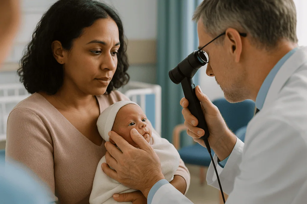
Feeding is often timed so the baby is not full right before the exam, which reduces the chance of spitting up during handling; the nurse will tell you if a feed is being shifted. Roughly an hour before the ophthalmologist arrives, the nurse instills dilating drops into each eye, sometimes in more than one round, and the baby rests while the pupils widen. Many units gently press on the inner corner of the eyes for a moment after the drops to limit how much drains into the nose and gets absorbed into the body.
When it is time, the baby is swaddled with arms contained, which most infants find calming, and placed where the examiner can reach the head easily. A numbing drop goes in. A small spring-loaded eyelid holder called a speculum keeps the lids open, because a baby cannot be asked to hold still. The ophthalmologist wears an indirect ophthalmoscope, a headband light with magnifying viewer, and holds a hand lens in front of the eye to see the retina. A slim, blunt instrument called a scleral depressor may gently press on the outside of the eye to bring the far periphery into view.
Each eye typically takes a few minutes. Throughout, a nurse watches the baby’s heart rate, breathing and oxygen saturation on the monitor and can pause the exam if the baby needs a break. The examiner then records what was seen using the zone and stage system described below and sets the date of the next check.
Some units use a wide-field digital retinal camera instead of, or alongside, the headset exam, with images read on site or remotely by an ophthalmologist. The AAP statement recognizes this as an acceptable screening approach when properly validated. Parents are usually welcome to be present; ask, and ask too whether you can hold the baby’s hand or offer a pacifier.
Pupil dilation for the retina check: how the drops work and what parents notice
The pupil is the opening in the colored iris, and in a baby it is small and quick to constrict in bright light. Looking through an undilated pupil at the retina is like looking into a room through a keyhole: you see the middle and miss the corners. Since ROP begins at the outer edge of the vascularized retina, the corners are exactly where the examiner needs to look.
Dilating drops are a class of eye medicines called mydriatics. Most regimens combine two actions. One ingredient temporarily relaxes the ring muscle that constricts the pupil; another stimulates the muscle that pulls it open. Together they produce a wide, fixed pupil for several hours. Neonatal units use formulations prepared for premature infants, because a tiny body absorbs a far greater proportion of any drop than an adult does, and the choice of preparation is a pharmacy and medical decision rather than something parents arrange.
What you may notice afterward is mostly harmless and short-lived. The pupils stay large, sometimes into the next day. The baby may squint at the lights and prefer dimmer surroundings. Some infants are sleepier or less interested in feeding for a few hours.
Less often, the medicine that reaches the bloodstream can nudge heart rate, blood pressure or gut activity, which is one reason the exam is done where monitoring is routine. Nurses are alert to a slowed heart rate, a pause in breathing, a drop in oxygen levels or a bloated, feed-intolerant tummy in the hours after dilation, and they adjust care accordingly. These effects are reported in the ophthalmology literature and are the reason the AAP guidance emphasizes careful monitoring during and after screening.
One practical point: if the pupils did not dilate well, perhaps because the baby’s dark irises are slow to respond, the examiner may ask for the drops to be repeated and return later. This is routine, not a sign that anything is wrong.
The retina check: zones, the ridge, and what the examiner is really looking for
Behind the headset, the ophthalmologist is answering three questions in a set order. How far have the vessels grown? Is there any abnormal growth at their leading edge? And do the vessels near the center look sick?
The first question is answered in zones, a map laid out in the International Classification of Retinopathy of Prematurity, now in its third edition (ICROP3), published in Ophthalmology and indexed on PubMed. Zone I is a circle at the back of the eye centered on the optic nerve, with a radius twice the distance from the nerve to the macula, the retina’s sharp-vision center. Zone II extends from the edge of zone I to the nasal edge of the retina. Zone III is the remaining crescent on the temporal side, farthest from the nose and the last to receive vessels. Disease in zone I is the most worrying because it means growth stalled very early, leaving a large area of retina without blood supply and a strong stimulus for abnormal vessels.
The second question concerns the boundary itself. In a healthy immature retina, the vessels fade gradually into the avascular area. In ROP, that boundary becomes a sharp line, then a raised ridge, then a ridge sprouting new vessels into the gel of the eye. The stage system, covered next, describes this progression.
The third question is about plus disease: dilated, twisting vessels around the optic nerve that signal very active disease. ICROP3 treats this as a continuum from normal through “pre-plus” to plus and provides reference images, because the judgment is partly visual. Plus disease is one of the strongest triggers for considering treatment.
ICROP3 also names aggressive ROP (A-ROP), a fast-moving pattern, usually in the most immature infants, in which flat abnormal vessels and plus disease develop without the orderly ridge, and which can advance within days. Recognizing this pattern quickly is a major reason the exams are performed by clinicians with specific training in ROP.
How retinopathy of prematurity stages are graded, from stage 1 to stage 5
Stages describe how far the abnormal process at the vessel boundary has gone. The language can sound alarming when written on a chart, so it helps to see the whole ladder at once. The definitions below follow ICROP3.
| Stage | What the examiner sees | What it usually means |
|---|---|---|
| Immature (no ROP) | Vessels fade gradually into unvascularized retina | Normal for the age; monitoring continues until vessels finish |
| Stage 1 | A thin, flat demarcation line between vascular and avascular retina | Mild; often resolves on its own |
| Stage 2 | The line has height and width, forming a ridge | Still mild to moderate; watched closely |
| Stage 3 | New abnormal vessels grow from the ridge into the vitreous gel | Treatment considered depending on zone and plus disease |
| Stage 4 | Partial retinal detachment (4A spares the macula; 4B involves it) | Surgical opinion typically sought |
| Stage 5 | Total retinal detachment | Most serious; surgery may be discussed, outcomes guarded |
| Plus disease | Dilated, tortuous vessels at the back of the eye, at any stage | Marker of active disease; a key factor in treatment decisions |
A complete record reads something like “zone II, stage 2, no plus, 2 clock hours.” Clock hours describe how much of the circumference is involved, as if the retina were a clock face. Zone, stage, plus status and extent together determine both urgency and follow-up interval.
Two things matter more than the stage number alone. The first is zone: stage 2 in zone I is more concerning than stage 3 in zone III. The second is plus disease, which signals that whatever stage is present is biologically active. This is why the influential Early Treatment for ROP trial defined “type 1” ROP, the category in which treatment is generally considered, by combinations of zone, stage and plus rather than by stage alone, a framework the AAP policy statement adopts.
What the result means in practice, and how often the exams are repeated
After each exam you are likely to hear one of a handful of phrases, and each carries an implied schedule. The AAP policy statement lays out typical follow-up intervals, which a unit may adapt to the individual baby.
“Immature, no ROP” is the most common early finding. If the vessels have reached zone II, a repeat exam in two to three weeks is typical; if they are still within zone I, the interval shortens to a week or less, because immature zone I retina carries the highest risk.
“Stage 1 or 2 in zone II” usually means review in one to two weeks. Many of these babies progress no further and their disease simply regresses as the vessels resume orderly growth.
“Stage 3 in zone II, no plus,” or any ROP in zone I, moves the interval to a week or less. “Type 1” findings, for example zone I disease with plus, or zone II stage 2 or 3 with plus, open the conversation about treatment; the AAP statement advises that treatment, when indicated, should generally be undertaken within about 72 hours of that determination.
The screening period ends when the examiner is satisfied the risk has passed. The AAP statement lists criteria that include full vascularization of the retina, vessels reaching zone III without prior zone I or II disease, reaching 45 weeks postmenstrual age without ever having had type 1 or prethreshold disease, or clear regression of ROP. Whichever applies, you should receive a plain statement that screening is complete and what longer-term eye follow-up is planned.
A note on honesty: the interval you are given is a plan, not a prophecy. ROP can speed up or stall, and the examiner may shorten or lengthen the gap after any exam. That flexibility is the system working as intended.
ROP exam side effects: does it hurt, and how is the baby kept comfortable?
Parents ask this more than any other question, and they deserve a straight answer. The exam is not painless. Studies measuring heart rate, oxygen levels, facial expression and crying show that premature infants experience stress and discomfort during the eyelid holder and scleral depression portions, even with numbing drops. The AAP policy statement acknowledges this and recommends measures to reduce it.
What those measures look like in practice: swaddling or “facilitated tucking,” where the baby’s limbs are held gently against the body, dampens the startle response. A pacifier gives the baby something to do with the discomfort. Many units offer a small amount of sweet-tasting solution on the tongue just before the exam, a well-studied non-drug comfort measure for brief procedures in newborns. Dimming the room, warming the hands, limiting the number of people and keeping the exam efficient all help. Some units involve parents directly, with a hand resting on the baby’s chest, and it is reasonable to ask whether that is possible.
Topical anesthetic drops numb the surface of the eye, which eases the eyelid holder, though they do not remove the sensation of pressure from the depressor. Any additional comfort medicine is a decision for the neonatal team, weighed against the baby’s breathing and feeding status.
Side effects afterward fall into two groups. The expected and minor: pink or slightly puffy eyelids for a day, large pupils, light sensitivity, extra sleepiness, a feed or two taken poorly. The less common but watched-for: a slowed heart rate, a pause in breathing, a drop in oxygen levels, or a swollen belly and vomiting in the day or so after dilation, effects linked both to handling and to absorption of the drops. In the unit these are monitored and managed. For a baby already home, the “When to call your doctor” section below covers what warrants a call.
Retinopathy of prematurity treatment: when it is considered and what the options are
Most babies screened for ROP are never treated, because their disease either never develops or regresses on its own. Treatment enters the conversation when the exam shows type 1 ROP, or A-ROP, the patterns in which the risk of progression to detachment is judged high enough to outweigh the risks of intervening. That judgment belongs to the ophthalmologist and neonatal team together, discussed with you.
Two main approaches exist for active disease, and both target the same problem: the avascular retina is sending out chemical distress signals, chiefly a protein called vascular endothelial growth factor (VEGF), which drives the abnormal vessel growth.
Laser photocoagulation applies hundreds of tiny burns to the peripheral avascular retina through the dilated pupil, under sedation or anesthesia arranged by the neonatal team. Destroying that tissue removes the source of the signal, and the abnormal vessels tend to regress over the following weeks. The trade-off is that the treated peripheral retina is permanently sacrificed, which can narrow side vision slightly and is associated with higher rates of nearsightedness later.
Anti-VEGF therapy involves a small injection into the gel of the eye of a medicine in the class that blocks VEGF directly. It preserves the peripheral retina and allows normal vessels to continue growing. The trade-offs are different: the disease can reactivate weeks or months later, so follow-up must be longer and more vigilant, and questions about the medicine’s effects on the rest of a developing body are still being studied. The National Eye Institute describes both laser and anti-VEGF injection as treatment options.
For stage 4 and 5, where the retina has begun to detach, surgery such as scleral buckling or vitrectomy may be discussed with a retinal surgeon. These are complex operations with guarded outcomes, and the evidence supports honest, individualized conversations rather than blanket promises.
No approach is “best” for every baby. Zone, stage, the baby’s overall health and the family’s ability to attend follow-up all shape the recommendation.
What the following weeks and months usually look like
For most families, ROP screening becomes a rhythm rather than an event. Exams land every one to three weeks, each one followed by a short conversation and a new date. If the baby goes home while screening is still underway, those dates move to an outpatient eye clinic, and the first appointment after discharge is the one families most often miss, simply because life at home with a premature baby is overwhelming. Treat that date as non-negotiable.
If treatment happens, the first exam afterward is typically within a week, then at intervals the ophthalmologist sets based on whether the abnormal vessels are regressing. After laser, regression is usually assessed over several weeks. After anti-VEGF injection, the AAP statement notes that follow-up needs to continue longer because late reactivation can occur, sometimes well after the baby would otherwise have graduated from screening.
Graduation from screening does not mean graduation from eye care. Babies who had ROP, and premature babies generally, have higher rates of nearsightedness, strabismus (misaligned eyes) and amblyopia (reduced vision in an eye that the brain has learned to ignore) through childhood, as the National Eye Institute and MedlinePlus both note. A plan for eye examinations in the first years of life, and for prompt treatment of glasses needs or alignment problems, is part of good follow-up.
The emotional timeline is real too. Research on parents of premature infants describes elevated rates of anxiety, depressive symptoms and post-traumatic stress during and after the neonatal stay; the Mayo Clinic’s overview of premature birth points to the strain on families as part of the picture. The weekly eye exam, with its wait and its verdict, can become a focus for that anxiety. Naming this to the team is not weakness. Many units have psychologists, social workers or parent peer programs precisely for this, and asking is a reasonable first step.
What people often get wrong about ROP and the eye exam
“They found something, so my baby will lose vision.” Most ROP found on screening is stage 1 or 2 and regresses on its own, as the National Eye Institute notes. A finding means monitoring, not a verdict.
“Oxygen caused it, so the unit did something wrong.” Oxygen is one of several contributors, and the relationship is complex: both too much and too little can be harmful, and modern units target saturation ranges to balance the lungs, the brain and the eyes. ROP occurs in carefully managed infants too, because immaturity itself is the main driver.
“If my baby’s eyes look normal, the exam is unnecessary.” ROP is invisible from the outside until it is advanced. The exam exists precisely because looking at the baby tells you nothing.
“The drops will hurt the baby’s eyes or heart.” Dilating drops are used in neonatal formulations with monitoring; transient effects on heart rate and digestion are watched for and are not the same as harm. The exam has been standard practice for decades.
“Laser means the baby will be blind in that part of the eye.” Laser treats the far periphery, where the retina contributes little to everyday vision. Some narrowing of side vision and a higher chance of needing glasses are the typical trade-offs, not blindness.
“Once screening is finished, the eyes are fine.” Premature babies carry a higher lifetime risk of nearsightedness, strabismus and amblyopia whether or not they had ROP, so childhood eye exams still matter.
“A missed appointment can be made up next month.” Active ROP can change within a week. If an appointment is missed, call the clinic the same day rather than waiting for the next routine slot.
“Bigger babies are automatically safe.” Screening cut-offs are generous for a reason, and larger infants with a stormy course are sometimes included. The decision rests with the neonatologist.
Questions to ask your care team about the ROP eye exam
Neonatal rounds move quickly, and the eye exam can be mentioned and gone before you have gathered your thoughts. Writing questions down beforehand changes that. The ones below are the ones experienced NICU parents say they wish they had asked earlier.
- Does my baby meet the screening criteria, and which criterion applies: birth weight, gestational age or clinical course?
- When is the first exam scheduled, and which clock, postmenstrual or chronological, set that date?
- Can I be present during the exam, and is there a comfort role I can take, such as holding a hand or offering a pacifier?
- What comfort measures does this unit use routinely, and are any additional options being considered for my baby?
- What did today’s exam show in terms of zone, stage, plus disease and clock hours, and can you write that down for me?
- What is the planned interval until the next exam, and what finding would shorten it?
- If my baby is discharged before screening ends, where will the next exam happen, who books it, and whom do I call if the appointment letter does not arrive?
- If treatment is ever recommended, what are the options being weighed, what are the trade-offs of each for a baby in this situation, and how quickly would it need to happen?
- How long after treatment would follow-up continue, and does that differ by treatment type?
- What eye follow-up is planned after screening finishes, and at what ages?
- What signs at home should prompt a call, and what number do I use out of hours?
- Is there a psychologist, social worker or parent support program attached to the unit?
Asking for the exam findings in writing deserves special emphasis. A line such as “zone II, stage 1, no plus, review in 2 weeks” on a card you keep is the single most useful record you can carry between units, clinics and clinicians, and it turns a stressful exchange into something you can reread calmly later.
When to call your doctor
While your baby is in the neonatal unit, the team watches for every effect of the exam and you do not need to monitor anything yourself. The red flags below apply once the baby is home, whether screening is still in progress or has finished.
Call emergency services immediately if your baby has a pause in breathing, turns blue or gray around the lips, goes limp and unresponsive, or has a seizure. These are emergencies regardless of any recent eye exam.
Call the clinic or your pediatrician the same day if, in the day or two after an outpatient exam, your baby is persistently refusing feeds, vomiting repeatedly, has a swollen or hard belly, is unusually floppy or hard to wake, or has eyes that remain markedly red, swollen or sticky with discharge beyond a day. A mild pinkness and large pupils are expected; worsening is not.
Call the eye clinic promptly, not at the next routine visit, if you have missed or are about to miss a scheduled ROP examination, if a promised appointment letter has not arrived, or if you were discharged without a clear next date. Active ROP can progress within a week, and the AAP statement stresses that the burden of tracking should not fall to families alone, but a phone call from you is the fastest fix when the system stumbles.
Book an eye assessment if, at any point in the first year, you notice a white or cloudy appearance in the pupil in photos or in light, one or both eyes that drift, cross or wobble consistently, an eye that looks larger or cloudier than the other, or a baby who does not seem to look at faces or follow a slowly moving object by about three months of corrected age. These signs are not specific to ROP, but all of them warrant a professional look.
Whatever you notice, the decision about what it means and what to do rests with the treating team. Your job is to report it clearly and early.
Frequently asked questions
When is ROP screening done for a premature baby?
The first exam is typically at 31 weeks postmenstrual age or four weeks after birth, whichever is later, according to the American Academy of Pediatrics screening statement. Exams then repeat every one to three weeks depending on findings, continuing until the retina has fully vascularized, the disease has clearly regressed, or the baby passes a defined postmenstrual age without high-risk findings. The ophthalmologist sets each interval.
What determines whether a baby is premature?
The World Health Organization defines a premature, or preterm, baby as one born before 37 completed weeks of pregnancy. Within that, extremely preterm means before 28 weeks, very preterm 28 to under 32 weeks, and moderate to late preterm 32 to under 37 weeks. ROP screening focuses on the earliest and smallest babies, because retinal blood vessels are least developed in that group.
Is the ROP eye exam painful for the baby?
The exam causes measurable discomfort and stress, especially during the eyelid holder and scleral depression steps, even with numbing drops. Units reduce this with swaddling, a pacifier, a sweet-tasting solution on the tongue, dim lighting and an efficient technique. A nurse monitors heart rate and breathing throughout and can pause the exam. Any additional comfort medicine is a decision for the neonatal team.
Does ROP go away on its own?
Often, yes. The National Eye Institute notes that most retinopathy of prematurity is mild and resolves without treatment as normal vessels resume orderly growth. Stage 1 and 2 disease in zone II frequently regresses. A minority of babies develop higher-risk patterns, defined by zone, stage and plus disease, for which treatment is considered. Regular exams exist to tell these two courses apart in time.
Will a baby who had ROP need glasses later?
Not necessarily, but the chance is higher. Premature babies, and particularly those who had ROP or received laser treatment, have elevated rates of nearsightedness, misaligned eyes (strabismus) and reduced vision in one eye (amblyopia) through childhood, as MedlinePlus and the National Eye Institute describe. Planned eye examinations in the early years allow glasses or alignment treatment to be started early when needed.
Can parents stay with the baby during the ROP exam?
In many units, yes, and some actively encourage it. Ask the nurse in advance whether you can be present and whether you can take a comfort role such as resting a hand on the baby’s chest or offering a pacifier. Practices vary with unit layout and the baby’s stability, so the team decides on the day. If you would rather not watch, that is equally reasonable.
What are the psychological effects of premature birth on parents?
Studies consistently describe higher rates of anxiety, depressive symptoms and post-traumatic stress among parents of premature infants, both during the neonatal stay and in the months after discharge. Repeated screening tests, including the weekly eye exam, can become a focus for that worry. Neonatal units commonly have psychologists, social workers or peer-support programs, and telling the team how you are coping is a reasonable and welcomed step.
What support groups exist for families of premature babies?
Most neonatal units can connect families with a parent peer program, a social worker or a hospital-based support group, and many national prematurity charities run online communities and helplines. Your unit’s family liaison or social worker is the best starting point, because they know which local and national resources are active. Peer support from parents who have been through ROP screening can be particularly steadying.
What happens if we miss an ROP appointment after discharge?
Call the eye clinic the same day rather than waiting for the next routine slot. Active ROP can progress within a week, so a missed exam needs to be rebooked promptly, not next month. The AAP screening statement stresses that discharge paperwork should state the next exam date and that units should have a tracking system; your call is the quickest way to catch a gap if a letter never arrives.
How do you keep a premature baby warm at home?
Premature babies lose heat quickly, so keep the room comfortably warm without overheating, dress the baby in one more layer than you would wear, use a hat in cool conditions, and avoid drafts and bare skin on cold surfaces. Skin-to-skin contact is an effective warming method the World Health Organization recommends for small babies. Your discharge team will advise on checking temperature and when a reading warrants a call.
References
- MedlinePlus: Retinopathy of prematurity
- National Eye Institute (NIH): Retinopathy of Prematurity
- World Health Organization: Preterm birth fact sheet
This article is for general information only and is not a substitute for professional medical advice. Please consult a qualified doctor about your individual situation.

More from the Blog

Braces, Splints and Seating in Pediatric Physical Therapy: Checking Fit as Your Child Grows
A child braces and splints fit check is a planned review of how an orthosis, splint or seating system sits on a growing body.…

What Happens in a Sensory Integration Therapy Session? Swings, Textures and Guided Play
A sensory integration therapy session is a structured, play-based occupational therapy visit, typically lasting about an hour, in which a trained therapist guides a…

Phototherapy for Newborn Jaundice: What Happens Under the Lights and How Feeding Continues
Phototherapy treats newborn jaundice by shining blue-green light onto the baby's skin. The light changes bilirubin, the yellow pigment that builds up when an…

Living With Febrile Seizures: Household Planning, Caregiver Briefings and Calm Fever Routines
Parents cope best with febrile seizures by preparing before the next fever: a one-page plan every caregiver can follow, a calm first-aid routine (time…

When Can a Child Bathe, Play and Return to School After Pediatric Surgery? Recovery Milestones
Most children can shower or bathe once the surgical team says the wound may get wet, often within one to two days of a…

Why Are Repeat Echocardiograms Scheduled After Kawasaki Disease and What Do They Track?
Repeat echocardiograms after Kawasaki disease track the coronary arteries, the vessels that supply the heart muscle, because inflammation can stretch them into aneurysms during…