

Transthoracic vs Transesophageal Echocardiogram: When the Probe Approach Is Chosen
Key Takeaways
- TTE images the heart through the chest wall and takes 15 to 60 minutes with no sedation or fasting, according to the NHS.
- TEE places the ultrasound probe in the esophagus, only millimeters from the left atrium, which is why it sees the left atrial appendage and mitral valve so clearly.
- TEE is usually ordered to find atrial clots before cardioversion, to look for valve infection, to assess prosthetic valves, or to search for a source of stroke such as a patent foramen ovale.
- Neither test images the coronary arteries directly; both can only show wall-motion changes that hint at a blockage.
- Outside surgery, TEE is done under moderate sedation, so most people are drowsy but breathing on their own and remember little.
- After TEE you cannot eat until throat numbness fades, and should not drive for 24 hours because of the sedative.
A transthoracic echocardiogram (TTE) images the heart with an ultrasound wand on the chest and is the standard first test. A transesophageal echocardiogram (TEE) passes a thin probe down the esophagus, placing it directly behind the heart for sharper views of the valves, the left atrial appendage and the aorta. Doctors usually choose TEE when TTE images are unclear or a specific question, such as a clot or a valve infection, needs closer detail.
The letter arrives on a Tuesday. Last month’s heart ultrasound, the one with the cold gel and the dimmed room, was apparently “technically limited,” and now the cardiologist wants a second scan, this time with a probe that goes down the throat under sedation. The first question most people ask is not about the heart at all. It is: why wasn’t the first test enough?
That question sits at the center of the transthoracic vs transesophageal echocardiogram decision. Both tests use the same physics, ordinary ultrasound, and both are read by the same kind of specialist. What differs is the vantage point. One looks at the heart through the ribs and lungs; the other looks at it from a few millimeters away, with nothing in between.
Understanding that single difference explains almost every choice your care team makes about which probe to use, when, and for whom.
What happens during a transthoracic echocardiogram?
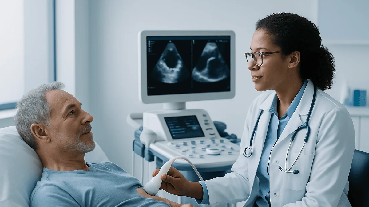
A transthoracic echocardiogram, usually shortened to TTE, is the heart ultrasound most people picture: a handheld transducer (a small device that sends and receives sound waves) pressed against the skin of the chest. The sound waves bounce off heart tissue and blood, and a computer turns the returning echoes into a moving picture in real time.
You lie on your left side on an examination couch, often with your left arm raised over your head, which spreads the ribs slightly and opens up an acoustic window. A few sticky electrodes on the chest record the heart rhythm alongside the images. The sonographer applies gel, then moves the probe between the ribs, under the breastbone and up toward the neck to capture the heart from several angles. Occasionally you will be asked to hold your breath or roll further onto your side, because lung tissue full of air scatters ultrasound and blurs the view.
The scan also measures blood flow using Doppler, a technique that detects the change in sound frequency as blood moves toward or away from the probe. That is where the whooshing noise comes from. Doppler tells the reader how fast blood is crossing a valve, whether it leaks backward, and roughly what pressure the right side of the heart is working against.
According to the NHS, a standard echocardiogram usually takes between 15 minutes and an hour, needs no sedation and no fasting, and you can go home and eat as normal afterward. There are no needles unless a contrast agent is added to sharpen the image of the heart’s inner lining, which the sonographer will explain beforehand. For most people this is the first, and often the only, echocardiogram they will ever need.
What is a transesophageal echocardiogram, and what actually happens?
A transesophageal echocardiogram, or TEE, uses the same ultrasound principle but moves the transducer to the tip of a flexible tube roughly the width of a finger. That tube is passed through the mouth and down the esophagus, the muscular pipe that carries food from the throat to the stomach. The esophagus runs directly behind the heart, separated from the left atrium by only a thin layer of tissue. From there the probe sees the back of the heart without ribs, lungs or chest fat in the way.
The process has a rhythm to it. First, the team places an intravenous line and monitors: blood pressure cuff, oxygen sensor, heart rhythm leads. A local anesthetic spray numbs the throat, and a sedative given through the line makes you drowsy and relaxed. A soft bite guard protects both your teeth and the probe. Lying on your left side, you are asked to swallow as the tip is guided into the esophagus, much as during an upper endoscopy. Once in place, the operator rotates and tilts the tip to sweep across the valves, the atria and the aorta.
MedlinePlus describes the whole examination taking around 30 to 60 minutes, and much of that is preparation and monitoring rather than time with the probe in place. Afterward you rest until the sedation fades and the throat numbness wears off, and someone drives you home.
Two clinical settings deserve a mention. TEE is often used in the operating room during heart surgery to check a valve repair before the chest is closed, and in the catheter lab to guide procedures inside the heart. In those cases you are already under general anesthesia and will not remember it.
Difference between TTE and TEE at a glance
People searching for the difference between TTE and TEE usually want one honest comparison rather than two separate brochures. The table below sets the tests side by side on the points that shape the decision in practice.
| Feature | Transthoracic (TTE) | Transesophageal (TEE) |
|---|---|---|
| Probe position | On the chest wall, outside the body | Inside the esophagus, directly behind the heart |
| Sedation | None | Throat numbing plus intravenous sedation; general anesthesia in surgery |
| Fasting | Not required | Required, typically for several hours beforehand |
| Image quality | Good for most people; limited by lungs, ribs, obesity, prior chest surgery | High resolution, especially of the back of the heart |
| Best for | Pumping function, chamber size, most valve questions, screening | Small clots, valve infection, prosthetic valves, holes between atria, aortic tears |
| Main risks | Essentially none from the scan itself | Sore throat common; esophageal injury rare; sedation effects |
| Recovery | Immediate | Rest until sedation clears; no driving that day |
| Where it happens | Clinic or bedside | Procedure room, operating room or catheter lab |
Two things stand out. The image advantage of TEE is real, but it is targeted: it matters most for structures at the back of the heart and for detail below what TTE can resolve. Meanwhile, TTE remains the better tool for the day-to-day questions, such as how strongly the ventricle pumps, because it can image from multiple chest windows and the reader can integrate them.
Neither test is a substitute for the other. In many cardiology reports the two appear together, the TTE giving the overview and the TEE answering the one question that overview could not settle.
Why would a cardiologist order a transesophageal echocardiogram?
Cardiologists reach for TEE when the clinical question is more precise than a chest scan can answer. Mayo Clinic, Cleveland Clinic and Johns Hopkins list a consistent set of reasons, and each maps onto a structure that sits close to the esophagus or requires fine detail.
The most common is the search for a blood clot in the left atrial appendage, a small pouch off the top-left chamber of the heart where blood can pool and clot in people with atrial fibrillation, an irregular heart rhythm. The appendage lies at the back of the heart, almost invisible to TTE but clearly seen by TEE. Before cardioversion, a controlled electrical or medication-based reset of the rhythm, a clot found there changes the plan entirely, because shocking the heart back into rhythm could dislodge it.
Suspected infective endocarditis is another. This is an infection of the heart valves or the inner lining, and it produces small clumps of bacteria and clot called vegetations. TTE can miss small ones; TEE resolves detail down to a few millimeters and can spot abscesses beside the valve.
Other frequent reasons include:
- Assessing a leaking or narrowed valve when TTE images are inconclusive, particularly the mitral valve or a prosthetic (replacement) valve, where the artificial material casts shadows on chest imaging.
- Looking for a source of stroke, including a patent foramen ovale, a small flap between the atria that fails to seal after birth in a substantial minority of adults.
- Examining the thoracic aorta for a tear (dissection) or an enlarging aneurysm.
- Guiding procedures such as valve repair, closure devices or appendage occlusion in real time.
In each case, the ordering doctor is weighing a specific benefit, a clearer answer to one question, against the small but real burden of sedation and an invasive probe.
Why is a TEE better than a TTE? Sharper, not superior
Searches for “why is a TEE better than TTE” carry a hidden assumption, and it is worth correcting early. TEE is not a better echocardiogram. It is a closer one, and closeness helps with some questions and not others.
The physics explains it. Ultrasound loses energy as it travels through tissue, and higher frequencies, which give finer detail, fade fastest. A chest probe has to work through skin, fat, muscle, rib and sometimes lung to reach a heart that may be 10 centimeters away, so it uses lower frequencies and accepts a softer picture. An esophageal probe sits a few millimeters from the left atrium, can use higher frequencies, and gets crisp images of the mitral valve, the atrial septum and the appendage as a result.
Turn that around, though. The structures at the front of the heart, including the tip of the left ventricle and parts of the right ventricle, are farther from the esophagus and can be harder to see with TEE. A TEE probe also cannot be freely repositioned across the whole chest the way a handheld transducer can. For measuring how much blood the left ventricle ejects with each beat, one of the most important numbers in cardiology, TTE is the standard tool and TEE adds little.
Then there is the cost that has nothing to do with money: fasting, an intravenous line, sedation, a sore throat and a lost day. Cardiologists accept that trade when the added clarity will change management. When it will not, they do not.
Better questions to ask are: “What does my doctor need to see that the chest scan could not show?” and “Would the answer change what happens next?” If both answers are clear, the choice of probe usually makes sense.
Who is usually offered a TTE first, and who is asked to wait for a TEE?
In almost every pathway, TTE comes first. It is quick, harmless and answers most questions, so guidelines from the major cardiology societies treat it as the default whenever a heart ultrasound is needed: a new murmur, breathlessness, swelling in the legs, an abnormal ECG, follow-up of a known valve problem, or checking the heart before certain cancer treatments.
People are typically moved on to TEE in three situations. First, when the TTE was technically limited. Chest wall shape, lung disease such as emphysema, obesity, breast implants, bandages after surgery or a person who cannot lie on their side for long can all leave the sonographer with windows too poor to answer the question. Second, when the TTE was fine but raised a question it cannot settle, for instance a suspicious shadow near a valve. Third, when the question was always a TEE question, such as clearing the appendage before cardioversion or mapping the mitral valve before repair.
Some people are asked to wait, or offered an alternative, because the esophageal route is not safe for them. Cleveland Clinic and Mayo Clinic describe caution or avoidance when there is a known esophageal stricture (narrowing), esophageal varices (swollen veins from liver disease), recent esophageal or stomach surgery, active bleeding in the upper gut, a tumor in the throat or esophagus, or neck instability that prevents safe positioning. Difficulty swallowing that has not been investigated is often checked first. Severe breathing problems or unstable blood pressure may also postpone a sedated procedure until the person is steadier.
Where TEE is off the table, the team may turn to cardiac CT, cardiac MRI, or a contrast-enhanced TTE, each with its own strengths. Which path fits you is a decision for the treating team, informed by your history.
Can a transthoracic echocardiogram show blocked arteries?
Not directly, and this is one of the most common misunderstandings about heart ultrasound. The coronary arteries, the vessels that feed the heart muscle, are only a few millimeters wide and run along the outer surface of the heart. Standard echocardiography, whether from the chest or the esophagus, does not image their insides well enough to see plaque or measure a narrowing. The American Heart Association is explicit that an echocardiogram evaluates the heart’s structure and function rather than the arteries themselves.
What TTE can do is show the consequences of a blockage. Heart muscle that has lost its blood supply stops contracting normally, so a region of the wall that moves weakly or not at all points toward a previous heart attack or ongoing lack of blood flow. That is why an echo is often performed during or shortly after a suspected heart attack. A stress echocardiogram takes this further: images are captured at rest and again straight after exercise or a medication that makes the heart work harder, and a segment that moves well at rest but poorly under stress suggests an artery that cannot keep up with demand.
Tests that actually visualize the arteries are different tools:
- Coronary CT angiography, a CT scan with contrast dye that shows plaque and narrowing.
- Invasive coronary angiography, a catheter procedure with X-ray contrast, which also allows treatment in the same sitting.
- Nuclear perfusion scans, which map blood flow to the muscle rather than the vessels.
So if you have had a normal TTE, that is reassuring about pumping strength and valves, but it does not on its own rule out coronary disease. Conversely, an abnormal wall motion finding usually prompts one of the artery-specific tests above. The echo raises the flag; other imaging confirms the cause.
Are you awake during a transesophageal echocardiogram? Sedation explained
Usually you are somewhere in between: not asleep, not fully alert. Outside the operating room, TEE is performed under what clinicians call conscious or moderate sedation. A sedative from the benzodiazepine class, sometimes combined with a short-acting pain reliever, is given through the intravenous line. You breathe on your own, can respond to instructions such as “swallow now,” and may drift in and out. Most people remember little or nothing of the probe itself. Mayo Clinic and Johns Hopkins both describe this state, and it differs from general anesthesia, where a breathing tube is placed and you are fully unconscious.
Before the sedative, the back of the throat is sprayed or gargled with a local anesthetic to suppress the gag reflex. The taste is bitter and the numbness feels strange, as if the throat has gone to sleep. That numbness is why you cannot eat or drink until it has worn off afterward.
Comfort during the procedure comes down to several small things:
- Positioning on the left side with a pillow, which helps the probe pass and lets saliva drain rather than pool.
- A suction device for saliva, since swallowing is discouraged while the probe is in place.
- Continuous monitoring of oxygen and blood pressure, with supplemental oxygen via a nasal tube if levels dip.
- An operator who talks through each step, even when you appear to be dozing.
Some people do choose, or are offered, a deeper level of sedation supported by an anesthesia specialist, especially if they have a strong gag reflex, severe anxiety or a previous difficult endoscopy. Tell the team about any earlier sedation experiences, sleep apnea, or reactions to anesthetics. How deep the sedation goes is decided by the clinicians on the day, based on your history and how you respond.
How to prepare: eating, medicines and what to bring
Preparation is where the two tests part ways most obviously. For a TTE there is essentially nothing to do. Wear a top that is easy to remove, avoid heavy lotion on the chest, and expect to be there for an hour or less. The NHS notes that you can eat, drink and take your usual medicines beforehand.
TEE requires more planning, because sedation and a probe in the esophagus carry an aspiration risk, meaning stomach contents could be inhaled if you vomit while drowsy. MedlinePlus advises no food or drink for about six hours before the test; your team may set a specific cutoff and may allow small sips of water earlier in that window. Follow their instruction rather than a general figure.
On medicines, the rule is simple: do not change anything without asking. Most people are told to take their usual morning tablets with a sip of water. People on anticoagulants (blood thinners) or diabetes medicines get specific instructions, because fasting affects blood sugar and some teams have preferences around bleeding risk. Whatever you are told, the decision belongs to the prescribing clinician, not to a leaflet or a website.
Practical points that make the day easier:
- Arrange a responsible adult to take you home; after sedation you will not be allowed to drive, and many centers will not start the procedure without a confirmed escort.
- Remove dentures, partial plates and any tongue or lip piercings before the probe goes in.
- Bring a list of medicines and allergies, and mention any swallowing difficulty, hiatal hernia, liver disease or prior throat surgery, even if it seems minor.
- Tell the team if you are or might be pregnant. Ultrasound itself is considered safe in pregnancy, but sedation choices differ.
Plan for the rest of the day to be quiet. Sedatives linger longer than people expect.
Transesophageal echo risks compared with TTE
A TTE carries no meaningful physical risk. The gel is cold, the probe can press uncomfortably between the ribs, and if a contrast agent is used there is a small chance of a reaction, which staff monitor for. That is essentially the full list.
TEE is different because it is invasive and sedated, and honest counseling names both categories. Cleveland Clinic and Mayo Clinic describe the expected minor effects as a sore throat, hoarseness or a scratchy feeling for a day or so, mild nausea, and grogginess from the sedative. A slightly bruised feeling at the intravenous site is common.
Uncommon but more serious events include:
- Injury to the esophagus, ranging from a small scrape to, very rarely, a tear or perforation. Major sources consistently describe this as rare, and the pre-procedure screening questions about swallowing, varices and prior surgery exist precisely to lower the odds further.
- Breathing problems during sedation, including low oxygen levels, which is why oxygen and continuous monitoring are standard.
- Abnormal heart rhythms triggered by the probe or the sedation, usually brief.
- Bleeding, more likely in people with esophageal varices or on anticoagulants.
- Dental damage, reduced by the bite guard and by removing dentures.
- Allergic reaction to the sedative or the numbing spray.
Context matters. The reason TEE remains widely used despite these risks is that the alternatives, such as proceeding to cardioversion without checking for a clot, or missing an infected valve, carry their own dangers. Your cardiologist is expected to weigh that balance for your specific situation, and a good consent conversation makes that weighing visible rather than hiding it behind the word “routine.”
If a risk factor applies to you, ask what the team does about it. The answer might be a different sedation plan, an anesthetist in the room, an endoscopy first, or a different test altogether.
What the hours and days after each test usually look like
After a TTE, nothing changes. You wipe off the gel, get dressed and carry on with your day. Results are reviewed by a cardiologist and typically reach the referring doctor within days to a couple of weeks, depending on the service; sometimes the sonographer or a doctor gives an initial impression on the spot, but the formal report is what counts.
The TEE timeline has three phases. In the first hour or two you rest in a recovery bay while the sedative wears off. Staff check blood pressure and oxygen and watch for any sign of trouble. MedlinePlus advises not eating or drinking until the throat numbness has gone, because swallowing reflexes are unreliable until then; a sip of water once feeling returns is the usual test.
For the rest of the day, expect to feel foggy. Johns Hopkins and Cleveland Clinic both advise against driving, operating machinery, drinking alcohol or signing important documents for 24 hours after sedation, and against being alone if you live by yourself. A soft diet, cool drinks and throat lozenges help the scratchy feeling. Many people nap.
Over the next day or two the sore throat and hoarseness settle. A small amount of blood-streaked saliva on the first day can occur from the probe brushing the throat and generally stops on its own.
Results follow the same route as a TTE, although TEE performed to answer an urgent question, such as clearing a clot before cardioversion, is often reported the same day because the next step depends on it. Ask before you leave how and when you will hear the result, and who will explain it. The imaging is only half the test; the conversation about what it means is the other half.
What people often get wrong about TTE and TEE
Myths gather around any test that involves a tube, so it helps to set a few straight.
“A TEE means my heart is in serious trouble.” Not necessarily. TEE is frequently ordered because the first scan was technically difficult, not because it showed something alarming. It is also a routine safety step before some rhythm procedures in people who feel entirely well.
“The probe goes into the lungs or the heart.” It stays in the esophagus and, at most, the upper stomach. It never enters the airway or the heart. The heart is imaged through the esophageal wall.
“Ultrasound uses radiation.” It does not. Both TTE and TEE use sound waves, with no X-rays involved. That is one reason echocardiography is repeated freely and used in pregnancy.
“A normal echo means my arteries are clear.” As covered earlier, neither test images the coronary arteries directly. A normal echo is reassuring about structure and pumping, not about plaque.
“I’ll be fully unconscious.” Outside surgery, most TEEs use moderate sedation. You may respond to instructions and later remember snatches of it, or nothing at all. Both are normal.
“TEE is always more accurate, so I should ask for it instead.” It is more detailed for the back of the heart and less useful for parts of the front. Choosing it when a TTE would answer the question adds sedation, fasting and risk for no gain.
“A sore throat afterward means something was damaged.” Mild soreness for a day or two is expected friction. What is not expected is severe or worsening pain, fever, or trouble swallowing, which are covered in the red-flag section below.
“I can drive myself home if I feel fine.” Sedatives impair judgment and reaction time long after you feel alert. The no-driving rule applies to how the medication behaves, not to how you feel.
Questions to ask your care team
A short, focused list gets better answers than a long anxious one. These questions are framed to surface the reasoning behind the test rather than just the logistics, and any of them is fair to ask the person who ordered the scan or the team performing it.
- What specific question is this test meant to answer, and why could the chest scan not answer it?
- Would the result change my treatment, and what would change if it were abnormal versus normal?
- Is there an alternative, such as cardiac CT or MRI, that could give the same information for me, and why was this one chosen?
- What level of sedation do you plan, who will give it, and how will I be monitored?
- Given my history, do any of the esophageal risk factors apply to me, and what are you doing about them?
- Which of my medicines should I take on the morning of the test, and which should I hold? Please tell me specifically rather than in general terms.
- How long should I fast, and does that include water?
- Who will explain the result to me, how soon, and will the report be shared with my other doctors?
- If a clot or infection is found, what happens next, and how quickly?
- What symptoms after the test should make me call, and what number do I call, including out of hours?
Write the answers down or bring someone to listen. After sedation, memory of the day is patchy, so anything discussed in recovery should also be given to you in writing or repeated to your companion.
One further question is worth asking yourself: do I understand why this probe, at this time? If the answer is no, say so. Good teams expect that question and would rather answer it beforehand than have you leave with the tube still a mystery.
When to call your doctor
After a transthoracic echocardiogram there is nothing to watch for from the test itself. If contrast was used and you develop a rash, wheeze, facial swelling or feel faint within the following hours, contact the service or seek urgent care, since delayed reactions, though uncommon, can occur.
After a transesophageal echocardiogram, mild throat soreness, hoarseness and tiredness are expected for a day or so. The following are not expected, and Cleveland Clinic, Mayo Clinic and MedlinePlus list them as reasons to seek medical attention without waiting:
- Severe, persistent or worsening pain in the throat, chest, neck or upper abdomen, especially pain on swallowing or pain that spreads to the back.
- Difficulty swallowing that is getting worse rather than better, or a feeling that food is sticking.
- Vomiting blood, coughing up blood, or passing black, tarry stools.
- Fever, chills or sweats in the days after the procedure.
- Shortness of breath, noisy breathing, or a sensation that air has collected under the skin of the neck or chest (a crackling feel).
- Fast, pounding or irregular heartbeat that does not settle, or fainting.
- Confusion or drowsiness that persists well beyond the day of sedation.
Severe chest pain, breathing difficulty, or vomiting blood are emergencies. Call emergency services rather than driving to a clinic.
For the underlying heart question, the test does not replace your existing safety net. Sudden weakness on one side of the body, facial drooping or slurred speech can signal a stroke and need emergency care regardless of any recent scan. New or worsening breathlessness, chest pressure, or swelling of the legs should be reported to the team managing your heart.
Every decision about repeating imaging, changing medication or scheduling a procedure sits with your treating clinicians, who have your full history and the actual images in front of them.
Frequently asked questions
Why is a TEE better than a TTE?
It is not better overall; it is closer. The esophageal probe sits directly behind the heart with no ribs or lung in the way, so it produces sharper images of the mitral valve, the atrial septum, the left atrial appendage and the aorta. For measuring how strongly the left ventricle pumps, or for a general first look, a transthoracic scan remains the standard and avoids sedation entirely.
Why would a cardiologist order a transesophageal echocardiogram?
Most often to answer one question the chest scan could not settle: whether there is a clot in the left atrial appendage before cardioversion, whether a valve is infected, how a replacement valve is working, or whether a small hole between the atria could explain a stroke. It is also used to examine the aorta and to guide procedures inside the heart.
Are you awake during a transesophageal echocardiogram?
Usually partly. Outside the operating room, TEE is done with a numbing throat spray and intravenous moderate sedation, so you are drowsy, breathing on your own and able to respond to simple instructions, but most people remember little. During heart surgery, TEE is performed under general anesthesia and you will not be aware of it at all.
Can a transthoracic echocardiogram show blocked arteries?
Not directly. The coronary arteries are too small and run along the heart’s surface, so ultrasound does not image the plaque inside them. A TTE can show a section of heart wall moving weakly, which suggests reduced blood supply, and a stress echo sharpens that clue. Seeing the arteries themselves needs CT angiography or invasive angiography.
What is the difference between TTE and TEE in terms of preparation?
A TTE needs no preparation: you can eat, drink and take your usual medicines. A TEE requires fasting for several hours because of sedation, an intravenous line, removal of dentures, and a companion to take you home. Your team will tell you which medicines to take on the morning; never change them on your own.
How long does a TEE take, and how long until I feel normal?
MedlinePlus describes the examination as roughly 30 to 60 minutes, much of it setup and monitoring, with the probe in place for a shorter period. Recovery in the unit usually lasts an hour or two until sedation clears and throat numbness fades. Grogginess can persist for the rest of the day, and a sore throat typically settles within a day or two.
What are the main transesophageal echo risks?
Common effects are a sore throat, hoarseness, mild nausea and sleepiness from the sedative. Uncommon but more serious risks include injury to the esophagus, which major sources describe as rare, low oxygen during sedation, brief heart rhythm disturbances, bleeding in people with esophageal varices, and dental damage. Screening questions before the test exist to reduce these risks.
Why do I need a TEE before cardioversion if I feel fine?
Because atrial fibrillation lets blood pool in the left atrial appendage, where a clot can form silently. Restoring normal rhythm could dislodge such a clot and cause a stroke. TEE is the test that sees the appendage clearly, so it is used as a safety check when the duration of the rhythm or the anticoagulation history leaves uncertainty. Feeling well does not rule out a clot.
Is a TTE ever done with an injection?
Sometimes. If the edges of the heart’s inner lining are hard to see, a contrast agent made of tiny gas-filled microbubbles can be injected into a vein to brighten the blood pool and sharpen the picture. This is still a transthoracic scan, needs no sedation, and reactions are uncommon. Staff will explain and watch for any reaction if it is used.
Who should not have a transesophageal echocardiogram?
People with a known esophageal narrowing, swollen esophageal veins from liver disease, recent surgery on the esophagus or stomach, active upper gut bleeding, or a tumor in the throat or esophagus are generally steered away from TEE. Unexplained swallowing difficulty is usually investigated first. In these cases cardiac CT, MRI or contrast TTE may be offered instead, decided by the treating team.
This article is for general information only and is not a substitute for professional medical advice. Please consult a qualified doctor about your individual situation.

More from the Blog

Hypertension and Your Other Medicines: What the Doctor Reviews Before Adjusting Treatment
Before adjusting blood pressure treatment, doctors review every medicine and supplement a person takes, because several common ones can raise blood pressure or blunt…

Palpitations After Eating: Why It Happens, Common Triggers and How to Stop Them
Palpitations after eating are usually harmless and often settle within minutes. To calm them, sit upright, breathe slowly with a longer exhale than inhale,…

Reading Your Heart Failure Treatment Plan: Questions to Ask at Every Cardiology Visit
The most useful questions to ask about heart failure treatment cover five areas: what your ejection fraction and heart failure type are, which medicine…

Living With Angina: Exercise, Travel, Work and the Habits Cardiologists Ask You to Keep
Most people with stable angina can work, exercise, travel and enjoy a full life once the condition is treated and their triggers are understood.…

Why Are Hypertension Targets and Medicines Adjusted for Older Adults?
Blood pressure targets and medicines are adjusted for older adults because arteries stiffen with age, the body's reflexes that steady pressure on standing weaken,…

Endocarditis Recovery Week by Week: Finishing Antibiotics and Regaining Your Energy
Endocarditis recovery time usually spans several weeks to a few months. Most people receive intravenous antibiotics for roughly two to six weeks, often starting…