

What a Panoramic X-Ray or 3D Scan Shows About an Impacted Tooth: Roots, Nerves and Angle
Key Takeaways
- A panoramic X-ray captures both jaws, all teeth, the sinuses and the lower-jaw nerve canals in one image, but flattens a curved, three-dimensional jaw into two dimensions, so it cannot show whether a structure sits toward the cheek or the tongue.
- What looks like a nerve on the film is the bony inferior alveolar canal; nerves themselves, including the lingual nerve beside the tongue, are invisible on every kind of X-ray.
- Clinicians read a wisdom tooth's tilt as vertical, mesioangular, horizontal or distoangular, and its depth relative to the second molar, because those readings predict how much bone and sectioning a removal would involve.
- Signs such as a dark band across the root, loss of the canal's white line or a kink in the canal raise the likelihood of root-to-nerve contact and are the usual trigger for adding a cone-beam CT scan.
- The NHS states that wisdom teeth are usually removed only when they are causing problems, and Mayo Clinic acknowledges genuine disagreement about removing symptom-free teeth, so monitoring is a recognized option.
- If a tooth is removed, the NHS describes recovery as taking up to two weeks, and Mayo Clinic notes that any nerve-related numbness is usually temporary but can rarely persist.
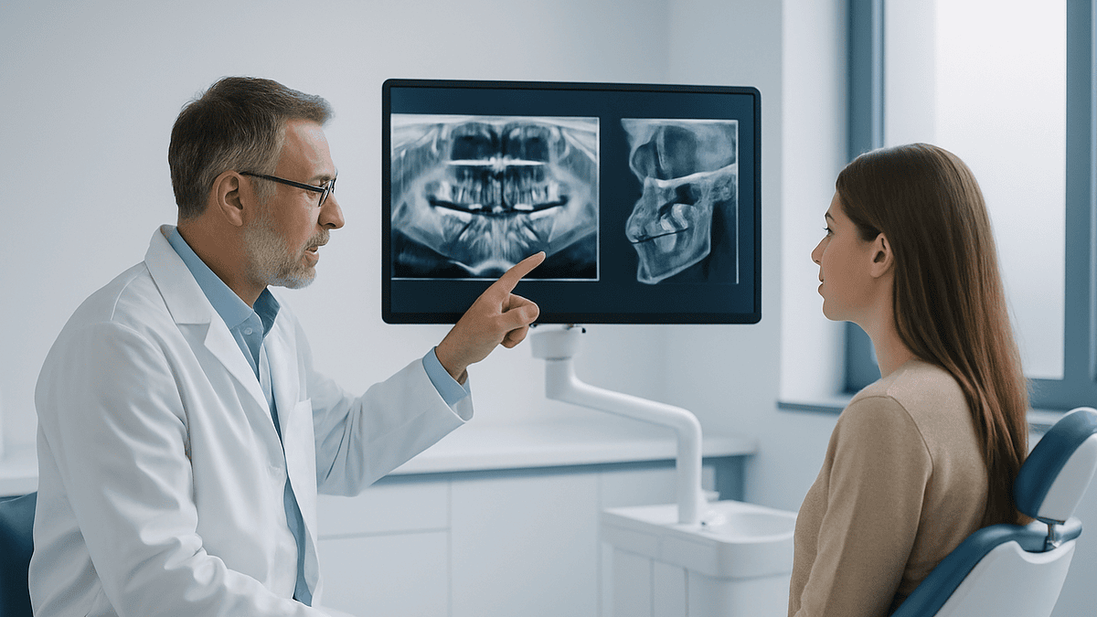
A panoramic X-ray of an impacted tooth shows, in one flat image, how many roots the tooth has and how they curve, how deeply it sits in the jaw, which way it tilts, and how close it lies to the canal carrying the nerve in the lower jaw or to the sinus above the upper teeth. Because the image is two-dimensional, a 3D cone-beam scan is sometimes added when those relationships look tight or unclear.
You bite gently on a small plastic tab, rest your forehead against a bar, and a machine glides in a slow half-circle around your head while a voice says, “Don’t swallow.” A minute later a wide, curved picture of your whole mouth appears on the screen. Somewhere at the back, lying almost on its side under a shelf of bone, is a tooth you have never seen and can barely feel.
That picture is a panoramic X-ray, and when the subject is an impacted tooth it is usually the first real evidence anyone has about what is going on. The dentist traces a fingertip along a faint white line and mentions “the nerve.” Suddenly the questions crowd in: Is it touching? Does it have to come out? Why is the image blurry at the edges? And why is a second, three-dimensional scan sometimes suggested?
This explainer walks through what that gray-and-white image can and cannot tell you, in the order a clinician actually reads it.
How a panoramic X-ray of an impacted tooth is taken, and what "impacted" means
An impacted tooth is one that cannot fully push through the gum because something blocks its path: bone, gum tissue, or a neighboring tooth. MedlinePlus lists wisdom teeth as the most common example, with the upper canines next, and notes that many impacted teeth cause no symptoms at all.
A panoramic X-ray, sometimes called an OPG or a pantomograph, is a single wide image that captures both jaws, every tooth, the jaw joints, the lower part of the sinuses and the nerve canals of the lower jaw. Rather than placing a small film inside the mouth, the X-ray source and the detector rotate around the outside of your head in an arc. As they travel, the machine records a thin strip at a time and stitches the strips into one continuous view. Cleveland Clinic describes this as the image dentists use to see the full mouth in one shot rather than piece by piece.
The machine is designed around an invisible, horseshoe-shaped zone of sharp focus that follows the average dental arch. Structures inside that zone appear crisp; anything in front of or behind it comes out blurred, stretched or shrunk. That is why the positioning fuss matters. The bite tab, the chin rest and the instruction to press your tongue against the roof of your mouth all exist to put your particular jaw into that zone of focus.
The result is a flattened map of a curved object, a little like a map of the globe printed on a rectangle. It is enormously useful for seeing the overall layout, and it is exactly the wrong tool for measuring precise distances or telling whether one thing sits in front of another. Both strengths and weaknesses follow from that single fact, and they run through every section below.
Why would a dentist order a panoramic X-ray?
The short answer is reach. The small bitewing and periapical films taken at check-ups show a few teeth at a time and often cannot get far enough back to capture a wisdom tooth buried behind the second molar. A panoramic image reaches all the way to the hinge of the jaw on both sides.
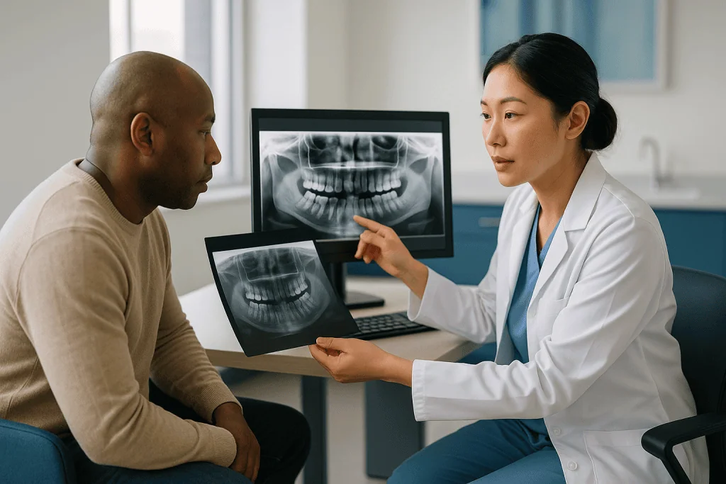
Wisdom teeth are the commonest trigger. Mayo Clinic notes they usually appear between the ages of 17 and 25, which is why a panoramic view is so often first suggested in the late teens, when the dentist wants to know whether there is room for them, which way they are pointing and whether the roots have finished forming.
Other common reasons include:
- A baby canine that has stayed put long past the age when its adult replacement should have arrived, raising the question of where the adult tooth has gone.
- Orthodontic planning, where the position of every unerupted tooth changes the treatment plan.
- Jaw pain, swelling or a lump that cannot be explained by what is visible in the mouth.
- A dark area or unusual shape noticed on a smaller film that needs a wider context.
- Trauma to the face or jaw.
- Assessment before implants, dentures or other procedures that depend on the shape of the bone.
Notice what is missing from that list: routine. A panoramic X-ray is not something taken at every visit. Cleveland Clinic points out that how often anyone needs dental X-rays depends on their age, oral health and risk of disease, and that the dentist tailors the interval to the person. The principle guiding every X-ray decision is simple: the picture should change what happens next. If it will not, there is no reason to take it.
What does a panoramic X-ray show about an impacted tooth's roots?
Roots are where the surgeon’s eye goes first, because roots determine how a tooth will behave if it ever has to be removed. On the image, the tooth is bright white, the surrounding bone is a mottled gray, and the roots taper down into the jaw like the prongs of a fork.
Several root features can be read straight off a good panoramic film:
- Number. A lower wisdom tooth usually has two roots, an upper one three, but fused single roots and extra roots are common and change the plan.
- Shape. Roots may be straight, splayed apart or hooked at the tip. A sharply bent root, called a dilaceration, is a warning that the tooth will not slide out along a single path.
- Stage of development. In a teenager the root tips may still be open and stubby. A tooth with roots only partly formed sits more loosely in the bone than one with long, complete roots, which is one reason age enters the conversation about timing.
- Relationship to the neighbor. If the crown of the impacted tooth presses against the root of the tooth in front, the film can show the neighbor’s root being worn away. Mayo Clinic lists damage to the adjacent tooth among the problems an impacted wisdom tooth can cause.
- Bone density around the root. Dense, brighter bone in older adults means more resistance; a dark halo around a crown may indicate a widened follicle or a cyst.
Here the flattening problem shows up. Two roots sitting one behind the other, cheek side and tongue side, are projected onto the same spot and look like a single thick root. A root that appears to overlap the nerve canal may in fact be sitting entirely to one side of it. The panoramic view tells you what is there; it is far weaker at telling you what is in front of what.
Reading the angle: what the tilt of a wisdom tooth tells the surgeon
Ask a surgeon to describe an impacted wisdom tooth in one word and the word will often be an angle. Clinicians borrow a classification devised by an oral surgeon named Winter, which describes the tooth’s long axis relative to the second molar beside it.
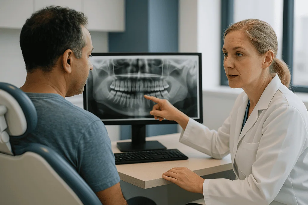
- Vertical. The tooth points straight up, in line with its neighbors, but has not cleared the bone or gum.
- Mesioangular. The crown leans forward toward the front of the mouth, tucking under the back edge of the second molar. This is the commonest pattern for lower wisdom teeth.
- Horizontal. The tooth lies flat on its side, crown pointing forward, roots pointing backward toward the hinge of the jaw.
- Distoangular. The crown leans backward, away from the other teeth and toward the bony ramus behind it.
A second system, from Pell and Gregory, adds two more readings from the same image: how deep the tooth sits compared with the chewing surface of the second molar, and how much space exists between that molar and the ramus, the vertical part of the lower jaw. A tooth whose crown is level with its neighbor and has open space behind it is a very different proposition from one buried below the neck of the molar with no room at all.
Why does the angle matter to you? It shapes what a removal would involve: how much bone would need to be lifted away, whether the tooth would likely be divided into pieces, and how close the instruments would come to the second molar and the nerve canal. Angle also influences the kind of trouble an impacted tooth can cause if left alone. A forward-leaning crown, for instance, creates a pocket between itself and the molar that traps food and is hard to clean, which is the setting for the gum infection called pericoronitis that the NHS describes as one of the reasons removal is considered.
What the angle cannot tell you is whether the tooth sits toward the cheek or toward the tongue. That third dimension is exactly what the panoramic view discards.
The nerve question: what the panoramic X-ray really shows about the inferior alveolar canal
Patients say “the nerve.” Radiologists say “the canal,” and the distinction is worth keeping. The inferior alveolar canal is a bony tunnel that runs through each side of the lower jaw carrying the nerve and blood vessels that give feeling to the lower lip, chin and lower teeth. Bone shows up on X-ray; nerves do not. What you see on a panoramic film is the tunnel, drawn as two thin, parallel white lines with a slightly darker band between them.
When a wisdom tooth root lies near that tunnel, clinicians look for a handful of classic signs, first described in a 1990 study by Rood and Shehab and still taught today:
- A dark band crossing the root where the canal passes over or through it.
- Interruption or loss of one of the canal’s white lines.
- A visible kink or diversion in the canal’s path as it meets the root.
- Narrowing of the canal at the point of contact.
- A root tip that appears deflected or notched.
Each of these raises the probability that root and canal are genuinely in contact. None proves it. The panoramic film cannot distinguish a canal that passes on the tongue side of a root from one that grooves the root itself, because both project onto the same spot. That uncertainty is the most common reason a three-dimensional scan is added.
The stakes explain the care. Mayo Clinic lists damage to nearby nerves, with numbness of the lower lip, tongue or chin, among the possible complications of wisdom tooth extraction, and notes that such changes are usually temporary but can occasionally be lasting. One more caution belongs here: the lingual nerve, which supplies feeling and taste to the side of the tongue, runs in soft tissue beside the jaw and is invisible on every kind of X-ray, panoramic or 3D. Its position can only be inferred, never seen.
Upper wisdom teeth, the sinus and the hidden canine
The upper jaw has its own set of relationships, and the panoramic view handles some of them well and others poorly.
Above the upper molars sit the maxillary sinuses, the air-filled spaces on either side of the nose. On the film, the floor of each sinus appears as a thin white curve, and the root tips of upper molars often seem to poke straight into the dark space above. Frequently that is an illusion of overlap; the roots may sit below or beside the sinus floor and simply project over it. Sometimes, though, only a wafer of bone separates the two, and Mayo Clinic lists damage to the sinuses among the risks of removing upper wisdom teeth. Knowing in advance how thin that floor is changes how gently a surgeon works and what they warn a patient about.
Upper wisdom teeth also tend to tilt outward toward the cheek and backward toward the bony hook behind the last molar. An upper tooth that sits high and far back can be surprisingly awkward to reach, even though the bone there is softer than in the lower jaw.
The other frequent upper-jaw puzzle is the impacted canine, the pointed tooth at the corner of the smile. MedlinePlus names it as the second most common impacted tooth after wisdom teeth. The question that decides treatment is whether the buried canine sits toward the palate or toward the lip. A single panoramic image cannot answer that; clinicians traditionally take a second film from a different angle and watch which way the tooth appears to shift, a trick called parallax. Increasingly, a small-volume 3D scan answers the question directly, and it can also show whether the canine’s crown is quietly wearing away the roots of the incisors beside it, a finding that changes urgency.
When a CBCT scan for wisdom teeth is added to the panoramic view
CBCT stands for cone beam computed tomography. It is a dental CT scanner: the machine still rotates around your head, but instead of a thin strip it captures a cone-shaped beam of data and rebuilds it into a three-dimensional block that can be sliced in any direction on screen. The clinician can scroll through the jaw front to back, side to side, or top to bottom, and view each slice at close to true size.
For an impacted lower wisdom tooth, that unlocks the answers the flat film could not give:
- Whether the nerve canal passes on the cheek side, the tongue side, between the roots or through a groove in the root.
- How many millimeters of bone, if any, separate root and canal, measured without the magnification error of a panoramic image.
- The true root count and the direction of any hooks.
- How thick the bone is on the tongue side of the jaw, which affects the risk of a fracture during removal.
- The real size and extent of any cyst or dark area seen on the panoramic film.
CBCT is not a routine first step, and the reason is principle rather than penny-pinching. Every X-ray decision rests on justification: the extra image should be expected to alter management. Cleveland Clinic makes the same point about dental X-rays generally, noting that they are taken when they are likely to help diagnose or plan care, at intervals tailored to the individual. In practice, a 3D scan tends to be added when the panoramic film shows one or more of the warning signs around the canal, when a tooth’s position is genuinely ambiguous, when a cyst or lesion needs mapping, or when a surgeon is planning to remove only the crown and leave the roots in place, a technique that depends on knowing the roots’ exact relationship to the canal.
If your dentist does not suggest a 3D scan, it usually means the flat film already answered the questions that matter. If they do, it is because something on that film left a question open.
Panoramic X-ray vs 3D scan: what each one is good at
The two tests are not rivals. One is a wide, quick survey; the other is a targeted, detailed map. Most impacted teeth are assessed and managed on the panoramic view alone, and the 3D scan earns its place only when the survey raises a question it cannot settle.
| Feature | Panoramic X-ray | Cone-beam CT (3D) |
|---|---|---|
| What you get | One flat, curved image of both jaws | A 3D block viewable as slices in any plane |
| Dimension | Two-dimensional; front-to-back position is lost | Three-dimensional; cheek-to-tongue position is visible |
| Measurement accuracy | Unreliable; magnification varies across the image | Close to true size when the scan is properly calibrated |
| Root-to-nerve-canal relationship | Suggests closeness through indirect signs | Shows the actual position of canal relative to root |
| Radiation | Low; MedlinePlus describes dental X-ray exposure as very small | Generally higher than a single panoramic image; kept low by using a small field of view |
| Typical role | First-line survey for wisdom teeth, canines, jaw pain, orthodontics | Added when anatomy is unclear or warning signs appear |
| Main weaknesses | Blur, overlap, ghost shadows, positioning errors | Higher dose, artifacts from metal, more expertise needed to read |
Two honest caveats belong under the table. First, “more detail” is not the same as “better care.” A perfectly clear 3D image of a symptom-free tooth that would have been monitored anyway adds dose without changing the plan. Second, even CBCT has limits: it shows bone and tooth superbly but soft tissue poorly, so it cannot display the lingual nerve or the thickness of the gum. Reading either image well depends far more on the experience of the person interpreting it than on the pixel count.
What are the disadvantages of a panoramic radiograph?
Every dentist who has squinted at a panoramic film knows its quirks. Understanding them helps you understand why a clinician sometimes says, “I need a better look,” even when the picture looks fine to you.
Uneven magnification. The image is enlarged, and the amount of enlargement differs from the front of the jaw to the back and from top to bottom. A distance measured with a ruler on the screen can be meaningfully wrong unless the software has been calibrated for that patient, which is why nobody plans surgery from panoramic measurements alone.
Overlap. Everything in the path of the beam is compressed into one plane. Roots stack on roots; the sinus floor drapes over molar tips; the nerve canal can appear to pierce a root it actually passes beside.
Ghost images. Dense objects outside the zone of focus, such as earrings, a hair clip or the opposite side of the jaw, can cast faint, enlarged shadows on the wrong side of the image. The spine often shows as a pale vertical band in the middle.
Positioning errors. A chin tipped too far up flattens the smile line and blurs the upper roots; too far down exaggerates the curve. A tongue not pressed against the palate leaves a dark band across the upper root tips that can hide or mimic disease. Slight head rotation makes one side of the jaw look wider than the other.
Lower sharpness than small films. For early cavities between teeth, a bitewing taken inside the mouth remains clearer. A panoramic image is a map, not a magnifying glass.
No third dimension. The single limitation that drives most follow-up imaging. Cheek-side or tongue-side is simply unknowable from this view.
What is not a meaningful disadvantage is radiation. MedlinePlus describes the exposure from dental X-rays as very low, and the same source notes that a lead apron or thyroid collar may be used to shield other parts of the body. If you are or might be pregnant, tell the team before any X-ray so they can weigh timing with you.
Who is usually imaged and who is usually asked to wait, and why experts no longer remove every impacted wisdom tooth
Here the conversation shifts from what the image shows to what should be done about it, and this is where opinion has genuinely changed over the past few decades.
The NHS states plainly that wisdom teeth are usually removed only when they are causing problems. It lists the kind of problems that count: tooth decay in the wisdom tooth or its neighbor because the area is hard to clean, repeated gum infection around a partly erupted crown, an abscess, cysts around the tooth, or damage to the second molar. Mayo Clinic describes the same list and adds that when an impacted wisdom tooth causes pain, infection or other dental problems, removal is generally recommended.
The disagreement concerns the impacted tooth that is doing nothing. Mayo Clinic is candid that some dentists and oral surgeons recommend removing symptom-free wisdom teeth to prevent future problems, while others prefer to monitor them, and that the evidence for routinely removing trouble-free teeth is not settled. Both sources describe the case for monitoring: symptom-free, disease-free teeth are often left in place with regular check-ups and periodic X-rays to catch change early. The panoramic film’s job in that scenario is comparison over time, not a one-off verdict.
Who is typically imaged? Anyone with symptoms at the back of the jaw, anyone whose dentist cannot see a wisdom tooth clinically and needs to know where it is, and young people in the age window Mayo Clinic describes, when roots are still forming and options are widest.
Who is typically asked to wait? Someone in the middle of an acute infection, where the team may want the inflammation to settle before any operation. Someone who is pregnant, where non-urgent imaging and elective surgery are commonly deferred and timing is discussed with the obstetric team. A younger teenager whose tooth is still high in the bone and may yet erupt normally. And, most importantly, anyone whose tooth is healthy and symptom-free, for whom watching is a legitimate choice rather than a failure to act.
None of this is a prescription. The people who have seen your film, examined your mouth and know your history are the ones who weigh it.
What the following days and weeks usually look like after the scan
The X-ray itself has no recovery. You can eat, drive and go back to work immediately, and there is no lingering effect from the exposure. What follows depends on what the image showed and which path you and your team choose.
If the plan is monitoring. Expect a conversation about warning signs to watch for and a suggested interval for the next review. Neither MedlinePlus nor the NHS fixes a universal schedule; the interval is chosen for you based on the tooth’s position, your age and your history of infection. Cleveland Clinic frames dental X-ray frequency the same way, as an individual decision rather than a calendar rule. Keep the panoramic image on file; a future film compared side by side with it is often more informative than either one alone.
If a 3D scan is recommended. This is often a separate appointment, sometimes in a different room or building. The scan takes a few seconds to a minute of staying still. Results are usually discussed at a planning consultation where the surgeon shows you the slices and explains what they change.
If removal is planned. The NHS describes what typically follows: swelling and some discomfort for a few days, possible bruising of the cheek, a stiff or sore jaw, and an unpleasant taste while the socket heals. It states that full recovery can take up to two weeks. Stitches, if used, usually dissolve or are removed at a check-up. Mayo Clinic notes that any numbness of lip, chin or tongue from nerve irritation is usually temporary, settling over weeks to months, though it can rarely persist. Pain relief is chosen and explained by the prescribing clinician; the image will have already told them how much bone was disturbed, which shapes their expectations for you.
Whatever the path, the film has done its job when the next step is clearer than it was before you bit down on that plastic tab.
What people often get wrong about impacted tooth X-rays
“The X-ray shows the nerve.” It shows the bony tunnel the nerve travels in. Nerves themselves are invisible on X-ray. The lingual nerve, beside the tongue, has no tunnel and never appears on any scan.
“The root is on top of the canal, so the nerve will be damaged.” Overlap on a flat image is not contact in real life. The canal may pass beside the root with a comfortable margin of bone. That is precisely why a 3D scan is added when the picture is ambiguous, and why an alarming-looking panoramic film often turns out to be reassuring in three dimensions.
“Every impacted wisdom tooth has to come out.” The NHS and Mayo Clinic both describe monitoring symptom-free, disease-free teeth as a recognized option. Removal is recommended when the tooth is causing or clearly threatening problems.
“Wisdom teeth push the front teeth crooked, so they should be removed to stop crowding.” Crowding does not appear among the reasons for removal the NHS lists, and neither MedlinePlus nor Mayo Clinic cites it as a justification. Front teeth drift for many reasons, and a buried third molar is a weak candidate for blame.
“3D is always better, so I should ask for it.” A 3D scan is better at one specific thing: showing position in the third dimension. When the flat film has already answered the clinical question, extra detail changes nothing and adds exposure.
“Dental X-rays are dangerous.” MedlinePlus describes the exposure as very low. The sensible posture is not fear but justification: each image should have a reason.
“A blurry panoramic image means a bad machine.” Blur at the edges and faint ghost shadows are built into the technique. What matters is whether the region in question sits within the zone of focus, and a clinician will re-take or supplement the image if it does not.
Questions to ask your care team about your impacted tooth X-ray
A panoramic film is a conversation starter, and the best conversations happen when you know what to ask. Bring a few of these to the appointment, or ask them while the image is still on the screen.
- Can you show me the impacted tooth and trace its roots for me? How many roots does it have, and are any of them hooked?
- Which way is the tooth tilted, and how deep does it sit compared with the tooth beside it?
- Where is the nerve canal on this image, and do you see any of the signs that suggest it is close to the root?
- Is anything about the position uncertain on this flat image that a 3D scan would settle? If so, what would the 3D result change about the plan?
- For an upper tooth: how does it relate to the sinus, and does that affect the approach?
- Is this tooth currently causing any damage: decay, gum infection, wear on the neighbor, or a widened space around the crown?
- If it is doing nothing right now, what would monitoring involve and how would you decide when to re-image?
- If removal were chosen, what does this image tell you about how involved the procedure would be, and what risks does it raise or lower for me specifically?
- Are there alternatives to full removal for this tooth, such as removing only the crown, and does my anatomy make them suitable or unsuitable?
- Do any of my medicines or medical conditions change how you read this image or what you would recommend?
- Can I have a copy of the image for my records?
You are entitled to answers in plain language. A good clinician will point, not just describe, and will be comfortable saying “I can’t tell from this view” when that is the honest reading. That admission is not a weakness; it is the reason 3D imaging exists.
When to call your doctor
An impacted tooth can stay quiet for years, and the image alone is never an emergency. Symptoms are what change the urgency, before or after any procedure.
Contact your dentist or doctor promptly if you notice:
- Pain, swelling or redness of the gum behind your last molar, especially if it flares repeatedly or makes it hard to bite down.
- A bad taste or pus coming from the back of the mouth.
- Difficulty opening your mouth fully, which can signal infection spreading into the jaw muscles.
- A tender lump or swelling in the jaw that was not there before.
- Numbness or tingling of the lip, chin or tongue that appears without an obvious cause, or that persists after a procedure beyond the timeframe your surgeon described.
After an extraction, Mayo Clinic advises calling the surgeon if you have difficulty swallowing or breathing, excessive bleeding, fever, severe pain not helped by the pain relief you were prescribed, swelling that worsens after the third day, a bad taste that does not improve with rinsing, pus in or oozing from the socket, or persistent numbness. The NHS adds that pain returning or worsening several days after removal, particularly with an exposed, empty-looking socket, may indicate a dry socket that needs to be seen.
Seek emergency care immediately, without waiting for a dental appointment, if swelling spreads toward the eye or down the neck, if you struggle to breathe or swallow, or if you develop a high fever with a rapidly enlarging face. Infections from the lower jaw can track into the neck, and these signs need same-day hospital assessment.
Every decision about imaging, monitoring or removal rests with the team who has examined you and seen your films. Use this article to understand the picture; use them to decide what to do about it.
Frequently asked questions
What does an impacted wisdom tooth X-ray actually show?
It shows the tooth’s position and depth in the jaw, the direction it tilts, how many roots it has and how they curve, and its relationship to the second molar, the sinus above upper teeth and the nerve canal in the lower jaw. It also reveals decay, cysts or bone loss around the crown. What it cannot show is whether the tooth sits toward the cheek or the tongue.
Why would a dentist order a panoramic dental X-ray instead of a small film?
Small films taken inside the mouth often cannot reach far enough back to capture a wisdom tooth buried behind the second molar. A panoramic image sweeps around the whole jaw from outside the mouth and includes the jaw joints, sinuses and nerve canals, giving the wide context needed to assess an impacted tooth, plan orthodontics or investigate unexplained jaw pain or swelling.
Can a panoramic X-ray show whether a wisdom tooth is touching the nerve?
Not with certainty. It shows the bony canal the nerve runs through, and signs such as a dark band across the root or a break in the canal’s outline suggest close contact. Because the image is flat, a root and canal that overlap on screen may be side by side in reality. A cone-beam CT scan is usually added when that relationship needs to be confirmed.
When is a CBCT scan for wisdom teeth recommended?
Typically when the panoramic film raises a question it cannot answer: warning signs near the nerve canal, an unclear root count or position, a cyst that needs mapping, or a planned technique such as removing only the crown. It is not a routine first step, because the extra exposure is justified only when the result is expected to change the plan.
What are the main panoramic radiograph disadvantages?
Uneven magnification makes measurements unreliable, overlapping structures hide detail, ghost shadows from earrings or the spine can confuse the picture, and small positioning errors blur or distort regions of the jaw. It is also less sharp than small intraoral films for early cavities. Its central limitation is the missing third dimension, which is what a 3D scan restores.
Why do experts now say not to remove every impacted wisdom tooth?
Because the evidence for removing teeth that are symptom-free and disease-free is unsettled. The NHS states wisdom teeth are usually removed only when they cause problems such as decay, repeated gum infection, cysts or damage to the neighboring tooth. Mayo Clinic notes that some clinicians still favor preventive removal while others prefer monitoring, and the choice is made individually.
What does the angle of an impacted tooth mean on the X-ray?
The angle describes which way the tooth’s long axis points relative to the tooth beside it: straight up, tilted forward, lying flat, or tilted backward. It matters because it predicts how much bone would need to be removed, whether the tooth would be divided, and how likely the tooth is to trap food and cause gum infection if left in place.
Is the radiation from a panoramic X-ray safe?
MedlinePlus describes the radiation exposure from dental X-rays as very low, and a lead apron or thyroid collar may be used for shielding. The guiding principle is that each image should have a clear clinical reason. If you are or could be pregnant, tell the team beforehand so timing can be discussed.
How long does recovery take if the impacted tooth is removed?
The NHS states that it can take up to two weeks to recover fully, with swelling, bruising, a stiff jaw and an unpleasant taste common in the first days. Mayo Clinic notes that any numbness of the lip, chin or tongue from nerve irritation is usually temporary, though it can rarely be lasting. Your surgeon will outline what to expect based on your own image.
Can I ask to keep a copy of my panoramic X-ray?
Yes. The image is part of your health record, and having a copy is useful if you move, change dentists or need a comparison in the future. Panoramic films are especially valuable side by side, because change over time in a monitored tooth often tells the team more than a single image ever could.
References
- MedlinePlus – Dental x-rays
- MedlinePlus – Impacted tooth
- NHS – Wisdom tooth removal
- Cleveland Clinic – Dental X-Rays
This article is for general information only and is not a substitute for professional medical advice. Please consult a qualified doctor about your individual situation.

More from the Blog

Impacted Tooth Treatment in Adults vs Teens: Why Timing Changes the Approach
An impacted tooth is one blocked from fully coming through the gum. In teens, roots are still forming and bone is more adaptable, so…

Keeping Up Orofacial Myology Exercises Between Visits: Why Daily Practice Shapes the Result
Daily myofunctional exercises matter because orofacial myology works through motor learning: the tongue, lips and jaw muscles only adopt a new resting posture and…

Bleeding Gums in Pregnancy and With Diabetes: Why Gum Care Is Planned Differently
Bleeding gums in pregnancy are usually pregnancy gingivitis: hormonal changes make gums react more strongly to plaque, so they swell and bleed with brushing.…
How Oral and Maxillofacial Surgeons Train, and Questions to Ask Before Your Procedure
Oral and maxillofacial surgeons first complete dental school, then a hospital-based surgical residency that typically runs four to six years and includes rotations in…

Recovery After Gummy Smile Treatment: Swelling, Sensitivity and the Reshaped Gum Line
Recovery after gummy smile treatment depends on the procedure. Gum contouring and crown lengthening usually bring mild swelling, tenderness and temporary tooth sensitivity that…

Is Sensitivity After Teeth Whitening Normal? What Helps and When to Call the Dentist
Yes. Temporary sensitivity is one of the most common side effects of peroxide-based teeth whitening. It usually appears during or shortly after treatment as…