

What Do EEG, Sleep-Deprived EEG and MRI Show in Pediatric Epilepsy? Each Test Explained

Key Takeaways
- A normal EEG does not rule out epilepsy, because seizure-related spikes are intermittent and a short recording can miss them.
- A standard EEG uses roughly 16 to 25 scalp electrodes that only record; nothing is delivered into the brain.
- Sleep-deprived EEG works because drowsiness and light sleep loosen the brain's inhibitory systems, bringing hidden epileptiform patterns to the surface.
- An epilepsy-protocol MRI uses thin slices and hippocampus-aligned coronal images to find abnormalities a rapid emergency scan is not designed to see.
- MRI involves no X-ray radiation; its main safety issue is metal, which is why the pre-scan questionnaire is taken so seriously.
- The most powerful diagnostic result is concordance, when the EEG focus, the MRI finding and the seizure description all point to the same brain region.
In pediatric epilepsy, an EEG records the brain's electrical activity through scalp sensors and can show seizure-prone patterns, though a normal result does not rule epilepsy out. A sleep-deprived EEG raises the chance of capturing those patterns because tiredness and drowsiness bring them out. An MRI shows brain structure, looking for a physical cause such as scarring or a malformation. The treating team combines all three with the child's history.
The waiting room has a fish tank, and your six-year-old is narrating the fish. Twenty minutes ago, she had paste combed into her hair and a bonnet of wires fitted over it, and she took it far better than you did. Now a technologist is asking whether she slept last night, because the plan was that she would not, much.
Families arrive at this appointment carrying two questions that sound similar but are not. One is: did the thing we saw on the kitchen floor happen because of epilepsy? The other is: what is going on inside her brain that made it happen? A routine EEG, a sleep-deprived EEG and a brain MRI are the standard tools for those questions, and each answers a different part.
This guide explains eeg and mri for childhood epilepsy the way a neurologist would if the clinic were quiet: what each test actually measures, what it cannot measure, and why a normal result is not the same as a clean bill of health.
Why do doctors order EEG and MRI for childhood epilepsy at all?
Epilepsy is not diagnosed by a machine. It is a clinical diagnosis, meaning the doctor decides based on what the child experienced and what witnesses saw. The NHS is blunt about this: the most useful information is a clear description of the event, ideally with a video from a phone, and the tests come afterward to support or refine that picture (see NHS epilepsy diagnosis page).
So why test at all? Because a seizure, a sudden burst of abnormal electrical activity in the brain, can look like several other things in a child: a faint, a breath-holding spell, a night terror, a tic. And because two children with identical-looking seizures may have very different reasons for them. One may have a genetic tendency that runs quietly in the family; another may have a small area of unusually formed brain tissue that has been there since before birth.
The two families of tests split neatly along that line. An EEG, short for electroencephalogram, listens to the brain’s electrical rhythm. An MRI, short for magnetic resonance imaging, photographs the brain’s physical structure. One is about function, the other about form. Neither replaces the other, which is why children are often sent for both.
Epilepsy is common enough that this pathway is well worn: the NHS estimates it affects around 1 in 100 people, and it frequently begins in childhood. The tests are routine to the people running them, even when nothing about the situation feels routine to you.
How does an EEG actually work?
Every thought, movement and dream runs on tiny electrical currents passing between brain cells. Those currents are faint, but millions of neurons firing in rhythm add up to a signal that can be picked up through the skull and scalp. An EEG measures that signal.
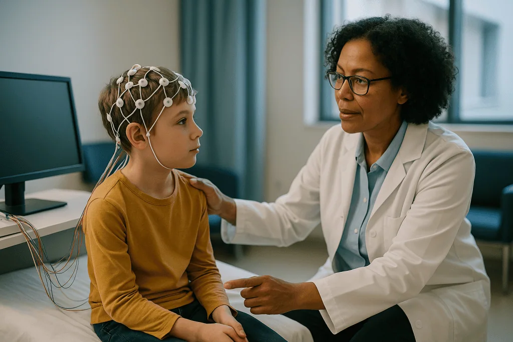
To do it, a technologist places small metal discs, called electrodes, at set points across the head. MedlinePlus describes between 16 and 25 of them for a standard study, fixed with a sticky conductive paste or fitted into a snug cap. Each electrode is a listening post; the wires run to an amplifier that boosts the signals thousands of times and draws them as wavy lines on a screen. Nothing goes into the child. The electrodes record; they do not deliver anything.
What the neurologist reads is rhythm. A relaxed, awake brain produces steady waves at a particular pace; a drowsy brain slows; deep sleep slows further. Against that backdrop, the reader looks for things that should not be there: sharp spikes, spike-and-wave bursts, or a region that stays sluggish when the rest of the brain is lively. These patterns are called epileptiform discharges, meaning electrical signatures typical of a brain prone to seizures.
The location matters as much as the shape. Because each electrode has a known address on the scalp, an abnormal pattern showing up under the left temporal electrodes points toward one part of the brain and toward particular seizure types. That geography is one reason EEG remains central to eeg and mri for childhood epilepsy work-ups decades after its invention: no other bedside tool maps electrical behavior in real time.
Can an EEG detect seizures, or only the risk of them?
Both, but mostly the second. If a seizure happens while the electrodes are on, the EEG records it directly, and that is the single most informative recording a neurologist can get. It shows where the seizure starts, how it spreads and how long it lasts. In practice, though, a routine study lasts under an hour, and most children do not have a seizure to order.
What the EEG usually catches instead are interictal discharges: brief abnormal spikes that occur between seizures, when the child looks entirely normal. Think of them as the brain clearing its throat. They suggest a lowered seizure threshold and often help classify the epilepsy type, which in turn shapes treatment choices made by the treating team.
Here is the part families most need to hear. A normal EEG does not exclude epilepsy. The NHS states plainly that an EEG cannot always show whether someone has epilepsy, because many people with the condition have normal readings between seizures. Mayo Clinic makes the same point. The recording is a snapshot, and the abnormality may simply not have shown up during that window, or it may arise from tissue too deep for scalp electrodes to register.
The reverse also holds, more gently. A small number of children who never have seizures show minor irregularities on EEG. That is why no responsible clinician diagnoses epilepsy from a printout alone, and why the description of what happened at home remains the anchor. The EEG adds weight to one side of the scale; it does not tip it by itself.
What happens during a routine EEG for a child?
The appointment usually runs about an hour, most of it setup. The NHS describes the recording itself as lasting around 20 to 40 minutes. Your child will sit in a reclining chair or lie on a bed while the technologist measures the head with a tape, marks points with a soft crayon, rubs each spot lightly with gritty gel to improve contact, and applies the electrodes. The paste is cool, the rubbing is a little scratchy, and the whole business is more tedious than uncomfortable. Small children often watch a cartoon through it.
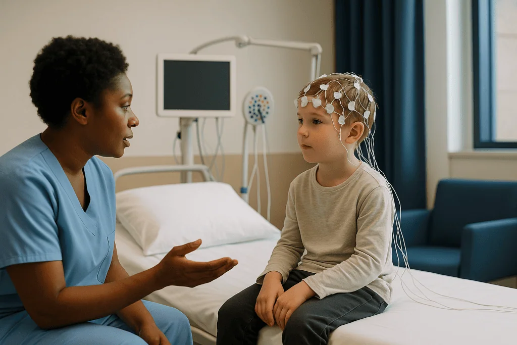
Once recording starts, the room goes quiet. The child is asked to lie still with eyes closed, then open, then closed again. Blinking and jaw clenching show up on the trace as big smudges, so the technologist may ask for stillness in short stretches rather than all at once.
Two extra steps are standard and worth explaining ahead of time. In hyperventilation, the child breathes deeply and quickly for a few minutes, as if blowing up a balloon or blowing out candles; the resulting shift in blood chemistry can bring out certain patterns, particularly in absence epilepsy. In photic stimulation, a lamp flashes at different speeds while the child keeps eyes closed or open. Only a small subset of children have light-sensitive epilepsy, but checking is routine, and the technologist stops immediately if anything appears.
Afterward the electrodes come off with warm water, and hair will feel gluey until a proper wash. There are no after-effects to watch for. Results go to the neurologist, not to you in the room, because interpretation takes a trained eye and a bit of time.
What is a sleep deprived EEG in children, and why does tiredness help?
A sleep-deprived EEG is the same recording done on a child who has been kept awake for much of the previous night. It sounds unkind. It is, in fact, one of the most useful tricks in pediatric neurology.
The logic is physiological. The transition from wakefulness into drowsiness and light sleep is a time when the brain’s inhibitory systems loosen. Epileptiform discharges that stay hidden during alert wakefulness frequently surface during this window, and some childhood epilepsy syndromes show their characteristic patterns almost exclusively in sleep. The NHS lists the sleep EEG, in which you may be asked to keep your child awake the night before, as a standard option when a routine study has not answered the question.
Practically, the department will give you a target: typically a later bedtime and a much earlier wake-up, adjusted for age, with no napping in the car on the way in. The exact instructions come from the clinic, and they vary, so follow theirs rather than any general rule. Caffeinated drinks are usually off the list for the child that morning.
At the appointment, the electrodes go on as usual and the lights go down. Most tired children fall asleep within minutes. The technologist records through drowsiness, light sleep and, if possible, waking, because the moment of waking is itself informative. The recording tends to run somewhat longer than a routine EEG to allow for this.
The following day is the price. Expect a cranky, clingy child and plan a soft afternoon. There is no evidence that a single night of missed sleep causes lasting harm, though it can, predictably, make a seizure slightly more likely that day, which is one reason the study is done under supervision.
What does a brain MRI show in pediatric epilepsy?
If an EEG is a microphone, an MRI is a camera, and a remarkably detailed one. It uses a powerful magnet and radio waves to build cross-sectional images of the brain, slice by slice, in any plane the radiologist chooses. There is no X-ray radiation involved, a point the NHS emphasizes and one that matters when the patient is a growing child who may need repeat scans over the years.
The question an epilepsy MRI answers is structural: is there a physical reason for these seizures? Mayo Clinic lists the kinds of findings that can be relevant, including scarring, tumors, cysts, areas of prior bleeding and lesions or abnormalities in brain development. In children specifically, radiologists pay close attention to malformations of cortical development, areas where the brain’s outer layer folded or layered unusually before birth. These can be subtle, sometimes only a slight blurring of the boundary between gray and white matter, which is why the scan technique matters so much.
They also look at the hippocampus, a seahorse-shaped structure deep in each temporal lobe that is involved in memory and is a frequent seizure source. Shrinkage or scarring there, called hippocampal sclerosis, is a recognized pattern.
Two honest caveats. Many children with epilepsy have completely normal MRIs; in genetic generalized epilepsies, that is the expected result and is reassuring rather than puzzling. And a finding on MRI is not automatically the cause of the seizures. Radiologists and neurologists compare the location of any abnormality with the EEG and the seizure description before drawing that line. Concordance, meaning the tests point to the same place, is what carries weight.
What is the epilepsy MRI protocol, and why is it different from a standard scan?
Parents sometimes learn their child had a brain MRI in the emergency department after a first seizure, and then hear the neurologist request another one. That is not duplication for its own sake. A general MRI and an epilepsy-protocol MRI are built for different questions.
An MRI protocol is simply the list of image sequences the scanner runs, each sequence a different way of tuning the magnet and radio pulses to highlight particular tissues. A standard emergency brain scan uses a handful of sequences designed to catch large, urgent problems quickly: bleeding, swelling, a big mass.
An epilepsy protocol trades speed for resolution. Typical elements include thin slices, often a few millimeters or less, so a small cortical malformation does not fall between images; high-resolution three-dimensional sequences that can be resliced in any direction after the scan; and coronal images, cut from ear to ear, angled along the axis of the hippocampus so both sides can be compared symmetrically. Fluid-sensitive sequences called FLAIR are included because subtle scarring often shows up as a faint bright signal on them. Where available, a stronger magnet improves detail further.
Contrast dye, a gadolinium-based injection that makes certain tissues stand out, is not routine for epilepsy imaging and is usually added only when a first look raises a specific question, such as a possible tumor or inflammation. Mayo Clinic notes contrast is used only in some MRI examinations.
The protocol also assumes stillness for longer than a quick scan does, which shapes the practical planning for young children discussed in the next section. If your child’s earlier scan was a rapid one, the second is looking for smaller things.
How long does an epilepsy MRI take, and how do young children stay still?
Stillness is the whole game. MRI images are built up over seconds to minutes per sequence, and movement blurs them the way a wobbling hand ruins a long-exposure photograph. The NHS gives a broad range for MRI scans of 15 to 90 minutes depending on the area and the number of images; an epilepsy protocol sits toward the longer end because of the additional detailed sequences. Ask the department for their typical time for this specific protocol so you can plan around it.
Three approaches are used to get a child through it.
- Awake with preparation. Older children, often from around school age upward, frequently manage the scan awake, especially after a practice session. Many departments run a mock scanner or a play-based walkthrough so the noise and the tunnel are familiar. Headphones with a film or music, a mirror to see a parent, and a squeeze-ball call button all help.
- Feed and wrap for infants. Babies under a few months can sometimes be fed, swaddled snugly and scanned during natural sleep. It takes patience and a quiet room, and it does not always work, but when it does it avoids medication altogether.
- Sedation or general anesthesia. For toddlers and preschoolers, and for older children who cannot tolerate the noise or the enclosed space, the team may recommend sedation or a brief general anesthetic. Mayo Clinic notes this is sometimes needed for young children. An anesthesiologist assesses the child beforehand, fasting instructions apply, and the child is monitored throughout and during recovery.
The scanner is loud, a rhythmic knocking and buzzing that ear protection dampens but does not silence. Nothing touches the child, and the table can be withdrawn at any time. Parents can usually stay in the room after their own metal-safety check.
What can an MRI show that an EEG cannot, and vice versa?
Families often ask which test is better. The honest answer is that they are not competing. Each sees something the other is blind to, and the useful information comes from laying them side by side.
| Feature | Routine EEG | Sleep-deprived EEG | Epilepsy-protocol MRI |
|---|---|---|---|
| What it measures | Electrical activity, awake | Electrical activity through drowsiness and sleep | Physical structure of brain tissue |
| Best at finding | Interictal spikes, background rhythm, photosensitivity | Sleep-activated discharges, syndrome-specific patterns | Malformations, scarring, tumors, hippocampal changes |
| Cannot show | Anatomy; deep or brief abnormalities | Anatomy; still a time-limited sample | Whether the brain is electrically prone to seizures |
| Typical duration | Recording about 20 to 40 minutes (NHS) | Somewhat longer, to capture sleep | Within the NHS range of 15 to 90 minutes, toward the upper end |
| Radiation | None | None | None |
| Preparation | Clean hair, normal sleep | Restricted sleep the night before | Metal check; fasting only if sedation planned |
| Discomfort | Sticky paste, sitting still | Tiredness beforehand | Noise, enclosed space; possible sedation |
Read across the second row and the logic of ordering both becomes clear. An MRI can reveal a small malformation and say nothing about whether it is electrically active. An EEG can show spikes over the right temporal region and say nothing about why. When the MRI abnormality and the EEG focus sit in the same spot, the team has a coherent story; when they do not, that mismatch is itself important information.
A CT scan, which uses X-rays and takes minutes, is faster and appears in emergency settings, but Mayo Clinic and the NHS both position MRI as the more detailed test for planned epilepsy evaluation.
Who is usually offered EEG and MRI for childhood epilepsy, and who is asked to wait?
Not every child who has a seizure needs every test, and not every child who needs a test needs it urgently. The pattern of ordering follows guidance rather than habit.
An EEG is generally arranged for a child who has had an unprovoked seizure, meaning one without an obvious immediate trigger such as a high fever in a young child or a head injury. The NHS describes EEG as a routine part of the epilepsy diagnostic pathway once a specialist suspects the condition. Where a first EEG is normal but suspicion remains, a sleep-deprived EEG, an ambulatory EEG worn at home over one or more days, or video-EEG monitoring in hospital over a few days may follow, again per NHS guidance. The escalation is driven by how much doubt remains, not by how worried a family feels.
MRI is usually recommended when the seizures may have a focal onset, meaning they begin in one region; when the child is very young at onset; when there are abnormal findings on examination or in development; or when seizures are not controlled as expected. Mayo Clinic frames imaging as the search for a structural cause.
Who is asked to wait, or not scanned at all? Two groups stand out. Children with a single simple febrile seizure, the brief convulsion some young children have with fever, generally do not need EEG or MRI, because the risk of epilepsy in that setting is low and imaging rarely changes management. And children whose history and EEG together fit a well-recognized genetic generalized epilepsy syndrome may not need MRI, since the scan is expected to be normal.
These are typical patterns, not rules. Your child’s neurologist weighs the individual picture and decides.
How do you prepare a child for these tests, and what helps with comfort?
Most of the distress children feel at these appointments comes from surprise, not pain. Preparation is therefore mostly information, pitched at the right age.
For an EEG, wash the hair the night before and skip conditioner, gel or braids, since clean dry hair helps the electrodes stick. Tell the truth about what will happen: a hat of stickers and wires, some scratchy rubbing, then lying still and watching a screen. Bring a comfort object, a tablet with downloaded content, and a snack for afterward. For a sleep-deprived study, follow the clinic’s specific wake-time instructions and resist the urge to let the child doze on the drive.
For an MRI, the metal check is the first and most serious step. Anything ferromagnetic in or on the body can be pulled by the magnet or heated, so the team will ask about implants, dental braces, hearing aids, and any metal fragments. Clothing without zips or studs saves changing time. If sedation or anesthesia is planned, fasting instructions are exact and non-negotiable; ask for them in writing.
Many children’s departments offer a preparation visit run by a child life specialist or play therapist, sometimes with a toy scanner and a doll. Where offered, take it. Children who have rehearsed lying still with headphones on tend to manage the real thing better, and that can be the difference between an awake scan and a sedated one.
A few small things help more than parents expect: letting the child pick the film, agreeing a hand signal for a break, and treating the appointment as a job the child is doing rather than something being done to them. Nurses and technologists have seen every kind of nervous child and are not easily flustered. Neither, it turns out, are most six-year-olds once the fish tank has been discussed.
What do the following days and weeks usually look like?
The tests themselves leave almost nothing behind. After an EEG, hair needs washing and that is the extent of it. After an awake MRI, children walk out and go to school if they wish. After sedation or anesthesia, the day is written off: expect grogginess, occasional nausea and wobbliness, and follow the recovery team’s instructions on food, supervision and activity for the rest of that day.
The harder part of the following weeks is the waiting. EEG traces are read by a neurologist or a neurophysiologist, and MRI images are reported by a radiologist, then both are reviewed together in the epilepsy clinic. Departments vary, and the report may take from days to a few weeks; ask at the appointment when and how you will hear. Results are generally discussed in person or by phone with the doctor who ordered them rather than posted cold to a portal, because context matters and questions are inevitable.
Four outcomes are common, and none should surprise you.
- Both tests normal: the diagnosis still rests on the clinical history, and further monitoring may or may not be suggested.
- EEG abnormal, MRI normal: often consistent with a genetic or unexplained epilepsy, and frequently the expected pattern.
- MRI shows a finding that matches the EEG: a focal epilepsy with a likely structural cause, which opens a different set of conversations.
- An incidental finding on MRI unrelated to seizures: not rare, usually benign, and explained by the team.
If treatment is being considered, anti-seizure medicines, a broad class of drugs that reduce the brain’s tendency to fire abnormally, are usually the first discussion. Choices, timing and any changes belong to the prescribing clinician, informed partly by what these tests showed. Repeat EEGs over time are common and are not a sign that something went wrong the first time.
What people often get wrong about EEG and MRI results
Some misunderstandings come up so often in clinic that they are worth correcting head-on.
“The EEG was normal, so it wasn’t epilepsy.” This is the most consequential error. The NHS and Mayo Clinic both state that a normal EEG does not rule out epilepsy. Discharges are intermittent, and a short recording may miss them. A normal trace narrows possibilities; it does not close the file.
“The EEG can read thoughts or measure intelligence.” It records rhythm and voltage, nothing more. It cannot tell what a child is thinking, whether they are lying, or how clever they are.
“The EEG sends electricity into the brain.” The electrodes are passive listeners. Nothing is delivered, and the child feels nothing from them.
“An abnormal MRI means surgery.” Most structural findings lead to conversations about medication first. Surgery is considered for a specific subset of children whose seizures are not controlled and whose tests point clearly to one removable area, and that assessment is lengthy and involves far more than one scan.
“MRI uses radiation.” It does not, as the NHS notes. It uses a magnet and radio waves. The safety issues are about metal, not dose.
“Sleep deprivation is dangerous for a child with seizures.” A single supervised night of reduced sleep is a long-established diagnostic method. It can modestly raise seizure likelihood that day, which is exactly why the recording is done with a technologist present.
“If the ER scan was fine, the neurologist’s scan will be too.” A rapid emergency scan and an epilepsy-protocol MRI look for different things at different resolutions. The second is not a repeat; it is a closer look.
“Results will be ready before we leave.” Expert interpretation takes time, and combining EEG, MRI and history is deliberately unhurried.
Questions to ask your care team
Appointments move quickly and questions evaporate under fluorescent lights. Write yours down. These are the ones that tend to matter most, phrased so the answers are useful rather than reassuring noise.
- What specific question is this test meant to answer for my child, and what would you do differently depending on the result?
- Is this a routine EEG or a sleep-deprived study, and exactly what sleep schedule do you want the night before, for a child this age?
- Will hyperventilation and flashing lights be part of the EEG, and can we explain them to our child in advance?
- Is the MRI being done on an epilepsy protocol, and is contrast dye planned?
- Roughly how long will this particular scan take in your department, and will my child need sedation or a general anesthetic?
- Is there a practice or play-preparation session available before the MRI?
- Can a parent stay in the room during the EEG and the MRI?
- If the tests are normal, what happens next? If they are abnormal, who explains the findings and when?
- When and how will we get the results?
- Should we keep a seizure diary or record events on a phone between now and the follow-up?
- If my child has a seizure on the day of the test, do we still come in?
- Are there any activities to avoid before or after, particularly on the day of a sleep-deprived study?
One more, often left unasked: how confident are you in the diagnosis at this stage? Good clinicians answer that honestly, and the answer tells you how to read whatever the tests show. Epilepsy work-ups are iterative. A team that says “we are not sure yet, and here is how we will find out” is doing the job properly, not stalling.
When to call your doctor
The tests themselves rarely cause problems, but the weeks around them are a period when families are watching closely, and some signs should not wait for the follow-up appointment.
Call emergency services immediately if a seizure lasts longer than five minutes, if one seizure follows another without the child recovering in between, if the child has serious trouble breathing or turns blue, if they are injured during the seizure, if the seizure happened in water, or if it is the first seizure the child has ever had. These are standard red flags across NHS and Mayo Clinic guidance for any person with seizures, and they apply regardless of where a child sits in the diagnostic pathway.
Contact your child’s doctor or the epilepsy team the same day if seizures become noticeably more frequent, longer, or different in character from before; if your child develops a persistent headache with vomiting, a stiff neck or fever; if there is new weakness on one side, slurred speech, confusion that does not clear, or a change in vision; or if your child is unusually drowsy or difficult to rouse well beyond the expected recovery time after sedation or anesthesia.
After an MRI with sedation, the recovery team will give a specific list of warning signs, typically covering breathing difficulty, repeated vomiting, or failure to wake as expected. Follow their instructions over anything general.
Less urgent but worth a phone call: a rash or swelling where contrast dye was injected, or distress about the procedure that is not settling and might affect a planned repeat study. None of these should be managed alone. The point of the team is that you do not have to interpret any of it by yourself.
Frequently asked questions
Can an EEG detect seizures directly?
Yes, if a seizure occurs during the recording, the EEG captures it and shows where it begins and how it spreads. In a short routine study that is uncommon, so the test more often finds interictal spikes, brief abnormal discharges between seizures that indicate a seizure-prone brain. For children whose events are frequent, longer video-EEG monitoring in hospital over a few days is sometimes used specifically to record an actual event.
What is the epilepsy MRI protocol for children?
It is a set of high-resolution image sequences designed to find small structural causes of seizures. Typical elements include thin slices, three-dimensional sequences, coronal images angled along the hippocampus, and fluid-sensitive FLAIR images that highlight subtle scarring. Contrast dye is usually added only if a specific question arises. Because the protocol takes longer than a quick scan, young children may need play preparation, natural sleep, or sedation to stay still.
How long does an epilepsy MRI take?
Longer than a basic brain scan, because more detailed sequences are run. The NHS gives an overall MRI range of 15 to 90 minutes, and an epilepsy protocol sits toward the upper part of that range. If sedation or general anesthesia is used, add time for assessment beforehand and recovery afterward. Your radiology department can give a specific estimate for its own protocol and scanner.
What can an MRI show that an EEG cannot?
An MRI shows physical structure: malformations of brain development, scarring, tumors, cysts, prior bleeding and changes in the hippocampus. An EEG cannot see any of these; it records only electrical activity. Conversely, the MRI cannot tell whether a structure is electrically active or whether the brain is prone to seizures. The two are complementary, and neurologists look for agreement between the MRI location and the EEG focus.
Is a sleep deprived EEG safe for children?
A single supervised night of restricted sleep is a long-established diagnostic method and is considered safe for most children. It can modestly raise the chance of a seizure that day, which is why the recording happens with a technologist present rather than at home. Expect a tired, irritable child afterward and plan a quiet day. Follow the clinic’s specific sleep instructions for your child’s age rather than general advice.
Why did my child need a second MRI after one in the emergency department?
Emergency scans are rapid studies designed to exclude urgent problems such as bleeding or a large mass. An epilepsy-protocol MRI uses thinner slices and extra sequences to look for small malformations or hippocampal changes that a quick scan is not built to detect. The second scan is a closer look at a different question, not a repeat of the first one.
Does a normal MRI mean my child does not have epilepsy?
No. Many children with epilepsy have entirely normal MRIs, and in genetic generalized epilepsies a normal scan is the expected finding. The diagnosis rests mainly on the description of the events, supported by the EEG. A normal MRI is still useful because it makes a structural cause less likely and shapes which treatments and further tests the team considers.
Does the EEG hurt or send electricity into the brain?
It does not hurt and delivers nothing. Electrodes are passive sensors that pick up the brain’s own tiny electrical signals through the scalp. The uncomfortable parts are minor: cool paste, light scratchy rubbing of the skin before each electrode, and the boredom of sitting still. Hyperventilation and flashing lights, if included, feel odd rather than painful. The paste washes out with warm water.
Will my child need sedation for an MRI?
It depends on age and temperament. Many school-age children manage awake after a practice session with headphones and a film. Small infants can sometimes be scanned during natural sleep after a feed. Toddlers and preschoolers, and older children who cannot tolerate the noise or enclosed space, are often offered sedation or a brief general anesthetic. The anesthesia and radiology teams assess each child and make the recommendation.
How soon will we get the EEG and MRI results?
Usually days to a few weeks, depending on the department, because a neurologist reads the EEG, a radiologist reports the MRI, and the epilepsy team then reviews both alongside the history. Results are normally explained in person or by phone rather than sent without context. Ask at the appointment exactly when and how you will hear, and whom to contact if the timeline passes.
References
This article is for general information only and is not a substitute for professional medical advice. Please consult a qualified doctor about your individual situation.

More from the Blog

Enlarged Adenoids in Children: Snoring, Mouth Breathing and When Surgery Is Considered
Adenoids are a small pad of immune tissue behind a child's nose that often swells between ages three and seven and shrinks by the…

Is It Kawasaki Disease or Scarlet Fever? How Doctors Tell Fever-and-Rash Illnesses Apart
Kawasaki disease and scarlet fever can both cause several days of fever, a red rash, a strawberry tongue and later peeling skin, but they…

Safe Sleep and Swaddling: The Rules That Reduce SIDS Risk
Swaddling itself has not been shown to lower SIDS risk; what reduces risk is the sleep setup around it. Place a swaddled baby on…

Pinworms: The Common Childhood Infection Explained
Pinworms are tiny white intestinal worms, about the length of a staple, that spread when microscopic eggs pass from hands to mouth. The classic…

Fasting Rules Before Pediatric Dental Sedation and the Health Questions Parents Answer
Most children are asked to stop solid food and formula about six hours before dental sedation, breast milk about four hours before, and clear…

Do Kids Need Vitamins? Iron, Vitamin D and the Evidence
Most children who eat a reasonably varied diet do not need a daily multivitamin, and trials in well-nourished kids show no measurable benefit. Two…