

What Does a Nasal Endoscopy Show in Sinusitis, and How Does It Shape Your Treatment?
Key Takeaways
- A nasal endoscopy views the nasal cavity and the sinus drainage corridors, not the inside of unoperated sinus cavities, so it reads sinus disease from what drains out and how swollen the doorways are.
- Chronic sinusitis is defined by symptoms lasting 12 weeks or more plus objective evidence of inflammation on endoscopy or CT, not by symptom duration alone.
- Discolored mucus seen on the screen does not by itself indicate bacteria; the CDC points to symptoms beyond 10 days or a worsening-after-improvement pattern as the more meaningful signals.
- The exam typically takes only a few minutes, needs no fasting or sedation, and feels like pressure and a bitter spray rather than pain for most people.
- Finding polyps places someone in a distinct form of chronic sinusitis that often travels with asthma and is more prone to regrowth after surgery, which changes long-term planning.
- Endoscopic sinus surgery is considered after medical treatment has not controlled disease; its uncommon but serious risks relate to the thin bone separating the sinuses from the eye socket and skull base.
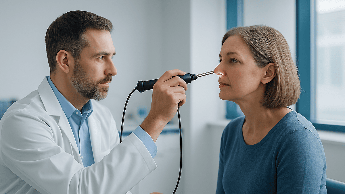
A nasal endoscopy in sinusitis lets an ear, nose and throat clinician look directly inside the nose with a thin, lighted camera, checking for swollen lining, pus draining from the sinus openings, polyps, crusting or a deviated septum. Those findings help separate viral, bacterial and chronic inflammatory disease, and guide whether saline rinses, anti-inflammatory sprays, antibiotics, imaging or a surgical opinion fit. The treating team makes the final decision.
The chair tilts back a few degrees, the lights dim, and a clinician holds something that looks like a slim metal straw with a lens on the end. For a lot of people, this is the moment the word “sinusitis” stops being a vague label and becomes something a person can actually look at. They have been living with pressure behind the cheekbones for months, a nose that never quite clears, a taste for coffee that faded somewhere along the way.
A nasal endoscopy for sinusitis takes only a few minutes, yet it changes the conversation. Instead of guessing from a list of complaints, the clinician can see whether the lining is swollen, whether anything is draining from the sinus openings, whether polyps are crowding the passages. The pictures on the screen are not pretty, but they are honest.
What follows is an attempt to be equally honest: what the scope can see, what it cannot, and how the view inside the nose tends to shape the plan that comes next.
What happens during a nasal endoscopy for sinusitis?
An endoscope is a narrow tube, rigid or flexible, with a light source and a tiny camera that sends images to a screen. The rigid version used for most adult sinus exams is thinner than a pencil; the flexible version bends around corners and is often chosen for children or for anyone with a very narrow nose.
You sit upright. The clinician usually sprays the inside of the nose with a decongestant, which shrinks the lining so the scope has room, and often a topical anesthetic, which numbs the surface. The spray tastes bitter as it trickles toward the throat, and that is the part most people remember.
Then the scope goes in. The clinician follows a set route: along the floor of the nose to the back, where the nose meets the throat; up under the middle turbinate, a curled shelf of bone and lining where the front sinuses drain; and higher still toward the openings of the sinuses at the back. Each pass takes seconds. The whole exam typically lasts only a few minutes, according to the Cleveland Clinic’s patient guide, and nothing about it requires sedation.
If pus is seen draining from a sinus opening, a swab can be taken through the scope for culture, a laboratory test that grows and identifies bacteria. Some clinics record still images so you can see what the clinician saw.
Afterward you walk out. The numbness fades over an hour or so, and the only common souvenir is a sneeze or a trace of blood on a tissue. Compared with the months of symptoms that often precede it, the exam is a remarkably small event.
Can a nasal endoscopy examine the sinuses themselves?
Mostly, no, and that distinction matters. The sinuses are air-filled rooms inside the bones of the face. In a nose that has never had surgery, their doorways are a few millimeters wide, far too small for a scope to pass through. What endoscopy examines is the hallway: the nasal cavity and the narrow corridors into which each sinus empties.
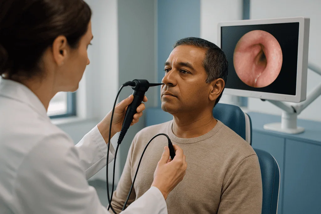
The most important of those corridors is the ostiomeatal complex, the shared drainage pathway beneath the middle turbinate where the maxillary (cheek), frontal (forehead) and front ethmoid (between-the-eyes) sinuses all open. Think of it as the landing at the top of a staircase onto which three bedroom doors open. If the landing is swollen shut, every room behind it fills with mucus. If pus is pooling on the landing, at least one room is infected.
Farther back sits the sphenoethmoidal recess, where the sphenoid sinus (deep behind the eyes) and the rear ethmoid cells drain. A good exam looks there too, because trouble in the back sinuses produces different symptoms, often pain at the top of the head or behind the eyes rather than in the cheeks.
So the scope reads the sinuses indirectly, from what spills out of them and from how swollen their doorways are. That indirect reading is surprisingly useful. The Mayo Clinic lists nasal endoscopy alongside imaging as a standard way to evaluate suspected chronic sinusitis precisely because the drainage pathways tell so much of the story.
After endoscopic sinus surgery, the picture changes. Once the doorways have been widened, the same scope can look directly into the cavities, which is why surgeons use it to monitor healing in the months that follow.
What the scope actually shows in sinusitis: the findings clinicians look for
A trained eye reads the nasal lining the way a dermatologist reads skin. Several findings carry weight.
Swelling, called edema, is the most common. Healthy lining is pink and moist with a visible gap between the turbinates and the septum, the wall dividing the two sides of the nose. Inflamed lining looks pale, boggy and crowded, and the gaps close up. Redness alone says little; the degree and location of swelling say more.
Discharge is the next clue. Clear, thin mucus leans toward allergy or a viral cold. Thick, cloudy or yellow-green discharge, called mucopus, draining from the middle meatus points to infection in the front sinuses; the same material in the recess at the back points to the sphenoid or rear ethmoids. The side matters too. One-sided drainage prompts a closer look for a localized cause.
Polyps are soft, pale, grape-like swellings of the lining that hang into the passages. They are not cancer, and they are a hallmark of one form of chronic sinusitis. Their size and whether they block the drainage corridors change the treatment conversation considerably.
Crusting, dried secretions stuck to the lining, suggests a dry or damaged surface and sometimes previous surgery. A deviated septum or an enlarged turbinate can narrow a corridor enough to trap mucus. In children, the scope may reveal enlarged adenoids, lymph tissue at the back of the nose that can mimic or worsen sinus symptoms.
Finally, there is the negative finding. A clean exam during active symptoms is informative in its own right. The NHS notes that facial pain and congestion have many causes, and a normal view inside the nose shifts attention toward migraine, dental problems or jaw joint pain rather than more sinus treatment.
Nasal endoscopy versus CT scan versus symptoms alone
People sometimes assume a scan is the “real” test and endoscopy is a warm-up. In practice the two answer different questions, and symptoms on their own answer a third.
| Approach | What it shows well | What it misses | Typical place in the pathway |
|---|---|---|---|
| Symptom history and exam without a scope | Pattern and duration, fever, triggers, response to past treatment | Cannot confirm inflammation or distinguish causes of facial pain | First step for most acute episodes in primary care |
| Nasal endoscopy | Live view of lining, pus, polyps, septum, drainage pathways; allows a targeted culture | Interior of unoperated sinus cavities; bone detail | Specialist assessment, especially for persistent, recurrent or one-sided symptoms |
| CT scan of the sinuses | Every cavity, bony anatomy, extent of disease, surgical road map | Cannot show pus versus thick mucus, or how tissue looks and behaves; involves radiation | When chronic disease is suspected, before surgery, or when complications are a concern |
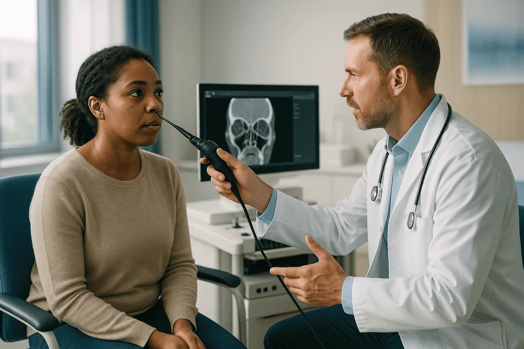
A CT scan uses X-rays from many angles to build cross-sectional pictures of the sinuses. It is excellent at showing which cavities are filled and whether bony walls are thin or breached. Yet a scan taken during a bad cold can look alarming and settle on its own, which is why guidelines reserve imaging for persistent or complicated cases. The Mayo Clinic describes imaging and endoscopy as complementary tools in chronic sinusitis, not rivals.
Endoscopy, meanwhile, is cheap in effort, repeatable and radiation-free. It can be done at every visit to track whether swelling is receding, something no one would do with serial scans.
The honest summary: symptoms tell you something is wrong, endoscopy tells you what the lining is doing today, and CT tells you how deep the problem runs.
Who is usually referred for nasal endoscopy, and who is usually asked to wait?
Not everyone with a stuffy nose needs a scope, and a good system is selective.
Referral typically follows symptoms that have outlasted a normal cold. The Mayo Clinic defines chronic sinusitis as inflammation lasting 12 weeks or longer despite treatment attempts, and that threshold is the clearest trigger for an endoscopic look. Recurrent acute episodes, several distinct infections a year with clear gaps between, also warrant one. So do features that do not fit ordinary sinusitis: symptoms on one side only, blood-tinged discharge, loss of smell that has not recovered, facial numbness, or a nose that feels blocked by something rather than swollen. Anyone being considered for sinus surgery will have endoscopy beforehand, because the surgeon needs to see the anatomy directly.
Children with persistent mouth breathing, snoring or recurrent infections are often scoped with a flexible instrument to check the adenoids as well as the sinuses.
Who waits? Most people in the first week or two of an acute episode. The NHS advises that sinusitis after a cold usually improves within 2 to 3 weeks and is commonly viral, so early endoscopy would show inflammation that is about to resolve anyway. The CDC’s guidance for clinicians likewise frames the first 10 days as a watch-and-support period unless symptoms are severe.
Waiting is not the same as being dismissed. Primary care can manage most acute sinusitis with saline rinses, rest and symptom relief, and the referral door opens if the picture drags on or changes. The judgment about timing sits with the clinician who knows your history, and it is reasonable to ask what would move you from the “wait” group to the “scope” group.
Can you have an endoscopy with a sinus infection?
Yes, and in many ways it is the most informative time to have one.
During an active infection the clinician can see exactly where pus is coming from and how much the lining has swollen. If treatment decisions hinge on whether bacteria are involved, a swab taken under direct vision from the middle meatus is more useful than a blind swab of the nostril, which mostly collects the ordinary bacteria that live at the front of the nose. The culture result, usually back within a few days, can confirm or redirect an antibiotic choice if one is being made.
There are practical caveats. A nose that is very swollen may limit how far the scope can travel, even after decongestant spray, so the exam may be partial. A clinician may also ask you to come back once the acute phase settles to see what the baseline lining looks like, because persistent findings between infections are what define chronic disease.
Having an infection does not make the procedure risky. The scope touches only the surface; it does not carry bacteria deeper. People with bleeding disorders, those taking blood-thinning medicines, or anyone with a recent facial injury should mention it, since the clinician may be gentler or defer the exam, but these are judgment calls rather than prohibitions.
One misunderstanding is worth clearing up. Some people fear that being scoped while infected will “spread” the infection to the brain or eye. The scope stays within the nasal cavity, and complications of sinusitis spreading beyond the sinuses are driven by the disease itself, not by looking at it. If anything, an exam that catches heavy pus or unexpected swelling is the thing that prompts timely action.
Is nasal endoscopy painful? What it really feels like
Uncomfortable, yes. Painful, rarely. That is the consistent report from patient guides, and it matches what most people say when the scope comes out.
The decongestant and anesthetic sprays go in first, and they are the part people dislike most: a sting for a moment, then a bitter drip down the back of the throat and a numb, slightly swollen feeling across the palate. Within a few minutes the nose feels wider and less sensitive.
When the scope enters, the sensation is pressure rather than sharpness, like a cotton swab pushed farther than you would push it yourself. Eyes water. There is an urge to sneeze, which usually passes if you breathe slowly through the mouth. A tickle at the back of the throat when the scope reaches the nasopharynx can trigger a brief gag in some people. Most of the exam involves the clinician angling the camera while you sit still, and the whole thing is over in a few minutes, as the Cleveland Clinic’s description notes.
A few situations make it harder. A badly deviated septum can create a tight spot where the scope rubs. Heavily inflamed lining is more sensitive and bleeds more easily. Anxiety tightens everything. Telling the clinician in advance, agreeing on a hand signal to pause, and keeping your eyes on a fixed point all help.
Children are examined with a thinner flexible scope and often tolerate it better than their parents expect, particularly if they are allowed to watch the screen.
Afterward, the lining may feel raw for an hour or two, and a pink streak on a tissue is normal. Significant pain or bleeding that does not stop with gentle pressure is uncommon and should be reported.
How does nasal endoscopy for sinusitis shape treatment decisions?
This is where the few minutes in the chair earn their keep. The pattern on the screen tends to sort people into recognizable paths, each handled differently by the treating team.
Pus draining from a sinus corridor, in someone whose symptoms have lasted beyond 10 days or who improved and then worsened again, raises the likelihood of bacterial infection. The CDC describes exactly those two patterns as the ones that may justify antibiotics, medicines that kill or stop the growth of bacteria. Even then, the choice to prescribe, the drug and the duration belong to the clinician; endoscopy only sharpens the odds.
Swelling without pus points toward inflammation rather than infection. Here the plan usually centers on saline irrigation, which physically washes mucus and irritants from the lining, and intranasal corticosteroid sprays, medicines that calm inflammation in the lining over days to weeks of regular use. The Mayo Clinic lists both as mainstays for chronic disease. The scope shows whether the swelling is receding at follow-up, which is how the team judges whether to continue, intensify or rethink.
Polyps shift the conversation again. Their presence places someone in the “with polyps” form of chronic sinusitis, where corticosteroid-based treatment is the foundation, where allergy and asthma are often evaluated in parallel, and where, for severe disease that persists despite medical care, the Mayo Clinic notes that injectable biologic medicines targeting specific inflammatory signals may be considered.
A fixed structural block, such as a severely deviated septum or scarring, suggests that medicines alone may not open the corridor, and a surgical opinion may be offered after a trial of medical therapy.
In each path the scope does not decide. It makes the decision better informed.
Chronic sinusitis diagnosis: why the scope is part of the definition
Here is a point that surprises many people. Chronic sinusitis is not diagnosed by symptoms alone, however long they have lasted. Modern definitions, reflected in the Mayo Clinic’s description, require both a symptom pattern persisting 12 weeks or more and objective evidence of inflammation, which means either an endoscopic finding or a CT finding. Without that evidence, months of facial pressure may be chronic rhinitis, migraine or something else wearing a sinus costume.
Endoscopy supplies that objective evidence in the office. Swollen lining in the middle meatus, mucopus, or polyps are each sufficient. A clean exam sends the clinician looking elsewhere, which spares the person repeated courses of treatment aimed at the wrong target.
The scope also divides chronic sinusitis into its two broad forms, with polyps and without. The distinction is not academic. Polyp disease tends to behave like other inflammatory conditions, often travels with asthma, and is more prone to recurrence after surgery. Disease without polyps more often relates to drainage obstruction or lingering infection. Treatment emphasis differs accordingly.
Follow-up endoscopy turns a single snapshot into a film. If the lining looks better at eight weeks, the team has a reason to stay the course even when the person feels only partly improved. If it looks the same or worse, that is a reason to add imaging, reconsider the diagnosis or discuss surgery sooner.
The quiet value of all this is accountability. Rather than asking “do you feel better?” and hoping the answer is reliable, the clinician can compare two images of the same corridor. Symptoms still matter most to the person living with them, but the scope keeps the diagnosis honest and the plan tethered to what is actually happening in the nose.
What the next days and weeks usually look like
The hour after the exam is unremarkable. The numbness in the palate and throat wears off, swallowing feels normal again, and most people are advised not to eat or drink hot things until it does, to avoid a burn they cannot feel. Sneezing and a runny nose for the rest of the day are common. A little blood on a tissue is expected; heavy bleeding is not.
What happens next depends on what was seen, but there is a familiar rhythm. If the findings suggested an acute bacterial infection and antibiotics were prescribed, the CDC notes that most people begin to feel better within a few days, and the full course is finished as directed even after improvement. If the plan is inflammation-focused, with saline rinses and an intranasal corticosteroid spray, the Mayo Clinic cautions that these work gradually; a fair trial is measured in weeks, not days, and daily consistency matters more than any single application.
A follow-up visit is often arranged in the range of several weeks to a few months, where the scope comes out again to compare. This is when the plan is adjusted. Allergy testing or a CT scan may be added. People with polyps may be asked about asthma or aspirin sensitivity, because those patterns cluster.
For those whose exam showed a structural problem or whose disease has not responded to a reasonable medical trial, this is also when a conversation about surgery may begin. That conversation is a beginning rather than a booking; it includes imaging, a review of what has and has not been tried, and time to ask questions.
The timeline is rarely dramatic. Chronic sinusitis is managed over seasons, and the first endoscopy is the opening chapter rather than the climax.
How safe is endoscopic sinus surgery, and when does the scope lead there?
Endoscopic sinus surgery uses the same camera technology, now with fine instruments alongside it, to widen the natural sinus openings and remove polyps or diseased tissue through the nostrils, without external cuts. The Mayo Clinic describes it as an option when medical treatment has not adequately controlled chronic sinusitis, and in practice it is offered after a reasonable trial of medicines rather than as a first move.
The scope leads there in a few circumstances: extensive polyps blocking drainage, a fixed structural obstruction, scarring from previous disease, or persistent infection confined to a cavity that cannot drain. Imaging confirms the map before anyone operates.
On safety, the honest framing is that this is widely performed surgery in which serious complications are uncommon but real, and the risks are tied to anatomy. The sinuses sit beside the eye socket and beneath the floor of the skull, separated in places by bone thinner than an eggshell. Rarely, instruments can injure structures around the eye, causing bruising or, very rarely, affecting vision, or breach the skull base and cause a leak of cerebrospinal fluid, the clear fluid surrounding the brain. Image-guided navigation systems exist to reduce these risks, and surgeons discuss them explicitly during consent.
Common, expected effects are bleeding in the first days, crusting that requires saline rinses and office cleaning for weeks, fatigue and temporary changes in smell. Polyps can regrow, so surgery is best understood as improving drainage and access for ongoing medical therapy rather than ending the condition.
Alternatives are usually continued or intensified medical treatment, and for polyp disease, biologic therapy may be discussed. Whether surgery is appropriate, and when, is a decision made with the treating team after weighing symptoms, findings and personal circumstances.
What people often get wrong about nasal endoscopy and sinusitis
Misunderstandings travel faster than sinus mucus, so a few corrections are in order.
The first is that endoscopy is a kind of scan. It is not. There is no radiation, no machine you lie inside, and no report that arrives days later. It is a direct look, and the clinician can usually describe the findings before the scope is even out.
The second is that green or yellow mucus seen on the screen means bacteria and therefore antibiotics. Color comes from immune cells and their enzymes, which appear in viral infections too. The CDC is explicit that discolored mucus alone does not indicate a bacterial infection; duration and the pattern of worsening matter more.
A third is the fear that the scope reaches the brain or the eye. It stays within the nasal cavity, separated from both by bone. The sensation of pressure “behind the eye” is the lining being touched near the back of the nose, nothing more.
People also confuse nasal endoscopy with the endoscopy used for the stomach or bowel. Those require fasting and often sedation. This one requires neither.
Then there is the assumption that a clean exam means the clinician thinks the symptoms are imaginary. It means the opposite: the symptoms are real and the sinuses are probably not the cause, which is useful information that redirects care.
Finally, the belief that surgery ends sinusitis for good. It can relieve blockage and make medicines reach farther, but the underlying tendency to inflammation often remains, especially with polyps. Ongoing care after surgery is the norm, not a sign that something went wrong.
None of these corrections should discourage anyone. They simply set expectations where the evidence places them.
Questions to ask your care team before and after the scope
A short exam leaves little time for discussion unless you arrive with questions. These are the ones that tend to pay off, grouped by moment.
Before the exam, it helps to ask how to prepare, whether to stop or continue any nasal sprays beforehand, and what to mention about blood thinners, previous nasal surgery or anxiety about the procedure. Ask whether the exam will be recorded so you can see the images yourself.
Immediately after, the useful questions are specific:
- What exactly did you see, and on which side?
- Was there pus, swelling, polyps or a structural narrowing, or was the exam normal?
- Does this look like acute infection, chronic inflammation or something else altogether?
- Do I need a culture, allergy testing or a CT scan, and what would each change?
When the plan is discussed, ask how long a fair trial of the proposed treatment is, how you will both know whether it is working, and whether a follow-up endoscopy is planned to compare. Ask which symptoms should prompt an earlier call rather than waiting for the scheduled visit.
If surgery is mentioned, slow down and ask what medical options remain untried, what the specific goals of an operation would be for your anatomy, what the realistic risks are given where your disease sits, and what aftercare involves in the first weeks.
One last question is often overlooked: “If this were your nose, what would you want to understand before deciding?” Clinicians tend to answer that one candidly, and the reply usually reveals what they consider the most important factor in your particular case.
When to call your doctor
Most sinusitis, and nearly every nasal endoscopy, passes without incident. A handful of signs mean the situation has changed and deserve a same-day call or urgent care.
The CDC and NHS both flag the following as reasons to seek prompt medical attention during a sinus infection: swelling, redness or pain around the eyes or eyelids; changes in vision, including double vision; a severe headache unlike your usual pattern; a stiff neck; confusion or unusual drowsiness; and a high fever that persists. These can signal infection spreading beyond the sinuses toward the eye socket or the lining of the brain, and they are emergencies regardless of what the scope showed last week.
Less dramatic but still important, the CDC advises contacting a clinician when symptoms last longer than 10 days without improving, when they get worse after initially getting better, or when fever and facial pain are severe for more than a few consecutive days. The NHS adds that symptoms persisting beyond about 3 weeks, or recurring frequently, warrant review.
After the endoscopy itself, call if bleeding does not stop after 15 minutes of steady pressure on the soft part of the nose while leaning forward, if you develop significant pain that worsens rather than fades, or if you notice a persistent clear, watery drip from one nostril, particularly when leaning forward, which should always be assessed.
If you have been started on a new medicine and develop a rash, swelling of the lips or face, or difficulty breathing, stop and seek emergency help; those are allergic reactions rather than side effects to tolerate.
Everything else, from lingering stuffiness to questions about whether the plan is working, belongs at the follow-up visit, and the people best placed to decide what to do next are the team who looked inside your nose.
Frequently asked questions
Can a nasal endoscopy examine the sinuses directly?
In a nose that has not had surgery, no; the natural sinus openings are too narrow for the scope. The exam inspects the nasal cavity and the corridors where each sinus drains, reading infection or inflammation from pus, swelling and polyps at those openings. After endoscopic sinus surgery widens the openings, the same scope can look directly into the cavities to monitor healing.
Is nasal endoscopy painful?
For most people it is uncomfortable rather than painful. The numbing and decongestant sprays sting briefly and taste bitter, then the scope produces pressure, watery eyes and an urge to sneeze. The exam lasts only a few minutes. A tight spot from a deviated septum or very inflamed lining can make it harder, and telling the clinician about anxiety or a sensitive nose beforehand helps.
Can you have an endoscopy with a sinus infection?
Yes. An active infection is often the most informative moment, because the clinician can see where pus is draining and take a targeted swab for culture. Swelling may limit how far the scope travels, so a repeat exam once things settle is sometimes arranged. The scope does not spread infection; it stays on the surface of the nasal lining.
How long does a nasal endoscopy procedure take?
The scope portion typically takes only a few minutes, according to patient guidance from major clinics, though you should allow extra time for the sprays to work and for discussion afterward. There is no fasting, no sedation and no recovery period. Most people drive themselves and return to normal activities immediately, with numbness fading within about an hour.
How safe is endoscopic sinus surgery?
It is widely performed, and serious complications are uncommon, but they are real because the sinuses border the eye socket and skull base. Rare risks include injury to structures around the eye or a leak of cerebrospinal fluid; common expected effects are bleeding, crusting and temporary smell changes. Your surgeon should discuss risks specific to your anatomy, and the decision rests with you and the treating team.
What should I know about endoscopic sinus surgery safety and recovery?
Recovery centers on saline rinsing and office visits where crusts are cleared from the healing sinuses over several weeks. Expect fatigue and some bleeding early on. Polyps can regrow, so continued medical therapy afterward is standard rather than a sign of failure. Ask what red flags, such as vision change or a clear watery drip, should prompt an urgent call.
What does chronic sinusitis diagnosis require besides symptoms?
Current definitions require symptoms persisting 12 weeks or longer and objective evidence of inflammation, found either on nasal endoscopy or a CT scan. Months of facial pressure without such evidence may reflect rhinitis, migraine or dental causes instead. This is why specialists scope people with long-standing symptoms rather than treating on history alone.
Does green mucus seen on the scope mean I need antibiotics?
No. Discoloration comes from immune cells and their enzymes, which appear in viral colds as well as bacterial infections. The CDC advises that color alone does not indicate bacteria. More telling are symptoms lasting beyond 10 days without improvement or a pattern of getting better and then worse. The decision about antibiotics belongs to your clinician, informed by the whole picture.
Will I need a CT scan if I have had a nasal endoscopy?
Not necessarily. Endoscopy often provides enough evidence to confirm chronic inflammation and start treatment. A CT scan is usually added when symptoms persist despite a fair medical trial, when surgery is being considered, when findings are one-sided or unusual, or when a complication is suspected. The scan shows the full extent of disease and bony anatomy that the scope cannot see.
What happens if my nasal endoscopy is completely normal?
A normal exam during symptoms is useful information: it suggests the sinuses are probably not the source of your pain or pressure. Attention then turns to other causes such as migraine, tension-type headache, dental or jaw joint problems, or nasal allergy without sinus involvement. It does not mean the symptoms are imagined; it means treatment can be redirected toward a more likely explanation.
References
This article is for general information only and is not a substitute for professional medical advice. Please consult a qualified doctor about your individual situation.

More from the Blog

What Results Does Endoscopic Sinus Surgery (ESS) Usually Bring, and Can Sinus Disease Return?
Endoscopic sinus surgery usually widens blocked sinus drainage pathways so that mucus can clear and medicines can reach inflamed lining. Most people report less…

How Long Does FESS Recovery Take? Crusting, Congestion and When Breathing Improves
Recovery from functional endoscopic sinus surgery (FESS) usually unfolds in stages rather than on one day. Most people feel congested and tired for the…

When Is an Intratympanic Steroid Injection Considered for Sudden Hearing Loss?
An intratympanic steroid injection is usually considered for sudden sensorineural hearing loss in two situations: as initial treatment when oral corticosteroids are unsuitable, unwanted…

Is FESS the Next Step? When Chronic Sinusitis Stops Responding to Medical Treatment
FESS (functional endoscopic sinus surgery) is usually considered only after chronic sinusitis, meaning symptoms lasting 12 weeks or more, has failed a full course…

SMR or Septoplasty: What Is the Difference, and When Do Surgeons Choose Submucous Resection?
SMR (submucous resection) and septoplasty are both operations that straighten the wall between the nostrils. SMR, the older technique, removes most of the deviated…

Sinusitis Warning Signs: High Fever, Facial Swelling and Vision Changes That Need Urgent Care
Most sinusitis improves on its own within about 7 to 10 days and can be managed at home. Seek urgent care the same day…