

What Does a Swallowing Study Show? Endoscopy and Imaging Tests Used in Dysphagia Care
Key Takeaways
- Silent aspiration, when liquid enters the airway without any cough, is a key reason clinicians order imaging rather than relying on watching someone drink.
- The modified barium swallow uses a moving X-ray to show the whole swallow in motion, including the exact moment material passes the vocal cords.
- FEES passes a slim camera through the nose and uses real food, so it can run longer and be repeated without radiation, though the image blanks briefly during the swallow itself.
- Throat-focused studies and esophagus-focused tests such as barium esophagram, endoscopy, and manometry answer different questions, so many people need one of each.
- A swallowing study is not pass or fail; the report maps which textures and postures were safe, and those findings become the diet and therapy plan.
- Results are a snapshot rather than a life sentence, and repeat studies after therapy are commonly used to ease restrictions as soon as the evidence supports it.
A swallowing study for dysphagia records what happens to food and liquid as you swallow, most often with a moving X-ray (videofluoroscopy) or a thin camera passed through the nose (FEES). It shows whether anything enters the airway, where material pools, how well the throat and esophagus move, and which textures are safest, so the care team can tailor therapy, diet, and any further testing.
It usually starts small. A pause mid-sentence at dinner, a cough that arrives a beat after the sip of water, a pill that seems to sit halfway down. Then one evening a daughter watches her father clear his throat for the fourth time in ten minutes and realizes she has been counting.
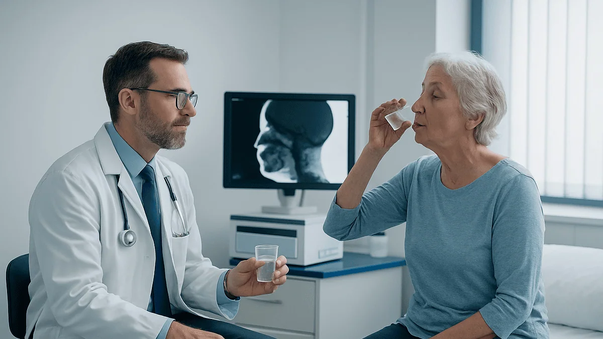
A few days later, a speech-language pathologist sits beside him in a radiology suite with a tray of cups and a spoonful of something that looks like pudding. A screen flickers to life, and for the first time anyone can actually see the swallow instead of guessing at it. That is what a swallowing study for dysphagia does: it turns a hidden, half-second event into something a team can watch, replay, and act on.
Most people have never heard the names of these tests until they need one. This guide walks through what each shows, what you swallow, what the results mean, and where the decisions are made.
Why a swallowing study for dysphagia is ordered in the first place
Dysphagia is the medical word for difficulty swallowing, whether the trouble is starting a swallow, moving food through the throat, or getting it down the esophagus, the muscular tube that carries food to the stomach. A normal swallow takes roughly a second and coordinates dozens of muscles with several cranial nerves, all while the airway seals itself shut. When any part of that sequence falters, food or liquid can head toward the lungs instead of the stomach.
Clinicians divide the problem into two broad territories. Oropharyngeal dysphagia lives in the mouth and throat and is often neurological: stroke, Parkinson disease, multiple sclerosis, and the muscle changes that follow head and neck cancer treatment are common causes, as MedlinePlus and Mayo Clinic describe. Esophageal dysphagia sits lower and tends to involve narrowing, inflammation, acid reflux, or muscle disorders of the esophagus itself. The two feel different to patients, and they call for different tests.
Here is the uncomfortable fact that drives most referrals: you cannot reliably tell from the outside whether a swallow is safe. A cough is a good sign in one sense, because it means the airway noticed the intrusion. Yet a substantial share of people who inhale liquid do so silently, with no cough and no visible distress. This is called silent aspiration, and it is one reason an exam that only watches a person drink can miss what an imaging study catches.
So the study is ordered not to confirm what everyone already suspects, but to answer questions the eye cannot: Where does the swallow break down? Does anything reach the airway? Do certain textures behave better than others? Are there simple posture or pacing changes that fix it on the spot? Those answers shape everything that follows.
What actually happens during a swallowing study, step by step
Most swallowing studies are run by a speech-language pathologist, a clinician trained in swallowing and communication, often working with a radiologist or an ear, nose and throat specialist. You are awake, usually seated upright in a chair or on a table tilted to sitting position, since lying flat changes how a swallow behaves and would not reflect real meals.
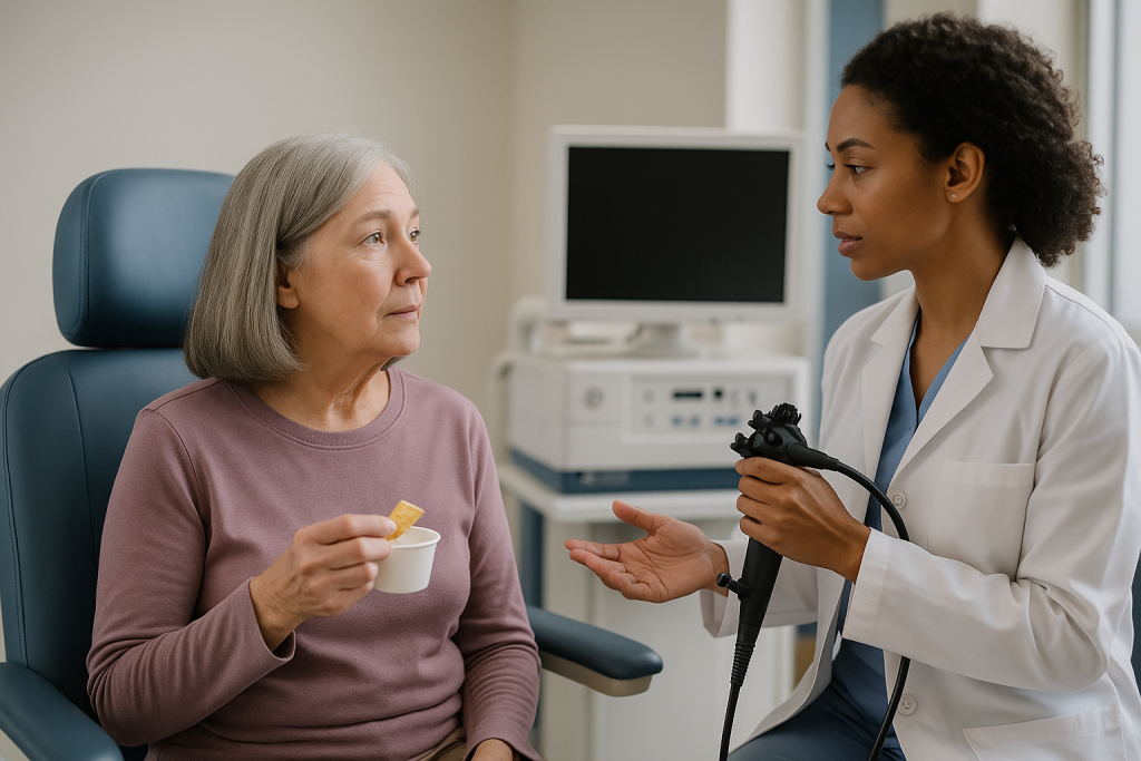
The test is built around trials. You take small, measured amounts of different consistencies, one at a time, while the clinician watches on a screen and records. A typical sequence might move from thin liquid to a thicker liquid, then a puree, then a soft solid, with the order and quantities adjusted to what looks safe. If a sip behaves poorly, the clinician may stop that texture and try another rather than push ahead.
Between trials the clinician may ask you to tuck your chin, turn your head, hold your breath before swallowing, or swallow twice. These are not tests of obedience; they are experiments. If a chin tuck keeps liquid out of the airway on screen, that becomes a practical strategy you can use at home the same day.
The recorded portion is usually brief. MedlinePlus notes that a barium swallow of the esophagus typically takes about 30 to 60 minutes including setup, and the throat-focused video study is often shorter than that because it concentrates on a handful of swallows. Afterward the clinician reviews the recording frame by frame. Some findings are discussed with you before you leave; a written report goes to the referring physician, who folds it into the wider picture of your diagnosis and care plan.
Bedside swallow evaluation: the exam that usually comes first
Before anyone books an imaging room, most people meet the swallow team at the bedside or in a clinic chair. A bedside swallow evaluation, sometimes called a clinical swallow examination, has no camera and no X-ray. The clinician asks about your history, looks at the strength and movement of your lips, tongue, palate, and jaw, listens to your voice, checks your cough, and then watches you swallow water and perhaps a few textures.
The clinician is looking for clues: a wet or gurgly voice after a sip, a delayed cough, food left in the mouth, throat clearing, a drop in breathing comfort. In hospitalized stroke patients this screening matters enough that the American Heart Association and American Stroke Association guideline recommends checking swallowing before a patient is given anything to eat, drink, or take by mouth, because the consequences of missing an unsafe swallow include pneumonia.
What a bedside exam cannot do is see inside. It cannot show whether liquid pooled above the airway, whether some slipped beneath the vocal cords without a cough, or how far a mouthful traveled before the swallow triggered. It is a screening and planning tool, and clinicians treat it that way. A reassuring bedside exam may be enough for someone whose history is benign. A worrying one, or a normal-looking one in a person with a high-risk condition, generally leads to instrumental testing.
Patients sometimes feel the bedside exam was casual because nothing was plugged in. In practice it decides which imaging test is most useful, which textures to try first, and how urgently to proceed. Think of it as the interview that determines what the cameras should look for.
Modified barium swallow test (videofluoroscopy): what the moving X-ray shows
The modified barium swallow test, also called a videofluoroscopic swallow study or VFSS, is the exam most people picture when they hear swallowing study. Barium is a chalky mineral compound that shows up bright white on X-ray and passes through the body unabsorbed. It is mixed into liquids and foods so that each mouthful becomes visible as it moves.
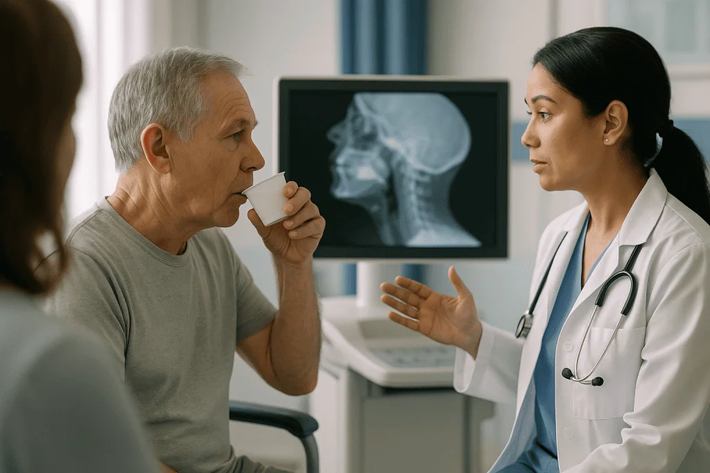
You sit sideways to a fluoroscopy machine, an X-ray unit that produces a live moving image rather than a single still picture. As you swallow, the clinician and radiologist watch the barium travel from the lips, over the tongue, through the throat, past the airway opening, and into the top of the esophagus. Everything is recorded so it can be slowed down and replayed.
The view answers questions with unusual precision. It shows whether the swallow reflex fires on time or lags, whether the tongue base and throat muscles squeeze fully, whether the airway closes, and whether the upper esophageal sphincter, the muscular valve at the throat’s exit, opens wide enough. Most importantly it shows penetration, when material dips toward the vocal cords, and aspiration, when it passes below them into the airway. It also reveals residue, the coating of barium left behind after the swallow, which can be inhaled later.
The trade-offs are real but modest. There is a small dose of ionizing radiation, which MedlinePlus describes as low for this type of study, and clinicians keep the recording time short for that reason. The barium taste is unremarkable to most people, and the images cannot show the actual color or wetness of tissues. For a view of the whole swallow in motion, though, nothing else offers quite the same picture.
FEES swallow test: watching the throat with a camera
The FEES swallow test stands for fiberoptic endoscopic evaluation of swallowing. A flexible endoscope, a thin tube no wider than a drinking straw with a camera at the tip, is passed gently through one nostril and positioned just above the throat. From there it looks straight down at the vocal cords and the entrance to the airway while you eat and drink ordinary foods, often tinted with a little food coloring so they stand out.
Where the X-ray shows shadows moving, FEES shows the tissue itself in full color. Clinicians can see swelling, secretions, how the vocal cords move, and how much food remains in the throat’s pockets after each swallow. Because no radiation is involved, the exam can run longer and can be repeated as often as needed, which makes it useful for tracking recovery or testing a full meal rather than a few sips.
It has one well-known blind spot. At the exact moment of the swallow, the throat squeezes around the camera and the image goes white for a fraction of a second, so the passage of food past the vocal cords is inferred from what is seen immediately before and after rather than watched directly. The X-ray study does not have this gap. Conversely, FEES sees residue and airway anatomy that fluoroscopy can only hint at, which is why Mayo Clinic lists both as standard tools rather than rivals.
A numbing spray is sometimes offered for the nostril, though many centers proceed without it so that throat sensation is not dulled. The exam is portable, which matters for people in intensive care or long-term facilities who cannot easily be moved to a radiology suite.
Barium swallow, upper endoscopy and esophageal manometry for dysphagia
When the problem seems to sit lower, in the chest rather than the throat, a different set of tests takes over. People with esophageal dysphagia often describe food sticking behind the breastbone several seconds after swallowing, and the throat-focused exams may look entirely normal.
A standard barium swallow, sometimes called an esophagram, follows barium down the full length of the esophagus with still and moving X-ray images. It outlines narrowings, pouches, hiatal hernias, and the shape of the lower esophageal sphincter, the valve into the stomach. MedlinePlus describes it as a roughly 30 to 60 minute test in which you may be asked to drink in several positions and occasionally swallow a barium-coated tablet or marshmallow to see where solids hang up.
Upper endoscopy, or EGD, uses a camera passed through the mouth under sedation to look directly at the lining of the esophagus and stomach. It can sample tissue to check for inflammation, eosinophilic esophagitis, or other changes, and in some cases the same procedure allows a narrowed segment to be stretched. Mayo Clinic lists it among the core investigations for esophageal symptoms.
Esophageal manometry for dysphagia measures pressure rather than pictures. A slim catheter with sensors is passed through the nose into the esophagus, and you take small swallows while it records how the muscles contract in sequence and how the lower valve relaxes. It is the test for motility disorders such as achalasia, in which the valve fails to open, and it is uncomfortable but brief. A related test, ambulatory pH monitoring, tracks acid exposure over about a day when reflux is suspected as the driver. Which of these is ordered, and in what order, depends on the story you tell your physician.
Which swallowing study is right for which problem? A comparison table
No single test does everything, and the choice is less about which is best than about which question needs answering. The table below summarizes what each exam sees and what it asks of you.
| Test | Best for | What you swallow | Radiation | Main limitation |
|---|---|---|---|---|
| Bedside swallow evaluation | Screening and planning; first check after stroke | Water, sometimes a few textures | None | Cannot see silent aspiration or inside the throat |
| Modified barium swallow (VFSS) | Watching the whole oral and throat swallow in motion | Barium-mixed liquids, puree, soft solid | Low dose | Short recording window; no tissue color or detail |
| FEES | Airway anatomy, residue, secretions, repeat testing | Real food and drink, often tinted | None | Image whites out at the moment of the swallow |
| Barium swallow (esophagram) | Narrowing, pouches, hernia along the esophagus | Barium liquid, sometimes a coated tablet | Low dose | Does not show throat safety in detail |
| Upper endoscopy (EGD) | Lining of the esophagus; biopsies; stretching | Nothing; done under sedation | None | Sedation and recovery time; does not show swallow function |
| Esophageal manometry | Muscle coordination and valve pressure | Small sips of water or gel | None | Nasal catheter is uncomfortable; no images |
A practical way to read this: if the symptom is coughing, choking, or a wet voice with meals, expect the top three rows. If food seems to stick lower and arrive late, expect the bottom three. Many people need one from each group, and the sequence is set by the referring clinician and the swallow team based on what the first exam shows. Mayo Clinic and the NHS both describe this stepwise approach, starting with the least invasive test that can plausibly answer the question.
What is the gold standard test for dysphagia?
Search engines love this question, and the honest answer is that it depends on which kind of dysphagia you mean. For swallowing problems in the mouth and throat, the modified barium swallow has traditionally been described as the reference standard because it shows the entire sequence, including the instant food passes the airway. FEES is widely regarded as its equal for detecting aspiration and residue, and many specialists now describe the two as complementary reference tests rather than ranking one above the other.
That framing matters more than it sounds. A test earns gold standard status by being the thing other tests are compared against, not by being right for every person. A frail patient who cannot be transported safely may get a more useful answer from FEES at the bedside than from a fluoroscopy appointment that never happens. Someone whose main worry is the exact timing of airway closure may be better served by the X-ray view.
For esophageal dysphagia the landscape shifts entirely. Upper endoscopy is usually the first-line test when a structural cause such as a stricture or inflammation is suspected, because it can both see and sample tissue. Manometry is the reference test for motility disorders, since only pressure recordings can show whether the muscle sequence is coordinated. Mayo Clinic’s overview lays out this division without naming one universal winner, and the NHS similarly describes a menu of tests chosen to fit the symptom.
So when someone asks for the gold standard, the more useful question back is: gold standard for what? The team’s job is to match the test to the suspected failure point, and a well-chosen second-tier exam often beats a poorly matched first-tier one.
Who is usually offered a swallowing study for dysphagia, and who is asked to wait
The clearest candidates are people whose bedside screen raised concern or whose underlying condition makes a hidden problem likely. After a stroke, the AHA/ASA guideline recommends swallow screening before any oral intake, and those who do not pass generally move on to instrumental testing. People with Parkinson disease, motor neuron disease, multiple sclerosis, or myasthenia gravis are often studied when symptoms emerge, because their swallowing can change gradually and silently. Anyone treated for head and neck cancer with surgery or radiation is another frequent referral, since scarring and reduced sensation can develop months after treatment ends.
Recurrent chest infections without an obvious cause, unexplained weight loss, a persistently wet or gurgly voice, food sticking with most meals, and difficulty swallowing pills that persists for weeks all commonly prompt an evaluation, according to Mayo Clinic and the NHS. Children with feeding difficulties or certain developmental conditions may also be studied, using the same tests adapted for smaller volumes and family-friendly settings.
Who tends to wait? Someone with a single episode of food going down the wrong way during a rushed meal, with no other symptoms, is usually reassured and observed rather than imaged. A person with a sore throat from a viral infection whose swallow hurts but works is generally managed for the infection first. Very acutely unwell patients may have testing deferred until they are alert enough to participate, since a study performed on a drowsy person can underestimate what they can do when awake. Pregnancy is a reason to favor FEES over the X-ray-based exam rather than to skip testing altogether.
None of these are rules that a patient applies to themselves. They are the kinds of judgments the referring clinician and swallow team make together, weighing risk, urgency, and what each test could realistically change.
What do you eat during a swallow study, and is it painful?
Two of the most searched questions, and both have reassuring answers. During a modified barium swallow you consume small amounts of liquid barium at different thicknesses, often a spoonful of barium-mixed pudding or applesauce, and sometimes a cracker or cookie coated in barium paste to test a chewed solid. The portions are measured in teaspoons and sips rather than meals. Barium tastes faintly chalky and slightly sweet in most preparations; people compare it to a thick, bland milkshake. It is not absorbed by the body and passes out in the stool over the following day or two, which may look pale or white, as MedlinePlus notes.
In a FEES exam you eat and drink real food, typically water, a thicker liquid, a puree, and something that requires chewing, sometimes with a drop of food coloring added so it shows clearly against the pink tissue. Some clinicians ask you to bring an item that gives you trouble at home so they can watch what actually happens.
As for pain: the X-ray study involves nothing entering the body except what you swallow, so discomfort is limited to the taste and the odd sensation of sipping on cue. FEES involves a slim scope through the nose, which most people describe as pressure, a tickle, or watering eyes for a few seconds while it passes, followed by little awareness of it once in place. Gagging is uncommon because the scope stays above the throat’s most sensitive zone. Manometry, with its nasal catheter that reaches the stomach, is the least comfortable of the group, though it is over within roughly half an hour.
You can usually eat normally beforehand unless told otherwise, and the team will tell you specifically if fasting is needed for a sedated endoscopy.
What happens if you "fail" a swallow test?
Clinicians wince a little at the word, because swallowing studies are not graded like exams. There is no single pass mark. Instead the report describes what was seen with each texture and each strategy, and the result is a map rather than a verdict. Still, the fear behind the question is legitimate: what changes if the study shows that swallowing is not safe?
Often the first change is texture. If thin liquids entered the airway but thicker ones did not, the team may recommend thickened drinks or a modified diet, described in a standardized way so that hospital kitchens and families prepare the same thing. Postural strategies that worked on screen, such as a chin tuck or head turn, become instructions for every meal. Pacing rules, smaller sips, and sitting fully upright for a period after eating are common.
The second change is therapy. Speech-language pathologists prescribe exercises that strengthen the tongue base, improve airway closure, or retrain the timing of the swallow, and these are practiced daily over weeks. Mayo Clinic and the NHS both describe swallowing therapy as a central treatment for oropharyngeal dysphagia.
Sometimes the study shows that no texture is currently safe. In that case the team may discuss temporary nutrition by a tube through the nose or, for longer-term needs, through the abdominal wall, while therapy and recovery continue. This is a conversation, not an automatic consequence, and it weighs the person’s wishes, prognosis, and quality of life. Some people choose to continue eating small amounts by mouth with known risk, and that choice is respected when it is informed.
What a concerning result does not do is close the door. Swallowing is frequently retested, and a result reflects one day, one set of conditions, and a body that may be changing.
The days and weeks after a swallowing study: what usually follows
Most people walk out of a swallowing study the same way they walked in, and can drive, work, and eat according to whatever plan was discussed. After a barium exam, MedlinePlus advises drinking extra fluids to help clear the barium, and warns that stools may be light-colored for a day or two. A little nasal tenderness after FEES or manometry usually fades within hours. After a sedated endoscopy you will need someone to take you home and should expect to rest for the remainder of the day.
The report typically reaches the referring clinician within days, and a follow-up visit or call is where the pieces come together: the study findings, the underlying condition, blood tests or scans already done, and your own account of meals at home. If diet changes were started on the day of the study, this is when they are confirmed, adjusted, or lifted.
When therapy is prescribed, the early weeks are about consistency. Exercises are often done several times daily and reviewed at intervals so the therapist can raise the difficulty. Progress is not linear; a person recovering from stroke may improve quickly in the first weeks and then plateau, while someone with a progressive neurological condition may be working to hold ground rather than gain it. The NHS describes rehabilitation as a gradual process that varies widely between individuals.
Repeat studies are common. A second look after a few weeks of therapy can show whether a chin tuck is still needed or whether thin liquids can safely return, and clinicians use these follow-ups to loosen restrictions as soon as the evidence supports it. If a structural cause was found in the esophagus, the timeline follows whatever procedure or medication plan the gastroenterology team recommends, with decisions resting with that team.
What people often get wrong about swallowing studies
The first myth is that a normal appetite and a clear voice mean the swallow is fine. Silent aspiration, by definition, produces no cough and no obvious sign, and it is a recognized reason clinicians rely on imaging rather than observation in high-risk patients. Eating comfortably is good news, but it is not proof.
The second is that the test itself is dangerous because it deliberately gives liquid to someone who might inhale it. Studies are run with tiny volumes, the clinician stops the moment a texture looks unsafe, and the whole point is to identify hazards under controlled conditions rather than discover them at a family dinner. The barium used in the X-ray study is not absorbed and is considered low risk, per MedlinePlus, and the radiation exposure is kept brief.
Third, people assume the result is permanent. A swallowing study is a snapshot. Fatigue, illness, medication timing, and time of day all affect performance, and a recommendation made one week is often revised the next after therapy or recovery.
Fourth, many believe that thickened liquids are a punishment or a sign of giving up. In fact they are a tool chosen because the study showed that a thicker consistency traveled more safely, and the target is nearly always to return to thinner drinks as soon as it is safe to do so.
Fifth, there is a widespread idea that FEES is a lesser test because it has no X-ray. Clinicians who run both describe them as answering different questions, and for tracking residue or examining the airway over a full meal, the camera often does better.
Finally, some assume only older adults need these studies. Dysphagia crosses every age, from infants with feeding difficulties to young adults after concussion or cancer treatment, and the tests are adapted accordingly.
Questions to ask your care team
A swallowing study is a good moment to become an active participant rather than a passive subject. Bringing a written list helps, because the discussion afterward can move quickly. These questions are a starting point, and the answers belong to your clinicians, who know your particular situation.
- Which test are you recommending, and what specifically are you hoping to see or rule out?
- Is there a reason to choose the X-ray study over the camera exam, or the other way around, in my case?
- Should I eat normally beforehand, and are there medications I should take at a different time on the day of the test?
- Can a family member or caregiver be in the room, and will you show us the recording afterward?
- If a texture or posture works on screen, will I get written instructions I can share with whoever prepares my meals?
- How soon will the full report reach my referring doctor, and who will explain it to me?
- If therapy is recommended, how often will I practice, and how will you measure whether it is working?
- When would you consider repeating the study, and what would need to change for restrictions to be eased?
- If the throat exam looks normal, what is the plan for investigating the esophagus?
- What warning signs at home should prompt me to call before my next scheduled visit?
Two further questions are worth asking even if they feel awkward. First, what happens if I choose not to follow a texture recommendation? An honest team will explain the risks without pressure, because informed choice is part of good care. Second, who is my point of contact if something changes between appointments? Swallowing problems evolve, and knowing whom to call closes the gap between a study on one day and a meal on another.
When to call your doctor
A swallowing study is a planned event, but the problem it investigates can flare between appointments, and some signs should not wait for the next scheduled visit. Call your doctor promptly if you notice a new or worsening cough during or right after meals, a voice that sounds wet or gurgly after drinking, unexplained fever or breathing that feels harder than usual, or a chest infection that keeps returning. These can be early markers of aspiration reaching the lungs, and Mayo Clinic and the NHS list them among the reasons to seek review.
Seek care the same day if food becomes stuck and will not go down or come up, especially if you cannot swallow your own saliva; if swallowing suddenly becomes painful; if you notice steady weight loss without trying; or if you find yourself avoiding meals or drinking less because of fear of choking, since dehydration develops quickly in older adults and in anyone with a neurological condition.
Call emergency services immediately if someone is choking and cannot speak, cough, or breathe; if lips or skin turn blue or gray; if there is sudden difficulty swallowing alongside facial drooping, arm weakness, slurred speech, or confusion, which can signal a stroke; or if breathing becomes rapid and distressed after a meal. These situations do not wait for a report to be written.
After the study itself, contact the team if nasal bleeding persists beyond a few minutes following FEES or manometry, if you have not passed the barium after several days or develop severe abdominal pain or constipation, or if a new diet or thickened-liquid plan is leaving you unable to take in enough fluid. None of these are reasons for alarm on their own, but each is a reason to check rather than guess. Your treating team, not a search result, decides what the next step should be.
Frequently asked questions
Is a swallow study painful?
Usually not. The X-ray-based modified barium swallow involves nothing entering the body except what you drink, so discomfort is limited to a chalky taste. FEES passes a slim scope through the nostril, which most people feel as brief pressure or a tickle with watery eyes for a few seconds. Esophageal manometry, with a catheter reaching the stomach, is the least comfortable but is over in about half an hour.
What is the gold standard test for dysphagia?
It depends on where the problem lies. For swallowing difficulty in the mouth and throat, the modified barium swallow has long been the reference standard, with FEES regarded as an equal, complementary test for aspiration and residue. For esophageal dysphagia, upper endoscopy is usually the first-line structural test and manometry is the reference for muscle disorders. Clinicians choose the test that matches the suspected failure point.
What do you eat during a swallow study?
Small, measured amounts of several textures. In a modified barium swallow that means barium-mixed thin and thick liquids, a spoonful of barium pudding or applesauce, and sometimes a cracker coated in barium paste. In a FEES exam you eat and drink real food and liquid, often tinted with food coloring so it shows clearly on camera. Portions are teaspoons and sips, not a meal.
What happens if you fail a swallow test?
There is no true pass or fail; the report describes what happened with each texture and strategy. If some consistencies were unsafe, the team may recommend thickened liquids, a modified diet, postural techniques such as a chin tuck, and swallowing exercises. When no texture is currently safe, temporary tube feeding may be discussed as one option, always as a shared decision, with retesting planned as recovery or therapy progresses.
How is a modified barium swallow test different from a regular barium swallow?
The modified version focuses on the mouth and throat, using small amounts of barium in several textures while a moving X-ray records the swallow from lips to upper esophagus, looking for aspiration and residue. A standard barium swallow, or esophagram, follows barium down the entire esophagus to outline narrowings, pouches, and the lower valve. One studies swallow safety; the other studies esophageal anatomy.
What does a bedside swallow evaluation involve?
A clinician reviews your history, examines the strength and movement of your lips, tongue, and palate, checks your voice and cough, and watches you swallow water and perhaps other textures, listening for a wet voice or delayed cough. It uses no imaging, so it cannot detect silent aspiration, but it guides whether and which instrumental study comes next. In stroke care it is recommended before any food or drink is given.
What can a FEES swallow test show that an X-ray cannot?
FEES shows the throat and airway in full color: swelling, pooled secretions, vocal cord movement, and exactly how much food is left in the throat’s pockets after each swallow. Because it uses no radiation, it can observe a full meal and be repeated often to track recovery. Its blind spot is the split second of the swallow itself, when the image whites out.
When is esophageal manometry for dysphagia recommended?
Manometry is usually considered when food seems to stick in the chest and endoscopy or barium studies have not found a structural cause, raising suspicion of a muscle coordination disorder such as achalasia. A thin pressure-sensing catheter is passed through the nose into the esophagus while you take small swallows, recording whether contractions occur in sequence and whether the lower valve relaxes properly.
Is the radiation from a swallowing study safe?
The modified barium swallow and esophagram use fluoroscopy, which delivers a low radiation dose that MedlinePlus describes as small for this type of exam, and clinicians keep recording time brief. FEES and manometry use no radiation at all, so they are typically favored during pregnancy or when repeated testing is needed. Your team can explain which option fits your situation.
How long after a swallowing study do you get results?
Immediate observations, such as a posture that kept liquid out of the airway, are often shared before you leave, and diet changes may begin the same day. The full written report, prepared after frame-by-frame review, typically reaches the referring clinician within days and is discussed at a follow-up visit or call, where it is combined with your history and other tests to set the plan.
References
This article is for general information only and is not a substitute for professional medical advice. Please consult a qualified doctor about your individual situation.

More from the Blog

How Long Does VNS Recovery Take? Incision Healing and the First Programming Visit
VNS recovery time is usually short compared with brain surgery. The operation typically takes one to two hours, most people go home the same…

Is Cerebral Palsy Care Different for Teens and Adults? Rehabilitation Goals as Needs Change
Yes. Cerebral palsy care for adults shifts away from the growth- and milestone-focused therapy of childhood toward protecting existing function, managing pain, spasticity and…

How Long Until Narcolepsy Treatment Settles? Follow-Up Visits and Adjustments Explained
Narcolepsy treatment usually settles in stages rather than all at once. Wake-promoting medicines are often noticeable within days, while medicines for cataplexy and night-time…

Epilepsy Myths Neurologists Still Hear: Flashing Lights, Tongue Swallowing and Driving
Most epilepsy myths do not survive contact with the evidence. Flashing lights trigger seizures in only a small minority of people with epilepsy, roughly…

What Happens in a Cognitive Assessment? Memory Tests, Imaging and Blood Work Explained
A cognitive assessment usually begins with a conversation about what has changed, a review of medicines and mood, and a short pencil-and-paper or verbal…

What Do Lumbar Puncture, Imaging and Blood Tests Show in Neuroinfectious Disease Care?
In neuroinfectious disease care, a lumbar puncture samples cerebrospinal fluid, the clear liquid around the brain and spinal cord, so laboratories can count cells,…