

What Happens During an Eye Laser Treatment: Drops, the Lens on Your Eye and the Flashes
Key Takeaways
- The drops-lens-flashes pattern belongs to clinic lasers for the retina, glaucoma and post-cataract clouding, not to LASIK, which reshapes the cornea under a different laser.
- Anesthetic drops wear off within roughly 15–20 minutes, so the gritty feeling on the way home is the numbing fading, not damage from the lens.
- The contact lens steadies the eye, holds the lids open, and neutralizes the eye's own focusing power so the beam lands precisely where it is aimed.
- NHS guidance puts a retinal laser session for diabetic retinopathy at about 20–40 minutes and notes it may need more than one visit, while capsulotomy and iridotomy usually take a few minutes.
- Extensive retinal laser can permanently reduce side vision and night vision, a trade made to protect central sight and a reason treatment is staged and targeted.
- Glaucoma laser trabeculoplasty lowers pressure gradually over weeks rather than on the day, so the follow-up visit, not how the eye feels, judges whether it worked.
During an in-office eye laser treatment, a clinician places numbing drops (and sometimes dilating or pressure-lowering drops) in your eye, rests a small contact lens against the eye's surface to steady it and focus the beam, then delivers a series of brief laser pulses you see as flashes. Most sessions take minutes, use no needles or incisions, and you go home the same day with blurry vision that usually settles within hours.
The letter says “laser.” That single word does more to a person’s imagination than the rest of the appointment paperwork combined. People picture a red beam, a surgical mask, maybe a scene from a film. What they usually get instead is a small, well-lit room, a clinician at a microscope, a chair you already recognize from your last eye exam, and a lot of gentle instructions to look at a green light.
If you have been asked to come in for a laser eye treatment, what happens is oddly undramatic once you know the sequence: drops, a lens, flashes. The drops numb and sometimes widen the pupil. The lens is a smooth glass or plastic disc that rests on the eye to hold it steady and aim the light. The flashes are the laser doing its work, one brief pulse at a time.
This explainer walks through that sequence for the common clinic-based eye lasers: retinal laser, glaucoma laser and the laser used to clear clouding after cataract surgery. It leaves every decision about whether you need one, and which one, with the team looking after your eyes.
Which eye laser treatments involve drops, a lens and flashes?
Not every “eye laser” is the same machine or the same experience, so it helps to name the family this article covers. The drops-lens-flashes pattern belongs to lasers delivered while you sit at a slit lamp, the upright microscope with a chin rest that eye clinics use for routine examinations. These are outpatient procedures, meaning you walk in, sit down, and walk out again, usually within the same hour.
Four treatments account for most of these appointments. Retinal laser photocoagulation uses heat to seal leaking blood vessels or to fence off a retinal tear; it is a mainstay for diabetic retinopathy and for tears that could otherwise lead to a retinal detachment, according to MedlinePlus and the National Eye Institute. Laser trabeculoplasty applies gentle pulses to the eye’s internal drainage meshwork to help fluid leave the eye and lower pressure in open-angle glaucoma. Laser peripheral iridotomy makes a pinpoint opening in the iris, the colored ring, so fluid can bypass a narrow drainage angle. YAG laser capsulotomy clears the thin membrane behind an artificial lens implant when it turns cloudy months or years after cataract surgery, a condition called posterior capsule opacification.
What this article does not cover is refractive laser surgery such as LASIK, which reshapes the cornea to reduce the need for glasses. That procedure happens lying down under a different laser, typically with a suction device rather than a hand-held lens, and it belongs to a separate conversation about elective vision correction. If your appointment letter mentions the retina, glaucoma, pressure, or a “membrane” behind your lens implant, you are in the right place. If it mentions your prescription or your glasses, ask the team which kind of laser they mean before reading on.
How does an eye laser actually work?
A laser is simply light of one precise color, marched in step so it can be focused to a spot smaller than a grain of salt. The trick in eye treatment is that different tissues absorb different colors. The retina’s pigment layer and the blood inside vessels soak up green and yellow light readily, converting it to heat. That heat, applied in a pulse lasting a fraction of a second, creates a tiny controlled burn that scars and seals. This is photocoagulation, the mechanism behind retinal laser for diabetic eye disease and retinal tears.
Glaucoma lasers exploit a gentler version of the same idea. In selective laser trabeculoplasty, very short pulses target pigmented cells in the drainage meshwork without cooking the tissue around them. The eye’s own repair response then improves outflow. Mayo Clinic describes laser trabeculoplasty as an option when drops alone are not enough, or as an early treatment in its own right, with pressure lowering that builds over weeks rather than instantly.
The YAG laser works on a completely different principle. Instead of heating, it uses an extremely brief, intense pulse to create a microscopic shock wave at a chosen point. Aimed at a cloudy capsule behind a lens implant, that shock wave punches a clean opening; aimed at the iris, it creates the tiny drainage hole of an iridotomy. Because the energy is delivered so fast, surrounding structures are barely disturbed. This is why a capsulotomy is often over in a couple of minutes and why you may hear soft clicks with each pulse.
The common thread is precision. The clinician sees your eye magnified through the microscope, chooses a spot, sets a tiny aiming beam on it, and fires. Nothing cuts. Nothing enters the eye. Light passes through the clear front structures and does its work exactly where it is aimed.
Laser eye treatment: what happens step by step
Picture the appointment as five short scenes. First comes the check-in and consent conversation. Someone reviews why the laser has been recommended, what it aims to do, and what it cannot do. You will be asked about allergies to drops, previous eye surgery, and whether you have anyone to travel home with you. Vision and eye pressure are usually measured so the team has a baseline.
Second, the drops. A nurse or technician tilts your head back and places one or more drops in the eye being treated. Depending on the procedure you may get an anesthetic drop, a dilating drop to open the pupil for retinal work, or a constricting drop to pull the iris taut for an iridotomy. Then you wait, often 15–30 minutes, for the pupil to respond. This is frequently the longest part of the visit.
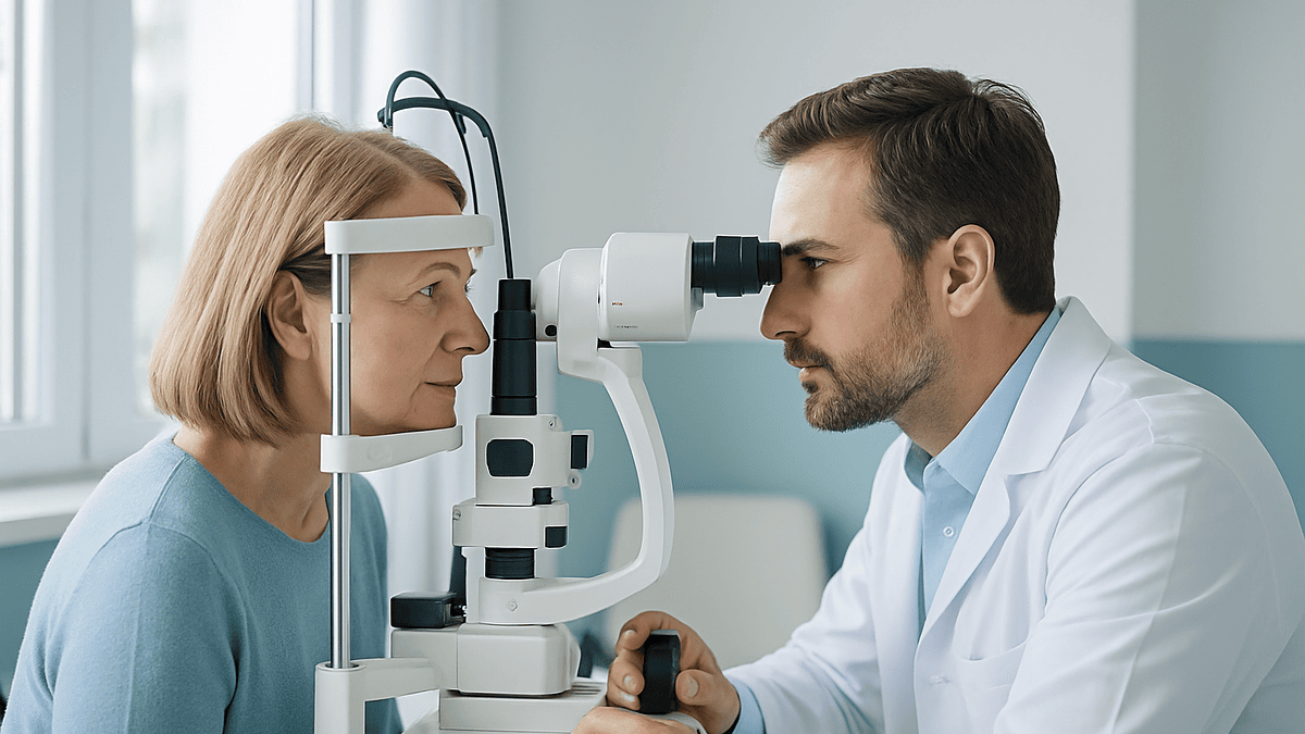
Third, positioning. You sit at the slit lamp, chin on the rest, forehead against the bar. The clinician explains where to look, usually at a small fixation light, and asks you to hold still. A clear gel is placed on a contact lens, and the lens is set gently against the numbed eye. It feels like cool pressure, nothing sharp.
Fourth, the flashes. The clinician steadies the lens with one hand, adjusts the microscope with the other, and begins. Each pulse arrives as a bright flash, sometimes with a click. Retinal sessions may involve dozens to several hundred pulses; NHS guidance notes that laser treatment for diabetic retinopathy typically takes about 20–40 minutes and may need more than one visit. A capsulotomy or iridotomy is often over in a few minutes.
Fifth, the wind-down. The lens comes off, the gel is wiped away, your pressure may be rechecked, and you receive written aftercare instructions and a follow-up date. Then you go home, blinking, with a slightly dazzled eye and a story that is far less dramatic than the one you rehearsed.
Why the drops? What each one is doing to your eye
The drops are not ceremony. Each has a job, and knowing which is which turns a slightly unpleasant few minutes into something you can follow along with.

Anesthetic drops numb the surface of the eye, specifically the cornea, the clear window at the front, and the conjunctiva, the thin skin over the white. They sting briefly, then leave the eye feeling faintly heavy. This is what allows a lens to rest on the eye without triggering a blink or a flood of tears. MedlinePlus lists numbing drops as a routine part of retinal laser photocoagulation. The effect is short-lived, wearing off within roughly 15–20 minutes, which is why your eye may feel gritty on the way home.
Dilating drops widen the pupil so the clinician can see and reach the retina at the back. They belong to two classes: one relaxes the muscle that narrows the pupil, the other stimulates the muscle that widens it. The trade-off is that a wide pupil lets in glare and blurs near vision for several hours. Sunglasses in your bag are a genuinely useful piece of preparation.
Constricting drops do the opposite and appear mainly before a laser iridotomy. A small, tight pupil stretches the iris thin, making the tiny opening easier and safer to create.
Pressure-lowering drops may be given before or after a glaucoma laser or a capsulotomy. Laser energy can cause a temporary rise in eye pressure in the first hours, and these drops blunt that spike. Some people already using pressure-lowering drops are asked to continue them unchanged; the clinician will say so explicitly. Whatever you are handed, it is a one-off part of the procedure, and any change to a regular prescription is a decision for the treating team, not a hint you should act on yourself.
Why is there a lens on your eye during laser treatment?
The lens surprises more people than the laser does. It is a smooth, disc-shaped piece of glass or plastic, sometimes with small mirrors built into its rim, and it sits directly on the numbed cornea with a layer of clear gel between. Three things depend on it.
Steadiness comes first. Eyes drift constantly; that is how they scan the world. A lens held against the surface damps those tiny movements so the aiming beam stays where the clinician put it. It also, quietly, holds the eyelids apart, so you do not have to fight the urge to blink through a hundred flashes.
Focus comes second. The cornea and your natural lens bend light strongly. A contact lens on the surface neutralizes most of that bending, letting the microscope and the laser reach a sharp focus on the target, whether that target is the retina at the back, the drainage angle at the edge of the iris, or the capsule just behind an implant. Some lenses magnify; others widen the field of view so the far periphery of the retina, where tears commonly form, becomes visible. Mirrored lenses angle the beam into the drainage angle for trabeculoplasty and iridotomy, spots that cannot be viewed head-on.
Protection comes third. Certain lenses spread the beam across the cornea so no single point receives concentrated energy, and the gel keeps the surface moist under the lens.
What it feels like: cool, slightly heavy pressure, as if someone is resting a fingertip on a closed eyelid. Because the surface is numb, there is no scratchiness during the procedure. Afterward, the gel can leave vision smeary for an hour or so, and the eye may feel as though something is in it. That sensation is the numbing wearing off, not the lens having done harm. The team should tell you what is normal for the rest of the day and what is not.
Is eye laser treatment painful? What the flashes really feel like
Ask ten people who have had retinal laser and you will get ten answers, which is itself the honest answer: sensation varies with the procedure, the part of the eye being treated, and how much energy is needed.
YAG capsulotomy and laser iridotomy are usually described as painless or as a brief pinch. The flash is startling, and the click that accompanies it can make you jump the first time, but the pulses are so short that the tissue has no time to register heat. The main discomfort is the brightness itself, a series of camera-flash moments while you concentrate on holding still.
Selective laser trabeculoplasty is similarly mild for most people. A dull ache or a feeling of pressure in the eye afterward is common and typically eases within a day.
Retinal photocoagulation sits at the other end. Pulses aimed at the far periphery of the retina, or many pulses delivered in one sitting for widespread diabetic changes, can produce a deep, aching sensation, sometimes described as a toothache behind the eye or a jab that arrives with each flash. NHS guidance on diabetic retinopathy treatment lists aching or discomfort during and after the procedure as a common experience, alongside blurred vision and light sensitivity. Clinicians can pause, lower the energy, space the pulses, or in some cases use an injection of local anesthetic around the eye if the discomfort is too much. Saying “that one hurt” is useful information, not a complaint.
The flashes themselves are seen as bright bursts, often white or green, followed by a fading afterimage. Because the pupil is dilated, everything seems brighter than it is. Many people find that focusing on the fixation light and on slow, steady breathing keeps the experience manageable. If you have had a bad experience with bright lights or with eye procedures before, mention it in advance; the team can adjust pace and explanation accordingly.
Who is eye laser treatment usually for, and who is usually asked to wait?
Lasers are chosen for a reason, never as a default, and the reason differs by disease.
Retinal laser is most often recommended when diabetic retinopathy has progressed to the point where new, fragile vessels are growing (proliferative retinopathy), or when leaking vessels are swelling the central retina in patterns where laser still adds value. It is also standard for a retinal tear or a small, localized detachment, where the National Eye Institute describes laser as a way to weld the retina to the tissue beneath before fluid can lift it further. Timing matters here; a fresh tear is often treated within days.
Laser trabeculoplasty is generally offered to people with open-angle glaucoma or raised eye pressure, either as an alternative to starting daily drops or when drops are not lowering pressure enough. Laser iridotomy is for eyes with a narrow or closed drainage angle, including the emergency of acute angle closure and the preventive treatment of the other eye afterward, as described in NHS glaucoma guidance.
YAG capsulotomy is for people whose vision has become cloudy after cataract surgery because the capsule holding the implant has thickened, a change the NHS notes can appear months or years after the original operation.
Who is asked to wait? Anyone whose media are too hazy to see through: dense cataract, blood in the vitreous gel, or an inflamed, red eye. Lasers need a clear path. People with very high eye pressure at the visit may be treated for the pressure first. Someone with a retinal detachment too large for laser is usually referred for surgery instead. Pregnancy is not a barrier to retinal laser for diabetic eye disease, and it is sometimes preferred over injected medicines during pregnancy, but that judgment belongs to the obstetric and eye teams together. In every case, the decision to proceed, delay, or choose a different route sits with the clinician who has examined your eye.
The main in-office eye lasers compared
Seeing the four common treatments side by side clarifies why your neighbor’s laser story may not match your own. The figures below are typical ranges drawn from the cited public sources; your team will give you numbers specific to your eye.
| Treatment | What it targets | Drops usually used | Typical time in the chair | What you notice afterward |
|---|---|---|---|---|
| Retinal laser photocoagulation | Leaking or new vessels in the retina; retinal tears | Anesthetic, dilating | About 20–40 minutes; may need more than one visit (NHS) | Blurred vision for a few hours, aching, light sensitivity; possible small blind spots or reduced night vision over time |
| Selective laser trabeculoplasty | Drainage meshwork in open-angle glaucoma | Anesthetic, pressure-lowering | A few minutes per eye | Mild ache, temporary blur; pressure effect builds over weeks (Mayo Clinic) |
| Laser peripheral iridotomy | Iris, to open a bypass in narrow angles | Anesthetic, constricting, pressure-lowering | A few minutes | Brief ache, glare or a faint line of light in some people |
| YAG laser capsulotomy | Cloudy capsule behind a lens implant | Anesthetic, dilating, sometimes pressure-lowering | A few minutes | Floaters for hours to days, clearer vision as dilation wears off |
Two patterns stand out. First, the retinal laser is the outlier in duration and discomfort, simply because it delivers many more pulses to a sensitive structure. Second, none of these require an incision, stitches, or a hospital bed, which is why the same room that hosts your routine examination can host the treatment.
The table also shows why the glaucoma lasers ask for patience. Trabeculoplasty does not lower pressure on the spot; it nudges the eye’s own drainage to improve gradually, and the treating team will decide at follow-up whether the response is sufficient or whether other options, including continuing drops, remain necessary.
Laser eye treatment: what happens in the hours and days after
The first hour belongs to the drops. Your pupil is wide, so daylight feels aggressive and reading is a smear. The numbing wears off, and the eye feels gritty and watery. Gel from the lens leaves a film that clears with a few blinks. This is why clinics ask you not to drive home; the NHS specifically advises arranging transport after laser for diabetic retinopathy because vision is blurred for several hours.
By evening, most people can see roughly as they did before, with a dull ache that a quiet room and a cool cloth over closed lids often ease. Over-the-counter pain relief is something to ask the team about rather than assume; they know what suits your other medicines. Sleep usually resets the eye.
The following days differ by treatment. After a YAG capsulotomy, fragments of the cleared capsule float in the gel behind the lens, so new floaters drifting across your vision are expected and typically settle over days to a few weeks. After trabeculoplasty, the eye may stay mildly sore or light-sensitive for a day or two; the pressure benefit, if it comes, is judged at a follow-up visit weeks later. After iridotomy, some people notice a horizontal streak of light or extra glare from the new opening, which often fades as the brain adapts.
Retinal laser has the longest tail. Small blind spots at the treated sites, a modest loss of side vision, and poorer night vision are recognized effects of extensive treatment, as NHS guidance lists, and they reflect the trade of some peripheral retina for the protection of central sight. Central vision itself may remain a little hazy for a week or so.
Follow-up is not optional housekeeping. It is where the team checks that a tear is sealed, that pressure has responded, or that further sessions are needed. Keep the appointment even if the eye feels perfectly fine.
Retinal laser treatment recovery: driving, work and screens
Practical questions dominate the ride home, so here is what the evidence and standard aftercare support, framed as typical ranges rather than promises.
Driving is off the table on the day of treatment. Dilation, glare and blur make it unsafe, and many clinics ask you to wait until the next day and until you feel your vision has returned to its usual level. If you hold a driving license and have had extensive retinal laser, side-vision loss can matter for legal fitness to drive; the treating team can advise whether your visual field needs formal testing before you return to the wheel. That is a question worth asking out loud before you leave.
Work and screens are a matter of comfort rather than danger. Nothing about a screen harms a lasered eye, but focusing on text with a dilated pupil is tiring and often pointless for the first evening. Many people take the rest of the day off and return to desk work the next morning. Physically demanding jobs, dusty environments, or roles that depend on sharp vision may warrant a day or two more; the team will tailor this.
Bathing, hair washing and gentle exercise are generally fine from the next day after in-office laser, because nothing has cut the eye. Swimming in pools and heavy lifting are commonly held back for a short period after some procedures, chiefly to avoid a pressure surge or exposure while the surface is irritated; follow the written instructions you were given rather than a general rule.
Flying is not restricted by laser photocoagulation itself, since no gas has been placed in the eye. Gas is a feature of certain retinal surgeries in the operating room, not of clinic laser. If any part of your treatment plan involved surgery as well as laser, confirm before booking travel.
The overriding principle: recovery is measured in comfort and in the follow-up examination, not in a fixed number of days on a leaflet.
What are the risks and side effects of eye laser treatment?
Neutral language matters here, because in-office lasers sit in an unusual place: they carry real, well-described side effects, yet they are used precisely because the condition left alone tends to be more dangerous than the treatment. Both halves of that sentence deserve equal weight.
Common and expected effects include temporary blurring, aching, light sensitivity and, after capsulotomy, floaters. A short-lived rise in eye pressure can follow any of these lasers in the first hours, which is why pressure is often rechecked before you leave and why pressure-lowering drops are sometimes given.
Retinal photocoagulation, especially when it covers a large area to control proliferative diabetic retinopathy, is associated with permanent small blind spots, reduced peripheral vision and worse vision in dim light; the NHS lists these plainly among its side effects. Central vision can occasionally be affected if swelling develops or if a burn lands nearer the center than intended. These are the reasons treatment is targeted and staged rather than delivered all at once.
Glaucoma lasers carry a small risk of inflammation inside the eye and of pressure spikes. Iridotomy openings can produce lasting glare or a line of light in a minority of people, and an opening can occasionally close over and need repeating.
YAG capsulotomy has a recognized but uncommon association with retinal detachment and with swelling of the central retina afterward, which is one reason surgeons check the back of the eye at follow-up and ask you to report new floaters, flashes or a shadow.
What laser cannot do is restore vision already lost to scarring or long-standing swelling. Its purpose is to stabilize, to protect what remains, or to clear a specific obstruction. Every one of these risks is weighed against the alternative of not treating, and that weighing is the substance of the consent conversation with your treating team.
What are the alternatives to eye laser treatment?
Laser is one tool on a shelf, and it helps to know what else sits beside it, because the existence of alternatives is often what reassures people that the recommendation was considered rather than automatic.
For diabetic macular swelling, medicines injected into the eye that block vascular endothelial growth factor, the signal that drives leaky and abnormal vessel growth, have become a first-line option in many settings, with laser used alongside or for particular patterns of leakage. For proliferative retinopathy, both injections and laser are recognized approaches, and the choice depends on how the disease looks, how reliably a person can attend repeat visits, and the state of the other eye. Steroid implants are another option in selected cases. Controlling blood glucose, blood pressure and cholesterol remains the foundation beneath all of them, as the National Eye Institute emphasizes.
For retinal tears, freezing treatment (cryotherapy) can seal a tear from the outside when laser cannot reach it, and a detachment beyond the reach of either is treated surgically.
For open-angle glaucoma, daily pressure-lowering drops are the long-standing alternative to laser, and the two are frequently used together. When neither suffices, surgical options that create new drainage pathways exist. For narrow angles, removing the natural lens by cataract surgery can widen the angle and is sometimes chosen instead of, or after, iridotomy.
For posterior capsule opacification there is no drop or tablet that clears the membrane; the alternative to YAG laser is simply to live with the cloudiness, which some people choose if it is mild and not affecting daily life.
Doing nothing is itself an alternative, and an honest clinician will describe what is likely to happen if you choose it. The point of laying these options out is not to steer you but to make clear that the treating team has weighed them, and that you are entitled to hear that weighing before you agree.
What people often get wrong about eye laser treatment
Some myths travel from waiting room to waiting room. Correcting them takes a paragraph each.
“The laser will fix my eyesight.” For the treatments in this article, no. Retinal laser aims to stop things getting worse; it does not sharpen vision and can slightly reduce peripheral or night vision. Glaucoma lasers lower pressure to protect the optic nerve; they do not reverse damage already done. Only the YAG capsulotomy tends to make vision clearer, and that is because it removes a specific obstruction, not because it improves the eye itself.
“It’s surgery, so I’ll be asleep.” These are awake procedures done with drops. Being awake is not a downgrade; it lets you follow instructions about where to look, which is how the clinician keeps the beam on target.
“One session and I’m done.” Sometimes. Retinal laser for widespread disease is often staged across visits, as the NHS notes, and glaucoma lasers may need repeating over the years. A capsulotomy is usually a single treatment.
“The flashes mean something went wrong.” The flashes are the treatment. Seeing them, and a fading afterimage, is expected.
“The lens on my eye could scratch it.” The lens sits on a cushion of gel over a numbed surface. Grittiness afterward is the anesthetic wearing off, not a scratch.
“Laser is the same as LASIK.” Different machine, different purpose, different room. LASIK reshapes the cornea for glasses independence; the lasers here treat disease.
“If my eye feels fine, I can skip the follow-up.” A sealed tear, a pressure response, or the need for a second session cannot be felt. It has to be looked at. The follow-up is part of the treatment, not an optional extra.
Questions to ask your care team before eye laser treatment
The consent conversation goes better when you arrive with questions rather than trying to think of them under bright lights. These are the ones experienced patients tend to wish they had asked.
- Which laser is this, exactly, and which part of my eye is it treating?
- What is the goal: to stop things worsening, to lower pressure, or to clear something in the way?
- What is likely to happen to this eye if I do not have the laser, and over what timescale?
- What are the alternatives for my particular situation, and why have you recommended laser over them?
- Will this be one session or several, and how will you decide?
- Which drops will I receive today, and should I use my regular eye drops or other medicines as normal?
- How long is the whole visit likely to take, including the wait for dilation?
- What will I feel during the flashes, and what can you do if it hurts?
- What is normal for the rest of today and this week, and what is not?
- When can I drive, work, read and exercise again, in your view for my eye?
- Could this affect my side vision or night vision, and does that have implications for my driving license?
- When is my follow-up, what will you check, and how do I reach the clinic if something worries me before then?
Write the answers down or ask a companion to; dilated eyes make reading a leaflet in the car park nearly impossible. If an answer is “it depends,” ask what it depends on. Clinicians generally welcome that follow-up, because it shows you understand that laser is a judgment made for one specific eye rather than a standard fitting. The decision to proceed, delay, or choose another path is one you make together, with the treating team holding responsibility for the clinical recommendation.
When to call your doctor after eye laser treatment
Most people leave a laser appointment with a sore, dazzled eye and nothing more, and the ordinary aftermath, blur for a few hours, mild ache, grittiness, a few new floaters after capsulotomy, needs no call. The signs below are different. They are uncommon, but they can indicate a pressure spike, inflammation, or a retinal problem that needs same-day assessment, and the safe response is to contact the clinic that treated you or an emergency eye service without waiting for the scheduled follow-up.
- Severe or steadily worsening pain in the eye, particularly with a headache, nausea or vomiting, which can signal a sharp rise in eye pressure.
- A sudden shower of new floaters, a burst of flashing lights that continues after you have left the clinic, or a dark curtain or shadow spreading across part of your vision; these are the warning signs of a retinal tear or detachment described by the National Eye Institute and the NHS.
- Vision that becomes markedly worse rather than gradually clearer over the first day, or a sudden drop in vision at any point in the following weeks.
- An eye that turns intensely red, produces sticky discharge, or becomes so light-sensitive you cannot open it.
- Any new distortion of straight lines or a gray patch in the center of your vision, which can indicate swelling of the central retina.
If the eye was treated for a tear or for diabetic disease, the same red flags apply to the other eye too, because the underlying condition affects both. Keep the clinic’s out-of-hours number somewhere you can find it with one eye half-closed. When in doubt, call; the team would rather examine a healthy eye than miss a problem that laser could have caught early.
Frequently asked questions
What should I expect at an eye laser procedure appointment?
Expect a clinic visit rather than an operating room: a consent conversation, drops in the treated eye, a wait of roughly 15–30 minutes if the pupil needs to dilate, a few minutes to 40 minutes at the slit lamp while the clinician holds a lens on your eye and delivers flashes, then a pressure check and aftercare instructions. You go home the same day with blurred vision, so arrange a lift.
Is eye laser treatment painful?
It ranges from painless to a deep ache depending on the procedure. YAG capsulotomy and iridotomy are usually felt as bright flashes with little or no pain. Retinal photocoagulation, especially many pulses to the far retina, can cause aching or sharp jabs, and NHS guidance lists discomfort as a common experience. Tell the clinician if it hurts; energy, pacing and anesthesia can be adjusted.
What does the lens on your eye do during laser treatment?
The contact lens holds the eye steady, keeps the eyelids apart, and cancels most of the eye’s own focusing power so the microscope and laser can reach a sharp focus on the retina, the drainage angle or the capsule behind a lens implant. Mirrored versions angle the beam into areas that cannot be seen head-on. A layer of gel cushions it on the numbed surface.
YAG laser capsulotomy: what to expect afterward?
Expect new floaters, small dark specks drifting across vision, for hours to a few weeks as fragments of the cleared capsule settle. Vision usually becomes clearer once dilation wears off over several hours. Your team may recheck eye pressure before you leave. Report a sudden shower of floaters, persistent flashes, or a shadow across vision promptly, because capsulotomy carries a small association with retinal detachment.
How long does retinal laser treatment recovery take?
Vision is typically blurred for a few hours from the dilating drops and gel, with aching and light sensitivity that usually ease by the next day, according to NHS guidance. Central vision may stay slightly hazy for about a week. Some effects of extensive treatment, such as small blind spots or reduced night vision, can be permanent. Return to driving and work is judged by comfort and your team’s advice.
Can I drive home after laser eye treatment?
No. Dilated pupils, glare and blurred vision make driving unsafe on the day, and clinics routinely ask you to arrange transport. Most people feel able to drive the next day once vision has returned to its usual level, but after extensive retinal laser your side vision may need formal assessment for driving fitness. Ask the treating team before you leave the clinic.
Do the flashes during eye laser treatment mean something is wrong?
The flashes are the treatment itself. Each laser pulse arrives as a bright burst, often white or green, sometimes with a click, followed by a fading afterimage. Seeing them is expected. What is not expected is flashing that continues long after you have left the clinic, or a new curtain or shadow in your vision, which should prompt a same-day call to the eye service.
Will laser eye treatment improve my vision?
For most disease-treating lasers, the aim is to stop things getting worse, not to sharpen sight. Retinal laser protects central vision but can reduce peripheral or night vision; glaucoma lasers lower pressure to protect the optic nerve without reversing existing damage. YAG capsulotomy is the exception, since removing a cloudy capsule often clears vision. Your team will tell you which goal applies to your eye.
Will I need more than one eye laser session?
Possibly, depending on the treatment. NHS guidance notes that laser for diabetic retinopathy may need more than one visit, because treating a large area is staged for safety and comfort. Glaucoma laser trabeculoplasty can be repeated if pressure rises again over the years. YAG capsulotomy and iridotomy are usually single treatments, though an iridotomy occasionally needs reopening. The follow-up examination decides.
Can I keep using my regular eye drops on the day of laser treatment?
Usually yes, but confirm with the treating team rather than assume. Many clinics ask people with glaucoma to continue their regular pressure-lowering drops unchanged and may add a one-off drop before or after the laser to blunt a temporary pressure rise. Never stop, start or alter a prescribed medicine on your own; ask at check-in and follow the written instructions you are given.
References
- MedlinePlus: Laser photocoagulation – eye
- NHS: Diabetic retinopathy – Treatment
- National Eye Institute (NIH): Retinal Detachment
- NHS: Cataract surgery
This article is for general information only and is not a substitute for professional medical advice. Please consult a qualified doctor about your individual situation.

More from the Blog

Microsurgery Risks Explained: Why Blood Flow Is Watched Hourly in the First Days
Microsurgery complications most often involve the tiny reconnected blood vessels that feed transplanted tissue, called a flap. A clot in the artery or vein…

Scar Treatment Results: What Realistic Improvement Looks Like and Why Sessions Repeat
Realistic scar treatment results mean a scar that is flatter, softer, paler and less noticeable, not a scar that vanishes. Scar tissue is permanent,…

Neck Lift Recovery Time: Healing Stages, Scars and Results That Settle
Most people look socially presentable about two to three weeks after a neck lift, once visible bruising fades and swelling starts to settle. Full…

How to Prepare for Microneedling: Sun, Retinoids and the Two Weeks Before Your Session
Preparing for microneedling usually means keeping the skin out of strong sun and free of sunburn or tan for the two weeks beforehand, pausing…
IPL Photorejuvenation Results: Why Spots Darken Before They Fade and How Sessions Build
IPL photorejuvenation results usually appear gradually. Treated brown spots often turn darker and coffee-ground-like for several days before flaking away over one to two…

Surface Reconstruction Recovery: Early Supervised Walking, Physical Therapy and Milestones
Surface reconstruction recovery after ocular surface surgery usually unfolds in stages: the corneal surface layer re-covers over days, comfort and light sensitivity settle over…