

What Happens During an Ophthalmic Laser Session: Drops, the Lens on Your Eye and the Flashes
Key Takeaways
- Most office-based eye lasers are outpatient procedures measured in minutes, and the longest part of the visit is usually waiting for drops to work.
- Three different drops may be used for three different jobs: numbing the surface, widening or narrowing the pupil, and preventing a short-lived pressure spike.
- The contact lens rests on a numbed cornea with clear gel to hold the lids, steady the eye, focus the beam and, in mirrored designs, let the doctor see around corners inside the eye.
- The flashes are the treatment pulses themselves, seen because the retina registers any light; a faint steady dot beforehand is the aiming beam and carries no treatment energy.
- Heat-based retinal laser can ache deeply because the tissue beneath the retina has pain fibers that anesthetic drops cannot reach, which is why heavy treatment is often split across visits.
- Laser treats a specific problem at a point in time; glaucoma laser can wear off and may be repeated, and diabetic retinopathy still depends on blood sugar and blood pressure control.
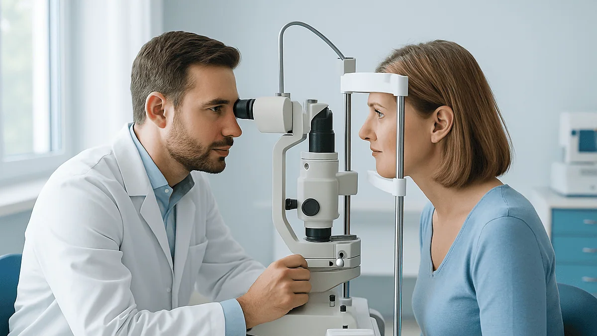
During an ophthalmic laser session you sit at a slit-lamp style machine while the eye is numbed with anesthetic drops and, for retinal work, dilated. The doctor rests a small contact lens with clear gel on the eye to steady it and focus the beam, then delivers short pulses that appear as bright flashes. Most sessions are outpatient and take minutes; vision is blurry for a few hours afterward.
The appointment letter said “laser,” and that single word did a lot of work in the days before the visit. It conjured surgery, masks, a blade. What actually greets most people is a small room, a chin rest, a bottle of drops and a doctor who says, “You’ll see some bright lights, and you may hear a click.”
If you are asking what happens during eye laser treatment, you are probably picturing something far more dramatic than the real thing. Office-based ophthalmic lasers, the kind used for glaucoma, diabetic retinopathy, retinal tears and the cloudy film that can form after cataract surgery, are closer to a long eye examination than to an operation. There is no cutting, no stitching and usually no gown.
Three moments tend to catch people off guard: the sting or blur of the drops, the odd sensation of a lens resting on the eye, and the flashes. This article walks through each one, explains why it happens, and sets out what the evidence says about comfort, timing and the days afterward.
What happens during eye laser treatment, step by step
The rhythm of a laser session is remarkably consistent, whichever condition is being treated. You check in, someone confirms which eye is being lasered, and a nurse or technician puts in drops. Then you wait. The wait is not idle time; it is the drops doing their job, either numbing the surface, widening the pupil, or gently lowering the pressure inside the eye.
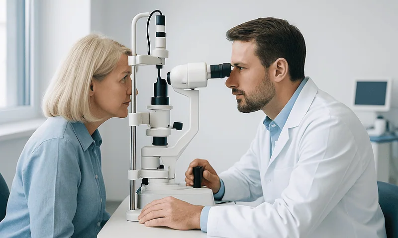
When you are called through, you sit at a machine that looks like the slit lamp used in routine eye checks: a chin rest, a forehead bar, a bright microscope on the doctor’s side. The laser is built into or attached to this microscope. You will be asked to press your forehead firmly forward and keep both eyes open, often fixing your gaze on a small light or on the doctor’s ear.
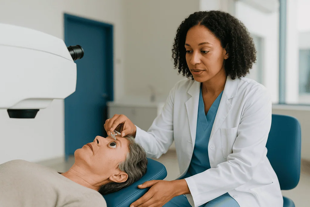
The doctor places a contact lens against the numbed eye. It is held there by hand, cushioned with a clear gel. Through the microscope the doctor lines up a faint aiming beam, then presses a foot pedal. Each press delivers one pulse or a short burst of pulses. You see a flash. For some treatments that is a handful of pulses; for others it is several hundred, applied in a pattern.
Afterward the lens comes off, any gel is rinsed away, and the pressure in the eye may be checked. Many people are given a further drop and asked to sit for a short while before going home with someone who can drive. According to the NHS, laser for diabetic retinopathy is usually an outpatient procedure lasting around 20 to 40 minutes, and more than one visit may be needed. Shorter treatments, such as the laser used for post-cataract clouding, are described by Mayo Clinic as taking about five minutes.
What happens during eye laser treatment depends on the condition being treated
“Laser” is a family of tools, not one procedure, and the experience differs with the target. It helps to know which of the common treatments you are having, because the drops, the lens and the number of flashes all change.
| Treatment | What it targets | How the laser acts | What you typically notice |
|---|---|---|---|
| Selective laser trabeculoplasty (SLT) | Drainage meshwork in open-angle glaucoma | Very short pulses absorbed by pigmented cells, prompting the tissue to drain better | A mirrored lens, dim green flashes, little or no pain |
| Laser peripheral iridotomy | Narrow or closed drainage angle | Creates a tiny opening in the iris so fluid can flow | A pupil-constricting drop first, a few bright flashes, sometimes a brief sharp sensation |
| YAG capsulotomy | Cloudy capsule behind a lens implant after cataract surgery | A focused spark clears a central window in the film | Dilating drops, soft clicks, floaters for a day or so |
| Retinal photocoagulation (focal or scatter) | Leaking or oxygen-starved retina, mostly in diabetes | Heat seals leaks or reduces the retina’s demand for oxygen | Dilating drops, many flashes, an ache toward the end of longer sessions |
| Laser retinopexy | A retinal tear or thin patch | Heat spots form a scar that bonds retina to the wall of the eye | Dilating drops, a ring of flashes around the tear |
Two broad mechanisms sit behind this table. Photocoagulation means using heat from light to make a controlled, tiny burn; MedlinePlus describes it as the basis for most retinal laser work. Photodisruption means using an extremely brief, intense pulse to create a microscopic spark that mechanically opens tissue; this is how a YAG laser clears the capsule or makes an iridotomy. SLT sits in a third category, delivering energy so briefly that it stimulates cells without visible burning. Knowing which of these you are having explains almost everything you will feel.
Why the drops come first: numbing, widening and pressure control
The drops are the least glamorous part of the visit and the most misunderstood. People often assume they are all anesthetic. In fact, three different jobs may be done by three different bottles.
The first job is comfort. A topical anesthetic numbs the cornea, the clear front window of the eye, so that a contact lens can rest on it without a blink reflex or a scratch sensation. It stings for a few seconds, then the surface goes pleasantly unaware. The NHS notes that local anesthetic drops are used before laser procedures for both glaucoma and diabetic retinopathy.
The second job is access. For anything at the back of the eye, the retina, the pupil must be widened so the doctor can see and aim. Dilating drops relax the muscle that closes the pupil. The cost is a few hours of blur and glare; the NHS advises against driving until this passes. For an iridotomy the opposite is needed: a drop that shrinks the pupil stretches the iris thin, making it easier to open a small channel.
The third job is protection. Any laser applied to the front drainage structures can cause a short-lived rise in the pressure inside the eye. A pressure-lowering drop, given before or immediately after, blunts that spike. It is a precaution built into standard practice, not a sign that anything has gone wrong.
Which drops you receive, and whether any are prescribed for the days afterward, is decided by the treating team based on your eye and the procedure. If you already use glaucoma drops, ask whether to take them as usual on the day; do not stop or change them on your own.
Why the doctor rests a lens on your eye
For many people the lens is the strangest moment of the session. A curved disc, a little wider than the colored part of the eye, is placed against the numbed cornea with a dab of clear gel and held there by the doctor’s fingers for the duration of the treatment. You feel pressure and a cool slipperiness, not pain.

The lens does four things at once. It holds the eyelids apart, which matters because you cannot blink the laser away. It steadies the eyeball, so tiny movements do not shift the aiming point. It focuses the beam precisely at the intended depth, whether that is the drainage meshwork a fraction of a millimeter inside the front of the eye or the retina at the back. And in some designs it contains angled mirrors, letting the doctor look around the corner into the drainage angle or out toward the far edge of the retina, places no straight view can reach.
The gel is a coupling fluid. Without it, the air gap between lens and cornea would scatter light and blur the view. It is the same principle as the gel used for an ultrasound scan. Any residue rinses away with saline at the end and may leave the vision slightly smeary for a short while.
Not every laser needs a lens. Some retinal systems and some capsulotomies are performed without one, and the doctor simply asks you to hold still and look at a target. If a lens is used, the most useful thing you can do is keep your forehead pressed to the bar and breathe normally. People often hold their breath, which makes the eye drift; a steady, ordinary breath keeps the picture still.
What the flashes are, and why you see them
The flashes are the laser itself, and the fact that you see them is reassuring rather than alarming. Light reaching the retina is registered as light, no matter where the beam is focused. The treatment pulse is intense, so each one appears as a brilliant white, green or red burst, followed by a fading afterimage, the same way a camera flash lingers.
Before the treatment pulse, most systems project a faint aiming beam, a small colored dot the doctor uses to position the shot. You may notice this as a steady red or green light drifting across your view. It carries no treatment energy.
The character of the flashes tells you what kind of laser you are having. Photocoagulation for the retina delivers many brief pulses, often in a grid, so the flashes come in a steady rhythm. Scatter treatment for advanced diabetic retinopathy can involve hundreds in one sitting; the NHS notes it may be split across more than one visit. A YAG laser fires only a handful of pulses, each accompanied by a distinct click as the spark forms; the click is audible, not felt. SLT pulses are gentle and the flashes softer.
Afterimages can outlast the session by minutes and colors may look washed out or tinted for a short while. This settles as the retina’s pigments recover. What should not happen is a sudden curtain or shadow across the vision, or a burst of new floaters with flashes that continue at home; those belong in the red-flag list later in this article.
The temptation is to flinch or to look at the flash. Neither helps. Looking at the fixation target, even when the flash is off to one side, keeps the eye where the doctor needs it and shortens the session.
Does laser eye treatment hurt?
The honest answer is that it depends on where the laser is aimed and how much is done, and that most sessions sit between painless and mildly uncomfortable.
Front-of-eye procedures with few pulses are the gentlest. Mayo Clinic describes YAG capsulotomy after cataract surgery as painless, and SLT is generally reported as no more than a mild ache. Laser iridotomy is brief but can produce a momentary sharp sensation as the iris opens, sometimes described as a pinprick from inside.
Retinal photocoagulation is where discomfort is most likely. The retina has no pain fibers, but the layer beneath it and the wall of the eye do, and heavier scatter treatment toward the periphery can produce a deep, dull ache. The NHS lists aching or discomfort during and shortly after diabetic retinopathy laser as a recognized side effect. Modern pattern systems deliver shorter pulses, and many doctors reduce discomfort by spacing treatment across sessions.
Several practical things influence how it feels:
- Keeping still shortens the session; a restless eye means more re-aiming and more pulses.
- Telling the doctor promptly if a spot is sharply painful lets them adjust settings or position.
- Anxiety amplifies sensation; a slow exhale before each cluster of pulses genuinely helps.
Anesthetic drops numb the surface but cannot numb deep tissue. For the small number of people who need extensive retinal treatment and find the ache hard to tolerate, an injection of anesthetic around the eye is an option the team may discuss. Whether that is appropriate is their call. Mild aching afterward usually eases within the day; persistent or worsening pain is a reason to call.
How long does laser eye treatment take?
People plan their day around the word “laser” and are often surprised that the laser itself is the shortest part. The visit is long because of the drops; the treatment is measured in minutes.
Published patient guidance gives a sense of the range. The NHS states that laser treatment for diabetic retinopathy usually takes around 20 to 40 minutes and is normally done as an outpatient, with more than one appointment sometimes required. Mayo Clinic describes YAG capsulotomy as roughly a five-minute procedure. SLT and iridotomy fall in a similar short bracket, with most of the appointment spent waiting for drops to act and for a post-laser pressure check.
A realistic timeline for the whole visit looks like this:
- Check-in, consent and confirmation of the correct eye: a few minutes.
- Drops and waiting for them to work: dilating drops need time to open the pupil fully, so this is often the longest stretch.
- The laser itself: from a couple of minutes to about forty, depending on the procedure.
- Post-treatment drop, pressure check and a short rest: variable, sometimes up to an hour when a pressure check is planned.
Add travel with a companion, since the treated eye will be blurry and light-sensitive. Bringing sunglasses and avoiding demanding close work that afternoon is sensible.
Timing also stretches when treatment is deliberately staged. Scatter photocoagulation for diabetic retinopathy is often split to reduce discomfort and swelling, and the National Eye Institute notes that more than one treatment may be needed. Retinal tears sometimes get a top-up if the first ring of spots looks incomplete at review. None of this signals failure; it reflects how these lasers are planned. Your team will tell you at the end of each session whether another is expected.
How the laser actually changes the eye: heat, spark or a tap on the cells
Three physical effects, chosen by wavelength and pulse length, account for nearly every ophthalmic laser. Understanding them explains both what you feel and what the treatment is trying to achieve.
Heat. Photocoagulation uses visible light, typically green or yellow, absorbed by pigment in the retina’s supporting layer. Absorbed light becomes heat, and heat coagulates a tiny disc of tissue. Around a retinal tear, a ring of these spots heals into a scar that bonds the retina to the wall behind it; Mayo Clinic describes the result as effectively welding the tear in place. In diabetic retinopathy, focal spots seal leaking vessels, while scatter spots reduce the oxygen demand of starved retina so that it stops sending the signals that grow fragile new vessels. The National Eye Institute notes that scatter treatment can reduce side vision and night vision in exchange for protecting the center.
Spark. A YAG laser fires an infrared pulse lasting nanoseconds and focused to a point. At the focus, energy density is high enough to strip electrons from atoms, creating a microscopic plasma and a shock wave. That mechanical pop, not heat, punches the opening in a cloudy capsule or in the iris. It is why you hear a click.
A tap on the cells. Selective laser trabeculoplasty delivers very short green pulses to the pigmented cells of the drainage meshwork at energies too low to burn. The pulses trigger a biological response, thought to involve cell turnover and signaling, that improves outflow over the following weeks. The NHS describes SLT as an option for open-angle glaucoma, and the National Eye Institute notes that its effect can wear off with time and that drops may still be needed. How much pressure falls, and for how long, varies between individuals and is assessed at follow-up rather than promised in advance.
Who eye laser treatment is usually for, and who is usually asked to wait
Ophthalmic lasers are offered when there is a specific, visible target and the evidence supports treating it. They are not a general-purpose fix for blurry vision, and understanding the typical candidates prevents disappointment.
Common reasons a team recommends laser include:
- Open-angle glaucoma or raised eye pressure, where SLT is an option alongside or instead of drops, according to the NHS.
- A narrow drainage angle at risk of closing, treated with iridotomy to prevent a sudden pressure attack.
- Diabetic retinopathy with leaking vessels near the center or with new abnormal vessels, where the National Eye Institute lists laser among the standard treatments.
- A fresh retinal tear or a thin area judged likely to progress, treated to reduce the risk of detachment.
- Clouding of the capsule behind a lens implant, which Mayo Clinic notes is a common late effect of cataract surgery and is treated with YAG capsulotomy.
People are often asked to wait, or offered something else, in several situations. If the eye is inflamed or infected, laser is generally deferred until it settles. If the view is too hazy, because of cataract, bleeding or corneal cloudiness, the beam cannot be aimed safely. If the cornea is very swollen, a lens may not sit properly. In diabetic retinopathy, a team may prefer to treat swelling with injections first and reserve laser for specific patterns of leakage; that decision is guided by imaging.
A few situations call for particular caution rather than exclusion: recent eye surgery, certain retinal conditions that make the retina more fragile, and difficulty keeping still at the machine. Pregnancy does not usually prevent retinal laser, since the treatment is local, but timing is discussed individually.
None of these lists replaces an examination. Whether laser is right, and when, is a judgment made by the treating ophthalmologist with the findings in front of them.
What the following hours, days and weeks usually look like
The first afternoon belongs to the drops rather than the laser. Expect blur, glare and a preference for dim rooms. The NHS lists blurred vision and increased sensitivity to light as common short-term effects after retinal laser and advises not driving until vision has cleared, usually within a few hours. A mild ache or a gritty feeling where the lens sat is common and fades the same day.
Over the next day or two, what you notice depends on the procedure. After a YAG capsulotomy, fragments of the cleared capsule float in the gel of the eye and appear as new floaters; these typically drift downward and out of the line of sight over days. Vision often seems brighter fairly quickly, though the team will tell you when to expect the change and whether your glasses prescription might need checking. After iridotomy, a faint extra line or arc of light in some lighting conditions is reported by a minority of people; it usually becomes less noticeable but should be mentioned at review.
After SLT, nothing dramatic happens, and that is expected. The pressure-lowering effect develops gradually and is measured at a follow-up visit; you continue whatever drops you have been told to continue.
After retinal photocoagulation, the treated spots take time to settle into scars. The NHS notes that some people notice small patches of reduced vision, reduced night vision or narrower side vision after scatter treatment, effects that reflect the treated retina and are weighed against protecting central sight. Swelling at the center can transiently blur reading vision.
Follow-up is part of the treatment. A retinal tear is re-examined to confirm the scar has sealed, and a pressure check after glaucoma laser confirms the eye has not spiked. Keep those appointments even if the eye feels fine.
What are the common eye laser side effects, and which are serious?
Neutral language matters here, because the same list can sound frightening or trivial depending on framing. Most side effects are short-lived and expected; a few are uncommon and important.
Commonly reported and usually temporary:
- Blurred vision and light sensitivity from dilating drops, lasting hours according to the NHS.
- A dull ache during and after retinal laser, listed by the NHS as a recognized effect.
- Afterimages and altered color perception for minutes after the flashes.
- Floaters after YAG capsulotomy, which usually fade over days.
- Redness or grittiness where the lens rested.
- A brief rise in eye pressure after front-of-eye lasers, which is why a pressure check is routine.
Less common, procedure-specific:
- Small permanent blind spots, reduced night vision or narrowed side vision after scatter photocoagulation; the NHS and the National Eye Institute both describe these as possible consequences of treating peripheral retina.
- A persistent line or glare from an iridotomy in certain lighting.
- Inflammation inside the eye after SLT or iridotomy, usually mild and managed by the team.
- Swelling at the center of the retina after capsulotomy or retinal laser, which can blur reading vision for a time.
Rare but serious:
- Retinal detachment, a risk that is discussed before capsulotomy and after which new flashes or a shadow need urgent assessment.
- A sustained pressure spike that does not settle with drops.
- An accidental burn at the center of vision during retinal laser, which is why fixation and stillness are emphasized.
Frequencies for these events vary by procedure, by the condition being treated and by the individual eye, and reputable patient sources describe them qualitatively rather than as single percentages. Your team can tell you how the risks apply to your eye.
Alternatives your care team may discuss instead of, or alongside, laser
Laser is rarely the only option on the table, and a good consultation lays the alternatives side by side without ranking one as universally better.
For open-angle glaucoma, the mainstays are pressure-lowering drops, laser trabeculoplasty and, when those are insufficient, surgery that creates a new drainage route. The NHS describes all three as standard, and notes that after laser some people still need drops. The choice depends on how high the pressure is, how much damage exists, and how well someone can manage a daily drop routine. For a narrow angle, iridotomy is the usual first step, though removing a bulky natural lens by cataract surgery is sometimes considered when a cataract is also present.
For diabetic retinopathy, injections into the eye of medicines that block a vessel-growth signal have become a first-line option for swelling at the center and for new vessels, with laser used for particular leakage patterns or when injections are not suitable. The National Eye Institute describes injections, laser and, for advanced disease, surgery to remove blood from the gel of the eye. Good blood sugar and blood pressure control remain the foundation under all of these.
For a retinal tear, laser and cryotherapy, freezing applied from outside the eye, achieve the same sealing scar; the choice depends on where the tear sits. Mayo Clinic lists both. For a detachment that has already occurred, laser alone is not enough and surgery is required.
For capsule clouding after cataract surgery, YAG capsulotomy is the standard treatment; the alternative is simply to wait if the clouding is mild and not troubling.
Each pathway carries its own trade-offs of convenience, repeatability and risk. The decision belongs with you and the treating team, informed by imaging and by what matters most in your daily life.
What people often get wrong about eye laser treatment
“It’s the same as laser vision correction.” It is not. Refractive procedures reshape the cornea to reduce the need for glasses and are elective. The lasers in this article treat disease: glaucoma, retinopathy, tears, capsule clouding. They do not remove the need for glasses, and a glasses prescription may even change slightly afterward.
“Once I’ve had laser, the condition is dealt with.” Laser controls or treats a specific problem at a point in time. Glaucoma laser can lose effect over months to years and may be repeated; the National Eye Institute is explicit that drops may still be needed. Diabetic retinopathy continues to depend on blood sugar and blood pressure, and new areas can need treatment. A retinal tear that is sealed does not prevent a new tear elsewhere.
“Seeing the flashes means the laser hit the wrong spot.” Seeing light is unavoidable; the retina is a light detector. Flashes confirm the machine is firing, nothing more.
“The lens on my eye could scratch it.” The cornea is numbed and the lens is cushioned in gel. Transient grittiness is common; a scratch is uncommon and heals quickly if it occurs.
“Laser will restore the vision I’ve lost.” Most retinal lasers aim to protect remaining sight, not to bring back what has gone. Capsulotomy is the exception, because it clears a physical obstruction. Setting expectations correctly before the session is one of the kindest things a team can do.
“I can drive myself home.” With dilated pupils and a blurry eye, the NHS advises against it. Arrange a lift.
“More laser is always better.” Every burn is a trade. Peripheral scatter treatment protects central vision at some cost to side and night vision, which is exactly why doctors treat in measured amounts and review.
Questions to ask your care team before the session
A laser appointment moves quickly, and it is easy to leave with questions unasked. Writing a few down beforehand, and bringing them along, turns a passive experience into a shared decision. These are the ones that tend to matter most.
- Which laser procedure am I having, and what exactly is it aiming at in my eye?
- What is the goal: lowering pressure, sealing a tear, protecting central vision, clearing a film? How will we know afterward whether it has worked, and when?
- Is this likely to be a single session or one of several?
- Which drops will I have on the day, and should I use my usual eye drops or other medicines as normal beforehand? (Do not change anything without asking.)
- Will a lens be placed on my eye, and roughly how many pulses should I expect?
- What will I feel, and what should I say if something is sharply painful?
- What side effects are expected in the first day, and which would you want to hear about straight away?
- Will my glasses prescription need rechecking, and when?
- What are the alternatives to laser for my situation, and why is laser being suggested now?
- When is my follow-up, and who do I contact out of hours if something changes?
- Are there activities I should avoid for the next few days?
Two further questions are worth raising if they apply. If you are pregnant, planning pregnancy or taking blood-thinning medicines, mention it; these rarely change the plan for laser, but the team should know. If you find it hard to keep still, whether from tremor, neck problems or anxiety, say so in advance. Positioning aids and pacing can be arranged, and knowing ahead makes the session smoother for everyone.
Finally, ask for the plan in writing if it is not offered. A short summary of what was done and what to watch for is a useful thing to have on the kitchen table that evening.
When to call your doctor after eye laser treatment
Most people go home, rest the eye and feel ordinary by the next morning. A small number develop symptoms that need same-day assessment, and knowing them in advance removes the guesswork. Contact your eye clinic, or emergency services if the clinic is closed, for any of the following.
- A sudden shower of new floaters, persistent flashing lights that continue after the session, or a shadow or curtain spreading across part of your vision. Mayo Clinic identifies these as warning signs of retinal detachment, which is a rare but recognized complication after capsulotomy and can also follow a tear that has not sealed.
- Pain that is severe, increasing, or accompanied by nausea and a hazy, rainbow-haloed view. This pattern can indicate a sharp rise in eye pressure and needs urgent measurement.
- Vision that is worse the next day than it was before the laser, rather than gradually clearing.
- A red, sticky or intensely light-sensitive eye developing over the following days, which may signal inflammation or infection.
- Any sudden loss of central vision, even if painless.
Symptoms that are usually expected, and can wait for the routine review unless they worsen, include mild aching on the day, blur from dilation, glare in bright light, a few floaters after capsulotomy and a gritty feeling where the lens sat.
If you are unsure which category a symptom falls into, call. Eye services would far rather check a settled eye than miss an unsettled one. Keep the clinic’s number and your appointment summary somewhere visible for the first week, and make sure whoever is with you knows the red-flag list too, since a blurry eye makes reading small print harder. Every decision about further treatment rests with the team who examined you; this list is a prompt to reach them, not a substitute for their assessment.
Frequently asked questions
Does laser eye treatment hurt?
For most procedures it ranges from painless to mildly uncomfortable. Mayo Clinic describes YAG capsulotomy as painless, and glaucoma lasers are usually felt as a brief sting or mild ache. Retinal photocoagulation is the exception; the NHS lists aching during and after treatment as a recognized effect, because tissue beneath the retina has pain fibers that surface anesthetic cannot reach.
How long does laser eye treatment take?
The laser itself usually takes minutes; the appointment takes longer because of drops and checks. The NHS states retinal laser for diabetic retinopathy usually takes around 20 to 40 minutes, and Mayo Clinic describes capsulotomy as about five minutes. Allow extra time for dilation to develop, a pressure check afterward, and travel with someone who can drive.
Why do I need someone to drive me home?
Because dilating drops leave the pupil wide open for hours, causing blur and glare, and a numbed or freshly treated eye is not reliable for judging distance. The NHS advises against driving until this has passed. Even for procedures without dilation, a post-laser check can leave the eye watery and light-sensitive, so a companion is the safe default.
What is the lens the doctor puts on my eye?
It is a small curved contact lens, cushioned with clear gel and held by hand against the numbed cornea. It keeps the lids apart, steadies the eye, focuses the beam at the correct depth and, in mirrored versions, lets the doctor view the drainage angle or far retina. It rinses off at the end and may leave brief grittiness.
Are the flashes I see during the laser dangerous?
No. You see flashes because the retina detects any light that enters the eye, including treatment pulses aimed elsewhere. Afterimages and washed-out colors for a few minutes are normal and fade as retinal pigments recover. Flashing lights that begin or persist at home after the session are different and should be reported, since they can signal a retinal problem.
What does laser treatment for a retinal tear involve?
After dilating and numbing drops, the doctor places a lens on the eye and applies a ring of small heat spots around the tear. Mayo Clinic describes the resulting scar as effectively welding the retina to the wall behind it. The session is short, the tear is re-examined at follow-up, and new flashes, floaters or a shadow afterward need urgent review.
What are the most common eye laser side effects?
The NHS lists blurred vision, light sensitivity and aching as the common short-term effects. Floaters after capsulotomy, a brief pressure rise after front-of-eye lasers and grittiness from the lens are also frequent and temporary. Less common effects include small blind spots or reduced night vision after scatter retinal treatment, and a faint glare line after iridotomy.
Will I still need my glaucoma drops after laser?
Possibly. The NHS and the National Eye Institute both note that laser trabeculoplasty lowers pressure in many people but its effect can fade and drops may still be required. Never stop or change drops on your own after laser; the team measures your pressure at follow-up and decides what, if anything, to adjust.
Can eye laser treatment fix my eyesight or remove the need for glasses?
The lasers described here treat disease rather than focusing errors, so they do not replace glasses. Most retinal and glaucoma lasers aim to protect remaining vision, not restore lost sight. Capsulotomy is the exception because it clears a physical film behind a lens implant. Laser vision correction is a separate, elective procedure on the cornea.
How soon can I return to work and normal activity?
Many people resume ordinary activities the next day once blur from the drops has cleared, which the NHS describes as taking a few hours. Reading-heavy or screen-heavy work may feel tiring that first evening. Your team may advise avoiding strenuous exertion or eye rubbing for a short period after certain procedures; follow their specific instructions rather than a general rule.
References
- NHS: Diabetic retinopathy, Treatment
- National Eye Institute (NIH): Diabetic retinopathy
- MedlinePlus: Laser photocoagulation, eye
This article is for general information only and is not a substitute for professional medical advice. Please consult a qualified doctor about your individual situation.

More from the Blog
How Glaucoma Damages the Optic Nerve Silently and Why Treatment Aims at Eye Pressure
Glaucoma damages the optic nerve by slowly killing the retinal nerve fibers that carry images from the eye to the brain, usually without pain…
Caring for Someone After Eye Surgery: Drop Schedules, Shields and the First Night Back
Caring for someone after eye surgery mostly means keeping to the written drop schedule without touching the eye, making sure the protective shield goes…

Macular Hole, Macular Pucker and Macular Edema: How the Main Macular Diseases Differ
The main types of macular disease damage the small patch of central retina in different ways. A macular hole is a small gap in…

Preparing for Cataract Surgery: Lens Measurements, Medication Review and Drop Schedules
Cataract surgery preparation usually involves a pre-assessment visit where the eye is measured to calculate the power of the replacement lens, a review of…

Does DCR Surgery Stop the Watering? Realistic Results and Why a Few Need a Second Look
For most adults with a confirmed blockage of the nasolacrimal duct, DCR surgery relieves or greatly reduces watering, because it creates a new drainage…
Ahead of Eye Angiography: Allergies, Pregnancy, Kidney History and Bringing a Driver
Eye angiography preparation is mostly about information, not fasting. Before the test, tell your eye team about any allergy or past dye reaction, whether…