

What Robotic Ophthalmology Changes in the Operating Room: Steadier Hands, Same Surgeon
Key Takeaways
- Ophthalmic surgical robots are teleoperated: the surgeon drives every movement in real time, and no system in human use has an autonomous mode.
- The core benefits are motion scaling, tremor filtering and a fixed pivot at the eye wall, not speed; early trials ran longer than manual surgery.
- The strongest human evidence is a twelve-patient randomized feasibility study of retinal membrane peeling, which showed no new safety signal but did not test whether vision outcomes improve.
- Laser-assisted cataract surgery and robotic surgery are different technologies; robotic cataract surgery is not in routine use.
- Recovery follows the underlying operation, for example around four to six weeks after cataract surgery per the NHS and weeks for a gas bubble to clear after vitrectomy per MedlinePlus.
- Worsening pain, falling vision, new flashes or floaters, or a curtain across vision after any eye surgery warrant a same-day call to the surgical team.
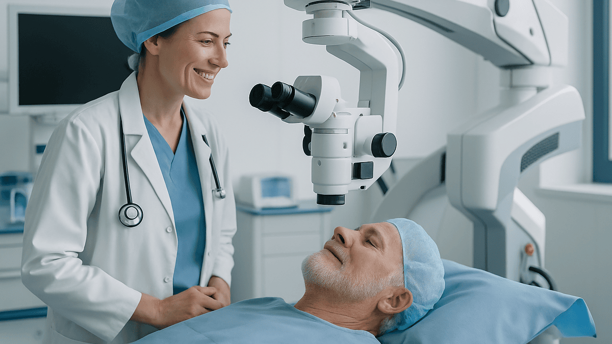
Robotic eye surgery uses a surgeon-controlled mechanical arm to hold and move instruments inside the eye with less tremor and finer motion than an unaided hand can achieve. The surgeon makes every decision; the machine translates hand movements into scaled, steadied movements. Most systems remain investigational or used in select centers, and evidence so far shows feasibility rather than proven better vision.
The letter arrives a week before the operation, and one line stops her: the surgery may be performed with robotic assistance. She has spent forty years trusting her optician, twenty minutes trusting the surgeon in clinic, and now a machine has joined the team without introducing itself. Her first question is not about lasers or lenses. It is simpler than that. Will a person still be holding the instrument?
That question sits at the heart of robotic eye surgery how it works, and the honest answer is more reassuring and less dramatic than the word robot suggests. Nothing in the operating room decides anything on its own. What changes is the path between the surgeon’s fingertips and the tip of a needle finer than an eyelash.
This explainer walks through what the technology actually does, where it is already used, where it is still being tested, and what a patient can reasonably expect before, during and after.
Robotic eye surgery how it works: the two-minute version
Strip away the science-fiction framing and a surgical robot is a very precise puppet. The surgeon sits at a console or beside the patient, holding a controller shaped a little like a pen or a joystick. A mechanical arm, mounted near the patient’s head, holds the actual instrument. When the surgeon moves the controller a few millimeters, the arm moves the instrument a fraction of that distance inside the eye.
Three things happen along that path that a bare hand cannot do on its own. The first is motion scaling: a large, comfortable hand movement becomes a tiny, controlled movement at the instrument tip. The second is tremor filtering: the small involuntary shake present in every human hand, even a rested and highly skilled one, is smoothed out by software before it reaches the eye. The third is a fixed pivot point, sometimes called a remote center of motion, which keeps the instrument rotating around the exact spot where it enters the eye wall so the entry wound is not tugged sideways.
Mayo Clinic describes robotic surgery in general terms as a way to give surgeons greater precision, flexibility and control through small incisions, with the surgeon directing the instruments at every moment. Ophthalmic robots follow the same principle at a far smaller scale.
What the robot does not do matters as much. It does not choose where to cut. It does not recognize tissue or decide a membrane is thick enough to peel. It has no independent judgment. If the surgeon lets go of the controller, the instrument stops. In the trials published so far, the machine is best understood as a stabilizer and translator of human intent, not a substitute for it.
Why the eye is such an unforgiving place for human hands
Consider the retina, the light-sensing layer at the back of the eye. It is thinner than a sheet of paper, and the surgeon operates on it from the outside in, through a fluid-filled globe, watching the whole thing through a microscope. There is no room to rest a wrist on the tissue. The instruments are long and fine, and the surgeon must hold them still while the heart beats and the lungs move.

The first-in-human randomized study of intraocular robotic surgery, published in Nature Biomedical Engineering and indexed on PubMed, framed the problem in numbers: the physiological tremor of an unaided hand and the fine retinal structures a surgeon targets both sit in a range of tens to roughly one hundred micrometers. A micrometer is one thousandth of a millimeter. In other words, the shake in a steady hand and the target on the retina are about the same size.
Experienced vitreoretinal surgeons compensate remarkably well. They brace their hands, slow their breathing, and time movements between heartbeats. Yet certain procedures push right up against the ceiling of what a hand can do. Peeling a membrane a few cells thick off the macula, or holding a needle motionless inside a retinal vein for a minute, sits at that ceiling.
This is the gap robotics tries to close. Not by being faster, and not by being smarter, but by removing the involuntary noise between intention and action. The National Eye Institute, part of NIH, notes that an epiretinal membrane (a thin scar-like layer on the macula, sometimes called a macular pucker) is treated by surgical peeling when it distorts vision. That single procedure is where much of the early robotic research has concentrated, precisely because the margin for error is so thin.
What actually happens in the operating room, step by step
From the patient’s side of the drape, a robot-assisted eye operation looks and feels much like a conventional one. The differences live above the head, out of sight.
The day begins as any eye surgery does. The team confirms identity and the correct eye, marks it, and reviews the plan. Anesthesia is most often local, with numbing drops or an injection around the eye and light sedation if needed, so the patient is comfortable but usually awake. The NHS notes that most cataract operations are done under local anesthetic and take around 30 to 45 minutes. Retinal procedures are typically longer.
The patient lies flat. The face is cleaned and a sterile drape placed, leaving only the eye exposed. A small lid holder keeps the eye open; blinking is not possible and not needed. The surgical microscope is positioned overhead.
Here the robotic arm enters. It is docked in a fixed position relative to the head. The surgeon makes the tiny entry ports in the eye wall by hand, as usual. The instrument is then attached to the robotic arm and guided in through one of those ports. The surgeon controls the instrument from the console while watching the same magnified view a conventional surgeon would use.
Inside the eye, the surgeon does the surgical work: peeling, injecting, or repositioning tissue. The robot filters tremor and scales movement but does not act independently. If a step is better done by hand, the instrument can be undocked and the surgeon continues manually. In the published trials, both approaches were available in the same room throughout.
At the end, the robotic arm withdraws, the surgeon closes or checks the ports by hand, and the eye is shielded. Recovery-room care is the same as for the equivalent manual operation.
Robot assisted cataract surgery: where lasers end and robots begin
Cataract surgery, the removal of a clouded natural lens and its replacement with a clear artificial one, is one of the most frequently performed operations in the world. The NHS describes it as a short procedure that usually improves vision within a few days, with full recovery over four to six weeks. Given its volume, people often assume it must already be robotic. Mostly, it is not.
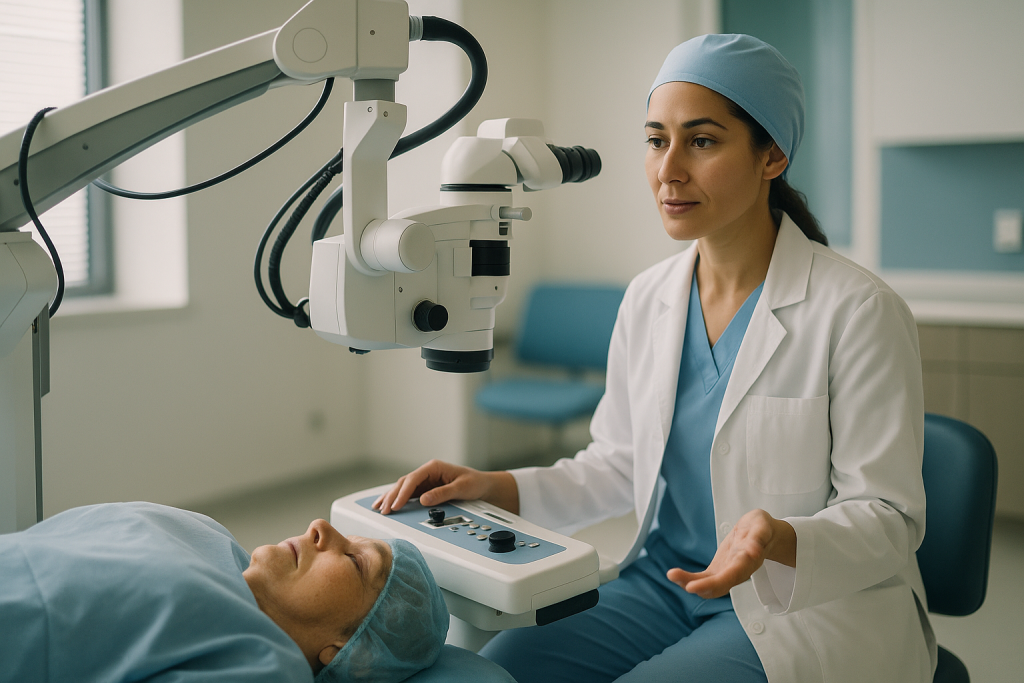
What many patients have heard of is laser-assisted cataract surgery. A femtosecond laser (a laser that fires ultra-short pulses to cut tissue with heat-free precision) can create the initial incisions, open the capsule around the lens, and soften the lens before removal. This is automation of specific steps, planned on imaging and executed by the laser, but it is not a robot in the sense of a surgeon-driven mechanical arm. The surgeon still performs the core removal and lens placement by hand.
Robot assisted cataract surgery, meaning a teleoperated arm holding the phacoemulsification probe (the ultrasound tip that breaks up the lens) or positioning the new lens, is at an earlier stage. Bench and animal studies exist, and a small number of human feasibility reports have appeared, but no robotic cataract platform is in routine mainstream use as of the evidence available through the sources cited here.
Why has robotics not swept into cataract surgery the way it has into some abdominal operations? Partly because manual cataract surgery is already fast, safe and refined over decades, leaving less room for a machine to add value. Partly because the cost and setup time of a robot are hard to justify for a fifteen-minute operation. The retina, with its longer, more delicate procedures, has been a more natural first home.
Robotic retina surgery: the membrane peel that started it all
The clearest human evidence for robotic eye surgery comes from a single, deliberately modest trial. Twelve patients needing a vitrectomy (removal of the gel inside the eye) and peeling of a membrane from the retinal surface were randomized: six had the peel done with robotic assistance, six by conventional hand-held technique. The study, published in Nature Biomedical Engineering and indexed on PubMed, was designed to answer one question: can this be done safely in a living human eye?
It could. All robotic procedures were completed. The robotic group took longer, which the authors attributed to the unfamiliarity of the new system and the cautious pace of a first-in-human study. The rate of retinal micro-injuries, small touches or hemorrhages visible under high magnification, was similar between the groups. The paper also described a second phase in which the robot was used to inject a clot-dissolving medicine under the retina in patients with bleeding beneath the macula, a task that demands the needle be held perfectly still for a sustained period.
Read that carefully for what it does and does not say. It shows feasibility and no obvious harm signal in a very small group. It does not show that robotic peeling produces better vision, fewer complications, or faster recovery. Twelve people cannot answer those questions, and the authors did not claim they could.
Since then, other groups have reported robot-assisted retinal vein cannulation (placing a micro-needle into a blocked retinal vein) and subretinal injections in small series. The direction of travel is toward tasks that are difficult or impossible by hand, rather than toward replacing routine manual surgery. MedlinePlus describes conventional vitrectomy as a well-established operation for conditions including retinal detachment and macular disorders; robotic tools are being tested as an addition to that toolkit, not a replacement for it.
Who is robotic eye surgery usually for, and who is asked to wait?
Because most ophthalmic robotic systems are investigational, the honest answer is that today it is mainly for people who are enrolled in research studies at the small number of centers running them, and who have the specific condition those studies are testing.
In the published work, that has meant adults with an epiretinal membrane or a related macular condition already scheduled for vitrectomy, adults with bleeding under the macula who might benefit from a precisely placed subretinal injection, and adults with retinal vein occlusion in feasibility studies of vein cannulation. Participants must be able to lie flat and still for a longer-than-usual procedure under local anesthetic and understand that the technique is new.
Who is usually asked to wait, or is not offered it?
- People whose condition can be treated well with standard manual surgery. If the hand already does the job safely, there is no clinical reason to add an untested layer.
- Those with medical or anatomical factors that make lying still difficult, such as uncontrolled tremor from other causes, severe breathing problems when flat, or significant cognitive impairment.
- Children. Pediatric eye surgery is almost always under general anesthesia and follows its own well-developed pathways; robotic tools have not been studied in this group.
- Anyone for whom a delay to join a study would risk vision. A detached retina, for example, is time-sensitive, and MedlinePlus is clear that prompt treatment matters.
Eligibility is not a judgment about the person. It reflects how young the evidence is. A surgeon who says no to a robotic approach is very often saying yes to the approach with the longest safety record. The decision, in every case, belongs to the treating team who can see the eye and the whole patient.
Is robotic eye surgery safe? What the evidence actually shows
Safety has two layers here, and it helps to separate them.
The first layer is the underlying operation. Vitrectomy and cataract surgery carry known risks whether or not a robot is involved: infection inside the eye, bleeding, retinal detachment, raised eye pressure, and, after cataract surgery, clouding of the capsule behind the new lens. MedlinePlus and the NHS both list these in plain terms. The NHS notes that serious complications from cataract surgery are uncommon and that most can be treated. A robot does not remove these risks. It changes only how the instruments are moved.
The second layer is the robot itself. Could it malfunction, move unexpectedly, or push too far? The systems studied in humans include software limits that stop the instrument at a preset depth, emergency stops that freeze all motion, and the ability to undock and continue by hand within seconds. In the twelve-patient randomized study indexed on PubMed, no device-related injuries were reported, and the micro-trauma rate was comparable to manual surgery. That is encouraging but thin; rare events cannot be ruled out in a dozen people.
Cleveland Clinic’s overview of robotic surgery in general notes that risks are broadly similar to those of the equivalent conventional operation, with the added consideration of a longer operating time while teams gain experience. That pattern held in the eye trial too.
What the evidence does not yet contain is any large, multi-center comparison showing fewer complications or better vision with robotic assistance in the eye. Until it does, is robotic eye surgery safe is best answered as: in small early studies, no new harm has appeared, and the standard risks of eye surgery remain. Anyone offered a robotic procedure should expect that framing from their surgeon, not a promise.
Robotic vs conventional eye surgery: an honest side-by-side
Comparisons in this field are often written by enthusiasts. The table below tries to be plainer, drawing on what has actually been published and on standard descriptions of the conventional operations from the NHS, MedlinePlus and the National Eye Institute.
| Aspect | Conventional hand-held surgery | Robot-assisted surgery |
|---|---|---|
| Who controls the instrument | Surgeon, directly | Surgeon, through a console; robot moves the tip |
| Tremor | Present; managed by skill, bracing and pacing | Filtered by software before reaching the eye |
| Finest reliable movement | Limited by hand physiology, roughly the same scale as tremor | Motion scaled down; sub-tremor movements possible |
| Operating time | Established norms; cataract about 30–45 minutes per the NHS | Longer in early trials, attributed to learning and caution |
| Anesthesia | Usually local, sometimes general | Same |
| Recovery and aftercare | Per the standard operation | No evidence of a different recovery pathway |
| Evidence base | Decades of outcome data | Small feasibility and pilot studies |
| Availability | Widespread | Select research centers |
Two rows deserve a second look. The operating-time row is not a criticism; nearly every surgical technology has started slower than the method it hoped to improve. The evidence-base row is the one that should shape expectations. When a technique has a mountain of data on one side and a handful of studies on the other, a cautious patient and a cautious surgeon will usually want a specific reason to choose the newer path, such as a task the hand genuinely cannot do.
Where robotics may eventually earn its place is not in the routine membrane peel but in procedures that do not exist yet because hands cannot perform them: prolonged, motionless delivery of gene or cell therapies under the retina, for instance. That is the frontier the early trials were built to open.
What the following days and weeks usually look like
Because the robot changes how the instrument moves and not what the operation is, recovery follows the timeline of the underlying procedure. Two typical patterns cover most cases.
After cataract surgery, the NHS describes a quick return of function: vision commonly begins improving within a few days, mild grittiness, watering and blurring are common early on, and full recovery takes around four to six weeks. Patients are usually given drops to prevent infection and calm inflammation and asked to avoid rubbing the eye, swimming, heavy lifting and dusty environments for a period the surgeon specifies. A protective shield at night is common in the first week.
After vitrectomy, the picture is slower and depends heavily on what was done inside. MedlinePlus explains that the eye may be filled with a gas bubble or, less often, silicone oil to hold the retina in place. If gas is used, the patient may be asked to keep their head in a particular position for a period, and must not fly or travel to high altitude until the bubble has absorbed, because pressure changes can expand the gas dangerously. Vision through a gas bubble is very poor at first and clears gradually as the bubble shrinks over weeks. Redness, soreness and a scratchy feeling in the first days are expected; increasing pain or worsening vision is not.
Follow-up appointments are typically scheduled within the first day or two, again at about a week, and then at intervals the surgeon sets. For anyone enrolled in a robotic study, extra visits and imaging are common, not because more went wrong but because researchers need to document the eye closely.
Driving, reading and screen use return on the surgeon’s advice, not a fixed calendar. The single most useful thing a patient can do in these weeks is use the drops as prescribed and ring the clinic about anything that worsens rather than waiting for the next appointment.
Will I be awake? Anesthesia, awareness and the console in the corner
Many people find the idea of being awake while a machine works inside their eye harder to accept than the surgery itself. It helps to know what awake really means here.
Most adult eye surgery, robotic or not, is done under local anesthesia. The NHS describes numbing drops or an injection around the eye so that the eye itself feels nothing, sometimes with a light sedative to ease anxiety. The patient can hear the team, may see shifting light and shadow, and can speak if needed, but cannot feel the instruments and cannot see them clearly. The eye is held open by a small device so there is no need to fight the urge to blink.
General anesthesia, where the patient is fully asleep, is chosen when a person cannot lie still, has severe anxiety, or when the procedure is expected to be very long. Robotic procedures, being longer in early studies, sometimes tip the balance toward deeper sedation, but that is a case-by-case anesthetic decision rather than a rule.
One practical difference in the robotic room is where the surgeon sits. With some systems the surgeon is at a console a step or two away from the table rather than leaning over the patient’s face. The voice a patient hears may come from slightly farther off. Teams that run these procedures generally explain this beforehand, and a nurse or anesthetist stays at the patient’s side throughout.
Movement is the main thing a patient is asked to help with. A cough or sudden shift is managed in conventional surgery by the surgeon lifting instruments away; in robotic surgery, the arm is anchored to a fixed point, so the team relies on warning systems and the ability to stop motion instantly. Telling the team before you need to cough or move is the most useful thing an awake patient can do.
What people often get wrong about robotic eye surgery
Some misunderstandings come up in almost every conversation about this topic. Each is worth correcting plainly.
The robot operates by itself. It does not. Every system studied in human eyes is teleoperated: the surgeon’s hand drives it in real time. There is no autonomous mode in clinical use. Artificial intelligence may one day help with imaging or tracking, but decision-making in the eye remains human.
Robotic means laser. These are different technologies. A femtosecond laser performs pre-programmed cuts based on imaging; a surgical robot moves an instrument under continuous human control. Laser-assisted cataract surgery is in use in many places; robotic cataract surgery is not.
Robots are already standard for eye surgery. They are not. Outside a handful of research centers, eye surgery is manual. Most people offered eye surgery this year will not encounter a robot, and that is entirely consistent with good care.
Robotic surgery is proven better. The published human evidence shows feasibility and no new safety signal in small numbers. It does not show better vision or fewer complications. Anyone who tells you otherwise is ahead of the data.
It is faster. In early trials it was slower. That may change with experience, but speed is not currently a reason to choose it.
Recovery is different. No evidence suggests the healing timeline changes. The NHS four-to-six-week cataract recovery and the weeks-long clearing of a gas bubble after vitrectomy described by MedlinePlus apply regardless of how the instrument was moved.
It is only about steadier hands. Tremor filtering gets the headlines, but the deeper promise is in tasks a hand cannot sustain at all, such as holding a needle motionless in a vessel or under the retina for a minute or more. That is where the research is actually pointed.
Who performs robotic eye surgery, and what does the training look like?
A robot does not turn a general eye surgeon into a retinal specialist. The people operating these systems in published studies are fully trained vitreoretinal or anterior-segment surgeons who have added device-specific training on top of years of conventional experience.
That training typically progresses in stages. Surgeons first practice on simulators and artificial eye models, then on animal or cadaveric tissue, and only then move to supervised human cases within a research protocol. The learning curve is real: the first-in-human study indexed on PubMed reported longer operating times in the robotic group and explicitly attributed part of that to the team’s unfamiliarity with a new way of moving.
Around the surgeon sits a wider team. A scrub nurse handles instruments and the docking of tools to the arm. An anesthetist manages sedation and monitors the patient, who is usually awake. A technician or engineer familiar with the device is often present in early adoption to troubleshoot. In research settings, a study coordinator tracks consent and follow-up.
Patients sometimes ask whether they should seek out a surgeon who uses a robot. The evidence does not support choosing a surgeon on that basis. What matters, for any eye operation, is the surgeon’s experience with the specific procedure, the team’s familiarity with the equipment they use, and honest counseling about alternatives. A surgeon who has performed a thousand manual membrane peels is offering something the data strongly supports.
For who performs robotic eye surgery, then, the practical answer is: the same specialists who would perform the operation by hand, working within a structured research or early-adoption program, with the manual technique always available in the same room. That last point is not a footnote. It is the safety net that made the first trials ethical.
Where the research is heading next
The first human trials were designed to prove that a robot could enter a living eye without causing harm. The next wave is about doing things that were previously out of reach.
Subretinal delivery is the leading example. Several emerging gene and cell therapies for inherited retinal diseases must be placed in a precise layer beneath the retina, and the needle must stay still while the fluid spreads. A hand can approximate this; a robot can hold position indefinitely. The published first-in-human work already included a phase testing exactly this kind of injection, using a clot-dissolving medicine under the macula. The mechanism, not the specific medicine, is the point: steady delivery to a layer a fraction of a millimeter thick.
Retinal vein cannulation is another. Blockage of a retinal vein is a common cause of sudden vision loss, and the idea of threading a micro-needle into the blocked vessel to deliver treatment has existed for decades but has been too difficult to do reliably by hand. Robotic assistance is being explored to make the sustained, motionless insertion possible.
Integration with imaging is a third front. Optical coherence tomography (a scan that produces cross-sectional images of the retina, like an ultrasound using light) can now be captured live during surgery. Linking that image feed to the robot’s depth limits could, in principle, prevent the instrument from going deeper than intended. This is still research.
Remote surgery, where the surgeon and patient are in different rooms or cities, is technically conceivable with teleoperated systems but faces safety, regulatory and connectivity questions far from settled. No mainstream guidance supports it for eye surgery at present.
Each of these directions shares a feature: the robot is being aimed at the edges of what hands can do, not at the center of what they already do well.
Questions to ask your care team
If a robotic approach has been mentioned, or if you simply want to understand what is being planned for your eye, these questions tend to produce the most useful answers. Take them written down; the clinic conversation goes quickly.
- Is the robotic system being offered part of a research study, and if so, what is the study trying to find out?
- What would you do for my eye if the robot were not available, and why is the robotic approach being considered instead?
- How many procedures has the team performed with this system, and how many of the same operation by hand?
- What happens if the robot cannot complete a step? Will the operation continue manually in the same session?
- Will I be awake, and how will I let you know if I need to cough or move?
- How long is the procedure expected to take compared with the conventional version?
- What are the specific risks of my underlying operation, and does the robot add any risks of its own?
- What will the first day, first week and first month of recovery look like, and what restrictions apply?
- If a gas bubble is used, how long must I avoid flying or altitude, and how will I know when it has gone?
- What follow-up is planned, and will there be extra visits because of the study?
- What symptoms should make me call the clinic immediately rather than wait for my next appointment?
- Are there non-surgical alternatives or a reasonable option of watchful waiting for my condition?
A good team welcomes these questions and will not be put off by them. If an answer feels vague, ask it again a different way. The decision about whether and how to operate rests with the surgeon who has examined your eye, but it should be a decision you understand.
When to call your doctor
The days after any eye operation, robotic or conventional, involve some discomfort that is expected and some signs that are not. Knowing which is which is the single most important piece of aftercare.
Expected in the first days, according to the NHS and MedlinePlus descriptions of cataract surgery and vitrectomy: mild aching, a gritty or scratchy sensation, watering, redness, some blurring, sensitivity to light, and, if a gas bubble was placed, very poor vision that slowly clears from the top down over weeks.
Call the clinic or surgeon the same day, or go to urgent eye care if the clinic is closed, for any of the following:
- Pain that is increasing rather than easing, especially if it is severe or accompanied by nausea or vomiting, which can signal dangerously high eye pressure.
- Vision that is getting worse after initially improving, or a sudden new drop in vision.
- A shower of new floaters, flashes of light, or a dark curtain or shadow moving across part of your vision. MedlinePlus lists these as warning signs of retinal detachment, which needs prompt treatment.
- Increasing redness, swelling of the lids, or a sticky yellow or green discharge.
- Any injury to the eye, or accidentally rubbing or pressing on it hard.
- Fever with eye symptoms.
If you have a gas bubble and must fly, travel to altitude, or have general anesthesia for another reason, tell every clinician involved beforehand; the surgeon will advise whether it is safe.
Never wait to see whether something settles when it involves worsening pain or vision. Serious complications after eye surgery are uncommon, and most can be treated, but the window for treating some of them is measured in hours. Your surgical team would far rather hear from you about a false alarm than not hear from you at all.
Frequently asked questions
Is robotic eye surgery safe?
In the small human studies published so far, robotic assistance has not produced any new type of harm, and rates of tiny retinal injuries were similar to manual surgery. The standard risks of the underlying operation, such as infection, bleeding, raised pressure and retinal detachment, remain. Large trials have not been done, so rare device-related events cannot be ruled out. Your surgeon should present it as early-stage rather than proven.
Who performs robotic eye surgery?
Fully trained eye surgeons, usually vitreoretinal specialists, who have completed additional device-specific training on simulators and models before supervised human cases. The robot adds a tool; it does not shorten the years of training needed to operate inside the eye. In every published study, the same surgeon could switch to conventional hand-held technique in the same room if needed.
Does robot assisted cataract surgery exist yet?
Not in routine practice. Laser-assisted cataract surgery, which automates certain cuts, is available in many places, but a surgeon-driven robotic arm performing lens removal has been studied mainly in laboratory and small feasibility settings. Because manual cataract surgery is already fast and refined, there has been less clinical pressure to add a robot than in longer, more delicate retinal operations.
What is robotic retina surgery used for?
In published research, mainly peeling thin membranes from the surface of the macula during vitrectomy, and delivering fluid precisely beneath the retina where the needle must stay perfectly still. Cannulating blocked retinal veins is also being explored. These are tasks at or beyond the limit of what an unaided hand can do steadily, which is why the retina has been the first focus.
Will I be awake during robotic eye surgery?
Usually yes, under local anesthesia with the eye fully numbed and often a light sedative, the same as most conventional eye surgery. You cannot feel the instruments or see them clearly. General anesthesia is chosen for people who cannot lie still, have severe anxiety, or face a very long procedure. The anesthetic plan is decided by the team based on your health and the operation.
Does the robot make the operation faster?
No, not at this stage. In the first randomized human study, robot-assisted procedures took longer than manual ones, which the researchers attributed to the team’s unfamiliarity with the new system and a deliberately cautious pace. Times may shorten with experience, but speed is not currently a reason to choose a robotic approach.
Is recovery different after robotic eye surgery?
There is no evidence that it is. Healing depends on the operation performed, not on how the instrument was moved. After cataract surgery the NHS describes vision improving within days and full recovery over four to six weeks; after vitrectomy with a gas bubble, MedlinePlus describes vision clearing over weeks as the bubble absorbs. Aftercare, drops and restrictions follow the standard pathway.
Can the robot move on its own or make mistakes?
The systems used in human eyes move only when the surgeon moves the controller, and stop when the surgeon stops. Software depth limits and an emergency stop are built in, and the instrument can be undocked so the surgeon continues by hand. Like any device, it could malfunction, which is why early cases are done within research protocols with manual backup in the room.
Should I look for a surgeon who uses a robot?
The evidence does not support choosing a surgeon on that basis. What matters for any eye operation is the surgeon’s experience with your specific procedure, honest counseling about alternatives, and a team familiar with its equipment. A conventional surgeon with extensive experience is offering the approach with the longest safety record. Any robotic option should be discussed as part of a research or early-adoption program.
What is the difference between laser eye surgery and robotic eye surgery?
A laser performs pre-programmed cuts based on imaging, for example creating incisions or softening a cataract, without a mechanical arm. A surgical robot holds a physical instrument and reproduces the surgeon’s hand movements in scaled, tremor-filtered form in real time. Laser-assisted cataract surgery is established in many centers; robotic eye surgery remains largely investigational.
References
- First-in-human study of the safety and viability of intraocular robotic surgery (PubMed)
- Robotic Surgery (Cleveland Clinic)
- Cataract surgery (NHS)
- Vitrectomy (MedlinePlus Medical Encyclopedia)
This article is for general information only and is not a substitute for professional medical advice. Please consult a qualified doctor about your individual situation.

More from the Blog

Is Super LASIK Only for Complex Prescriptions? Who Actually Benefits From Custom Treatment
No. Custom or wavefront-guided LASIK, often marketed as Super LASIK, is not reserved for complex prescriptions. It uses a detailed optical map of each…

When Does a Drooping Eyelid Need Oculoplastic Surgery? Vision, Comfort and Timing
A drooping eyelid usually needs surgery when the lid margin sits low enough to block the upper or central field of vision, when it…

Who Is Steered Toward LASEK PRK? Thin Corneas, Contact Sports and Dry Eye Explained
LASEK and PRK are surface laser treatments that reshape the cornea without cutting a flap, so eye surgeons often suggest them for people whose…

Preparing for Vitrectomy: Fasting, Medication Review and Arranging Help for the First Days
Preparing for vitrectomy usually means attending a pre-operative assessment, following the fasting instructions your surgical team gives you, reviewing every medicine and supplement with…

Booking an Eye Examination Abroad: Which Records to Carry and How Results Travel Home
For an eye exam for international patients, carry your current glasses or contact lens prescription, recent visual acuity and eye pressure readings, any retinal…

Conjunctivitis in Contact Lens Wearers: Why Your Doctor Treats It More Cautiously
Doctors treat conjunctivitis in contact lens wearers more cautiously because a lens can trap germs against the eye and can mask an early corneal…