

What Staging Tests for Head Cancer Reveal: Biopsy, Imaging and Endoscopic Examination
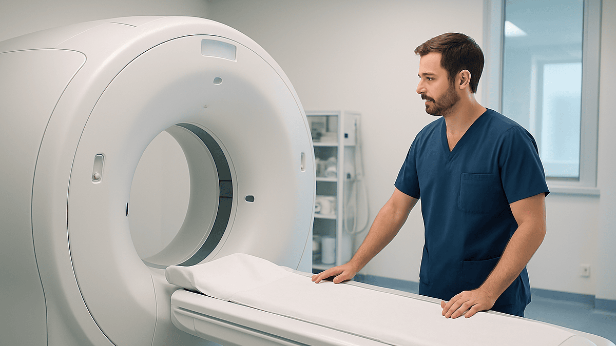
Key Takeaways
- Only a biopsy can confirm head and neck cancer; CT, MRI, PET-CT and ultrasound map its extent but cannot prove the diagnosis.
- TNM staging combines tumor size and invasion (T), neck node involvement (N) and distant spread (M) into an overall stage from 0 to IV.
- HPV-related cancers of the tonsil and tongue base have their own staging table, so a p16-positive cancer with several involved nodes may be stage I or II rather than IV.
- Stage IVA and IVB describe locally advanced disease without distant spread and are routinely treated with the aim of long-term control; only IVC indicates metastasis.
- Panendoscopy under general anesthesia lets the surgeon measure the tumor, take targeted biopsies and search for a second primary cancer along the same lining.
- A sore patch, hoarseness or neck lump lasting more than three weeks is the pattern that national guidance flags for urgent specialist review.
Head and neck cancer staging tests combine three things: a biopsy, which confirms what the cells are; an endoscopic examination, which lets the team see the lining of the nose, throat and voice box directly; and imaging such as CT, MRI, PET-CT or ultrasound, which maps how far the tumor extends. Together they produce a TNM stage that guides every treatment discussion.
The envelope says “appointment with the ear, nose and throat team” and nothing else. A lump under the jaw that would not settle, a hoarse voice that outlasted the cold, a sore patch on the tongue that the dentist frowned at. Now there is a referral, a growing list of tests, and a new vocabulary that includes words like nasendoscopy and PET-CT. Most people in this chair want one thing before anything else: to understand what each test is for, and what the results will and will not tell them.
That is what this explainer sets out to do. Head and neck cancer staging tests are not a single scan but a sequence, and each step answers a different question. Is this cancer at all? Which type? How large is it, has it reached the lymph nodes, and has it traveled anywhere else?
Nothing here replaces the conversation with your own team, who know your scans and your history. It should, though, make that conversation easier to follow.
What counts as head and neck cancer, and where does it most often start?
“Head and neck cancer” is an umbrella term rather than one disease. It covers cancers that begin in the moist lining of the mouth, nose, sinuses, throat (pharynx), voice box (larynx) and the salivary glands. Cancers of the brain, eye, thyroid and skin of the scalp are usually counted separately, even though they sit in the same region.
Most of these cancers are squamous cell carcinomas, meaning they arise from the flat cells that line these surfaces. According to the National Cancer Institute, head and neck cancers make up nearly 4 percent of all cancers in the United States, and tobacco and alcohol use are linked to at least 75 percent of cases. Human papillomavirus (HPV), a common virus, is now the leading cause of cancer in the oropharynx, which is the part of the throat behind the mouth that includes the tonsils and base of the tongue.
People often ask which site is the most common. The oral cavity, which includes the lips, tongue, gums, floor of the mouth and inner cheeks, has traditionally been the most frequent starting point, with the oropharynx and the larynx close behind. The balance has been shifting as smoking rates fall and HPV-related throat cancers rise, so the answer depends partly on the population being counted.
Why does the starting site matter so much? Because every structure in this region does a job you notice daily: speaking, swallowing, breathing, tasting. A tumor a few millimeters wide on a vocal cord changes the voice quickly; a tumor of the same size at the base of the tongue may cause no symptoms until it is larger. Staging tests exist to pin down exactly where a cancer sits and how far it has spread, so that treatment can protect as much function as possible.
Why head and neck cancer staging tests matter: what TNM actually tells your team
Staging is the shared language oncologists use to describe how advanced a cancer is. The system used for head and neck cancer is called TNM, and each letter answers a separate question.

- T (tumor) describes the size of the primary cancer and whether it has grown into nearby structures such as bone, cartilage or muscle.
- N (nodes) describes whether cancer has reached the lymph nodes of the neck, how many, how large, and whether it has broken through the node’s capsule.
- M (metastasis) records whether cancer has spread to distant organs, most often the lungs.
These three findings are combined into an overall stage from 0 to IV. Stage 0 means abnormal cells have not yet invaded deeper tissue. Stages I and II are generally smaller cancers confined to the starting site. Stage III and IV describe larger tumors, node involvement or distant spread, with stage IV subdivided into A, B and C to capture how extensive that involvement is.
Two further distinctions are worth knowing. Clinical staging is assigned before treatment, based on examination, endoscopy, imaging and biopsy. Pathological staging is assigned after surgery, when the pathologist has examined the removed tissue and nodes under the microscope. The pathological stage can differ from the clinical one, because a scan cannot see a microscopic deposit that a slide can.
Stage drives nearly every decision that follows: whether surgery, radiotherapy, systemic therapy or a combination is proposed, how wide a surgical margin is planned, and how many neck nodes need treatment. It is also how trials and guidelines are written, so a correct stage means your team is comparing your situation with the right evidence. That is why thoroughness at this step is not bureaucracy. It is the foundation of the plan.
How a head and neck cancer biopsy works and what actually happens
A biopsy is the removal of a small sample of tissue or cells so a pathologist can examine it under a microscope. It is the only test that can confirm cancer; scans suggest, biopsies prove.
The method depends on where the suspicious area is. For a lump in the neck, the usual first step is fine-needle aspiration: a thin needle, often guided by ultrasound, draws out cells in a few seconds. It feels similar to having blood taken, and most people go straight home. A core biopsy uses a slightly wider needle under local anesthetic to remove a sliver of tissue, which gives the pathologist more to work with when cells alone are not enough.
For a visible lesion in the mouth, a punch or incisional biopsy can be done in clinic under local anesthetic. For areas deeper in the throat, the sample is usually taken during an endoscopic examination under general anesthesia, described in the next section.
What the pathologist reports goes well beyond “cancer, yes or no.” The report typically states the cell type, how abnormal the cells look (the grade), and for tumors of the oropharynx, whether a marker called p16 is present, which indicates HPV-related disease. Depth of invasion may also be measured for oral tongue cancers because it influences the T stage.
Results take time. Tissue has to be fixed, sliced, stained and sometimes sent for additional tests, so a report usually arrives in days rather than hours. Ask your team for a realistic estimate and how you will be told.
Risks are modest: bruising, minor bleeding, soreness, and a small chance of infection. A result labeled “insufficient” or “inconclusive” is not a bad sign in itself; it simply means the sample did not contain enough of the right cells, and the test is repeated.
Endoscopic examination: nasendoscopy in clinic and panendoscopy for throat cancer
Much of the throat cannot be seen with a torch and a tongue depressor. Endoscopy solves that by passing a thin tube with a camera and light into the airway so the team can inspect the lining directly.

The first look is usually a flexible nasendoscopy in the outpatient clinic. A soft scope about the width of a drinking straw is passed through the nostril after a numbing spray. It is uncomfortable rather than painful, lasts a minute or two, and you can talk and swallow on request so the clinician can watch the vocal cords move. The NHS describes this as a routine first assessment for persistent hoarseness, throat lumps and swallowing difficulty.
If something suspicious is found, or if the primary site is still unknown, the next step is often a panendoscopy. This is a thorough examination of the nose, mouth, throat, voice box and upper esophagus under general anesthesia, using rigid and flexible scopes. Because you are asleep, the surgeon can look into corners that gag reflexes would otherwise block, take biopsies from several sites, and measure the tumor against fixed landmarks. It is typically a day-case procedure.
Panendoscopy serves three purposes in staging. It establishes exactly where the tumor begins and ends, which sets the T stage. It allows a search for a second primary cancer, because the same risk factors that caused the first can cause another along the same lining. And when a neck node contains cancer but no primary tumor is visible, targeted biopsies of the tonsils and tongue base can locate the source.
Afterward, a sore throat and mild hoarseness for a day or two are common. The team will tell you when it is safe to eat, drink and drive home, and what bleeding would be more than expected.
Imaging for head and neck cancer staging: CT, MRI, ultrasound and PET scan
Endoscopy shows the surface; imaging shows what lies beneath. No single scan does everything, so teams combine them according to the site and the question.
CT (computed tomography) uses X-rays to build cross-sectional pictures. It is quick, widely available, and particularly good at showing bone and cartilage involvement, for example whether a laryngeal tumor has eroded the thyroid cartilage or an oral tumor has entered the jaw. An iodine-based contrast dye is usually injected to make blood vessels and nodes stand out; tell the team about kidney problems or previous contrast reactions.
MRI (magnetic resonance imaging) uses magnets and radio waves rather than radiation and is better at distinguishing soft tissues from one another. It is often preferred for the tongue, tongue base, nasopharynx and sinuses, and for checking whether a tumor is tracking along a nerve. Scans take longer, typically 30 to 45 minutes, and some people find the enclosed tube difficult; say so in advance, because positioning and support can help.
Ultrasound of the neck is painless, involves no radiation and is the usual guide for needle biopsies of nodes.
PET-CT (positron emission tomography) combines a CT scan with a small injection of a radioactive sugar. Because most cancer cells take up sugar faster than normal tissue, they light up. PET-CT is used to look for distant spread, to hunt for an unknown primary, and to assess response after treatment. It can show false positives in inflamed or infected tissue, which is one reason a bright spot is not the same as a diagnosis.
A chest CT is often added, because the lungs are the most common site of distant spread and the most common place for a second primary cancer in people who have smoked.
Staging tests compared: what each one reveals and what it cannot
People often ask why they need so many tests when they have “already had a scan.” The table below shows why: each test answers a different part of the TNM question, and the gaps in one are covered by another.
| Test | What it mainly answers | What it cannot do | What it feels like |
|---|---|---|---|
| Fine-needle aspiration or core biopsy | Are the cells cancer, and what type? HPV status for oropharynx | Cannot show tumor size or spread | Brief needle sting; home the same day |
| Flexible nasendoscopy | Surface appearance of nose, throat, voice box; vocal cord movement | Cannot see beneath the lining or into nodes | Numbing spray, 1–2 minutes of pressure |
| Panendoscopy under anesthesia | Exact tumor extent (T stage); biopsies; search for second primary | Cannot assess distant spread | Day case; sore throat for a day or two |
| CT with contrast | Bone and cartilage invasion; node size; chest | Less precise for soft tissue; small nodes may be missed | Minutes; contrast gives a warm flush |
| MRI | Soft-tissue extent; nerve involvement; tongue base and nasopharynx | Slower; motion from swallowing can blur images | 30–45 minutes in a scanner; noisy |
| Ultrasound of neck | Node structure; guides needle biopsy | Cannot see deep or behind bone | Gel and probe; painless |
| PET-CT | Distant spread (M stage); unknown primary; treatment response | Inflammation can light up; small deposits can be missed | Injection, quiet rest, then a scan |
Two observations from this table deserve emphasis. First, no imaging test replaces the biopsy; a pathologist’s report is the only confirmation of cancer. Second, scans of the neck and the chest are doing different jobs, which is why both may be ordered even when symptoms are confined to the mouth.
The sequence is not fixed. Your team will choose tests based on the suspected site, the findings so far, your other medical conditions and what will actually change the plan.
Who is usually offered staging tests, and who is asked to wait?
Staging tests are offered to anyone in whom a head and neck cancer has been confirmed or is strongly suspected. In practice, the pathway usually starts with a referral from a dentist, family doctor or emergency department for a symptom that has not settled. In the United Kingdom, NHS guidance means a suspected cancer referral should lead to a specialist appointment within two weeks; equivalent fast-track pathways exist elsewhere.
A few groups are typically moved through quickly: people with a persistent neck lump, an ulcer in the mouth lasting more than three weeks, hoarseness lasting more than three weeks, or one-sided symptoms such as a blocked nostril with bleeding or earache without an ear problem. These patterns are not a diagnosis, but they are the ones that guidelines flag for urgent assessment.
Who is asked to wait? Sometimes the honest answer is the person whose first test has not yet come back. A nasendoscopy showing an obvious lesion will lead to a biopsy, and imaging is usually arranged before or alongside it so results arrive together. Ordering a PET-CT before a biopsy has confirmed anything can muddy the picture, because recent biopsy sites light up with inflammation. Teams therefore sequence tests rather than firing them all at once, and a short pause is often a sign of planning rather than neglect.
Other reasons to defer or adapt a test include pregnancy (MRI and ultrasound are preferred over CT and PET), kidney impairment that makes contrast dye riskier, metal implants that are not MRI-compatible, uncontrolled diabetes that affects the sugar-based PET tracer, and frailty that would make general anesthesia for panendoscopy unwise without preparation. None of these stops staging; they change how it is done.
If you are unsure why a particular test has been chosen, or why one has not, ask. The answer is nearly always specific to your situation.
What the days and weeks after staging tests usually look like
Staging rarely happens in one visit. A typical path runs from the first clinic appointment with nasendoscopy, to a needle biopsy and ultrasound, to a CT or MRI, to a panendoscopy if needed, and then to a multidisciplinary team meeting where surgeons, radiation and medical oncologists, radiologists, pathologists, dentists, dietitians and speech and language therapists review everything together. The NHS and Mayo Clinic both describe this team review as standard practice for head and neck cancer, because the trade-offs between removing a tumor and preserving speech and swallowing are genuinely complex.
What you may notice in the meantime:
- A sore throat or small amount of blood-streaked saliva for a day or two after biopsies or panendoscopy.
- A bruise at the needle site in the neck.
- Fatigue from a run of early starts, fasting for anesthesia and long scanning sessions.
- A dental check, because teeth in poor condition may need attention before radiotherapy to the jaw can be planned safely.
- Appointments with a dietitian or speech and language therapist, who assess swallowing and nutrition at baseline so changes can be measured.
The waiting is often the hardest part. Scan reports and pathology each take several days, and the team meeting follows on a fixed weekly rhythm, so a gap of a couple of weeks between first appointment and treatment discussion is common. That gap is not time lost; it is time spent making sure the stage is right before anything irreversible begins.
Practical steps help. Keep a single notebook or phone note of every appointment, question and result. Bring someone with you to the results discussion, because people remember roughly half of what is said in a stressful consultation. Ask for a named contact, usually a specialist nurse, who can be called between appointments. And if you smoke, tell the team; support to stop improves how tissues heal and how well treatment works, and the team can arrange it without judgment.
How HPV status changes the stages of head and neck cancer in the throat
One of the biggest changes in staging over the past decade concerns the oropharynx. The National Cancer Institute reports that HPV is now responsible for most oropharyngeal cancers in the United States, and these cancers behave differently from those driven by tobacco and alcohol. They tend to occur in younger people, often with little or no smoking history, and they respond better to treatment.
Because of this, the current TNM edition gives HPV-related oropharyngeal cancer its own staging table. The practical effect is striking. A cancer that would have been labeled stage IV under the old rules, typically because of several involved neck nodes, may now be classed as stage I or II if it is HPV-positive. The tumor has not changed; the stage now reflects what is known about its likely course.
HPV status is determined on the biopsy sample, most often by a stain for the protein p16, sometimes confirmed by a direct test for viral DNA. It is reported for cancers of the tonsil and tongue base, not for every head and neck site, because the evidence that HPV changes outlook is specific to the oropharynx.
Why this matters beyond the label: a lower stage means the guideline evidence being applied is the evidence for HPV-related disease, and some clinical trials are exploring whether treatment intensity can be reduced for selected HPV-positive cancers. Those trials are ongoing; outside of a trial, standard treatment is still recommended, and no reduction should be assumed.
If you have been told your cancer is p16-positive, three questions are worth asking: which staging table is being used, what the stage would have been under the older system (useful when reading older material), and whether any trial is relevant to you. Your team decides; the questions simply make the decision transparent.
Which head and neck cancers are most aggressive, and what does stage 4 really mean?
Two of the most searched questions are “what is the most aggressive head and neck cancer” and “how long can you live with stage 4.” Both deserve an honest answer rather than a number.
Aggressiveness is not a property of a site alone. It is a combination of cell type, grade, site, HPV status and stage. That said, certain patterns are recognized in guidelines. Cancers of the hypopharynx, the lowest part of the throat beside the voice box, are often diagnosed late because early symptoms are vague, and are generally regarded as having a less favorable outlook than laryngeal cancers found early. Rare types such as mucosal melanoma and some high-grade salivary gland cancers also tend to behave more aggressively. Within squamous cell carcinoma, HPV-negative disease in someone who continues to smoke is generally more difficult to control than HPV-positive disease.
Stage IV is often misread as “terminal.” In head and neck cancer it is not a single category. Stage IVA and IVB describe locally advanced disease, where the tumor is large or several nodes are involved but there is no distant spread; many such cancers are treated with the intention of long-term control, usually with combined surgery, radiotherapy and systemic therapy. Stage IVC means distant metastasis, most often to the lungs, and here treatment aims to control the disease and protect quality of life for as long as possible.
How long someone can live with stage IV disease therefore depends on which sub-stage they have, their HPV status, their general health, their ability to tolerate treatment, and how the cancer responds. Population statistics exist, but they describe groups treated years ago, not an individual being treated now. The only person who can give you a meaningful estimate is the oncologist who knows your scans, your pathology and you, and it is reasonable to ask them directly.
What people often get wrong about head and neck cancer staging
“If the scan was clear, there is no cancer.” Imaging shows shape and metabolism, not cell type. Small surface cancers of the mouth or vocal cord can be invisible on CT and still be confirmed on biopsy. The reverse also happens: a bright spot on PET-CT can be an infected tooth socket or a reactive node.
“A needle biopsy can spread the cancer.” For head and neck cancers sampled with fine-needle or core techniques under image guidance, this is not supported by the evidence that guides routine practice, and the information gained outweighs the theoretical risk. Teams avoid open surgical biopsy of a neck node before planning, because scarring complicates later surgery, which is a different concern.
“Stage IV means nothing can be done.” As the previous section explains, stage IVA and IVB are locally advanced and are routinely treated with the aim of long-term control.
“HPV-related throat cancer means I did something wrong.” HPV is extremely common; the CDC notes that most sexually active adults are infected at some point, and the vast majority clear it. A cancer decades later reflects bad luck, not behavior.
“The stage is final.” Clinical stage can be revised after surgery once the pathologist examines the removed tissue, and this is normal.
“Panendoscopy is major surgery.” It is a look-and-sample procedure under anesthesia, usually a day case, with a sore throat as the main aftereffect.
“Neck pain means neck cancer.” Most neck pain comes from muscles, joints and posture, and most neck lumps in adults under 40 are benign. The features that prompt urgent referral are persistence beyond three weeks, a painless enlarging lump, and one-sided symptoms, not pain on its own.
Correcting these ideas early tends to make the testing weeks less frightening and the results conversation more useful.
Risks, limitations and alternatives: what staging tests cannot guarantee
A good explainer should be candid about what tests cannot do.
Every test has a false-negative rate. A fine-needle aspiration can miss cancer if the needle samples the wrong part of a node; this is why a negative needle result in a persistently suspicious lump leads to a repeat or a core biopsy, not reassurance. CT and MRI can miss deposits smaller than a few millimeters in lymph nodes, so a neck reported as clear on imaging is sometimes still treated with surgery or radiotherapy if the primary tumor carries a known risk of hidden node spread. PET-CT can miss very small or low-metabolism tumors and can flag inflammation as suspicious.
Physical risks are small but real. Contrast dye carries a low chance of allergic reaction and can strain already impaired kidneys. CT and PET-CT involve radiation, which is why they are reserved for situations where the information changes management. General anesthesia for panendoscopy carries the usual anesthetic risks, higher in people with heart or lung disease, which the anesthetic team will assess beforehand. Biopsy sites can bleed, particularly in people on blood-thinning medication; never stop such a medicine on your own, but tell the team you take it so they can plan.
Alternatives are usually about sequencing rather than substitution. Someone who cannot have MRI because of a non-compatible implant will be staged with CT and ultrasound. Someone unfit for general anesthesia may have the throat assessed with office-based flexible endoscopy and image-guided biopsy instead of panendoscopy. Someone who is pregnant will be imaged with ultrasound and MRI without contrast where possible.
There is also the option of declining a test, which is always yours. The responsible way to use that option is to ask what decision the test would inform and what the plan would look like without it, then decide with the team rather than alone.
Questions to ask your care team about head and neck cancer staging tests
Consultations move quickly, and the questions that matter often surface in the car park. Writing them down in advance helps. These are the ones patients and specialist nurses most often find useful.
- Exactly where is the cancer, and which structures does it touch? Could you show me on the scan?
- What is the cell type and grade, and for throat cancer, what is the HPV or p16 status?
- What is my clinical TNM stage, and which staging table is being used?
- Have the lymph nodes been assessed, and is there any sign of distant spread?
- Were all the tests you would normally expect done? Is anything still pending?
- Was a second primary cancer looked for, including the lungs and esophagus?
- How confident is the team in this stage, and what might change it after surgery?
- Which treatment options does this stage make possible, and what are the trade-offs for speech, swallowing and appearance?
- Will I see a dentist, dietitian and speech and language therapist before treatment starts?
- Is there a clinical trial relevant to my stage and HPV status?
- Who is my named contact between appointments, and how do I reach them?
- What symptoms in the coming weeks should make me call rather than wait?
A few framing questions also help you understand the decision rather than just receive it. “What would you want to know in my position?” invites a clinician to prioritize. “What are you most uncertain about?” surfaces the honest limits of the evidence. And “If I did nothing for a month, what would change?” clarifies urgency without drama.
Bring someone to listen and take notes. Ask whether the consultation can be recorded on your phone; many teams are comfortable with this. Then ask for the written summary of the multidisciplinary team meeting, which lays out stage and recommended options in plain terms and is a document you can reread when the room has emptied.
When to call your doctor: red-flag signs before, during and after staging
Staging is a period of waiting, and waiting is when symptoms can change. These are the signs that warrant a call to your team, or emergency care, rather than the next scheduled appointment.
Before a diagnosis is made, the symptoms that national guidance flags for urgent assessment include: a mouth ulcer or white or red patch lasting more than three weeks; hoarseness or a voice change lasting more than three weeks; a lump in the neck that is persistent, painless or growing; difficulty or pain on swallowing that is getting worse; a blocked nostril with blood-stained discharge on one side; earache without an ear infection; and unexplained weight loss. The NHS lists these as reasons for prompt medical review.
During the testing weeks, call the same day if you notice: bleeding from the mouth, nose or a biopsy site that does not stop with gentle pressure within a few minutes; a fever, spreading redness or increasing pain at a biopsy site; difficulty swallowing saliva; or a rapidly enlarging neck swelling.
Seek emergency care immediately for any difficulty breathing, noisy breathing (stridor), inability to swallow liquids at all, heavy or continuous bleeding from the mouth or throat, or a sudden severe headache or confusion after a procedure. Tumors near the airway can swell after biopsy, and this is one situation where waiting is unsafe.
Your team should give you a direct number, usually for a specialist nurse, together with out-of-hours instructions. Keep it in your phone and use it; these services exist precisely so that people do not sit at home weighing whether a symptom “counts.” If you cannot reach anyone and a symptom concerns you, use emergency services.
Every decision about what these tests mean and what to do next belongs with your treating team. This article describes the territory; they hold the map of your particular case.
Frequently asked questions
What is the most common location of head and neck cancer?
The oral cavity, which includes the tongue, gums, floor of the mouth, lips and inner cheeks, has traditionally been the most common starting point, followed by the oropharynx and the larynx. The balance is shifting as smoking declines and HPV-related cancers of the tonsil and tongue base increase. Most cancers at all of these sites are squamous cell carcinomas arising from the lining cells.
How long can you live with stage 4 head and neck cancer?
There is no single answer, because stage IV covers three very different situations. Stage IVA and IVB mean locally advanced disease without distant spread, often treated with combined therapy aimed at long-term control. Stage IVC means spread to distant organs, where treatment aims to control the disease and protect quality of life. HPV status, general health and response to treatment all matter, so ask your oncologist for an estimate specific to you.
What is the most aggressive head and neck cancer?
Aggressiveness depends on site, cell type, grade, HPV status and stage rather than one factor. Hypopharyngeal cancers are often found late and generally carry a less favorable outlook, and rare types such as mucosal melanoma and high-grade salivary gland cancers tend to behave aggressively. HPV-negative squamous cell cancer in someone who continues to smoke is usually harder to control than HPV-positive disease.
What are some red flag symptoms for head and neck cancer?
Guidance flags a mouth ulcer or patch lasting more than three weeks, hoarseness lasting more than three weeks, a persistent or growing neck lump, worsening pain or difficulty on swallowing, one-sided nasal blockage with bleeding, earache without an ear problem, and unexplained weight loss. These signs do not mean cancer, but they are the pattern that justifies prompt specialist assessment rather than watchful waiting.
Does a head and neck cancer biopsy hurt?
A fine-needle aspiration of a neck lump feels much like a blood test and takes seconds. A punch biopsy in the mouth is done under local anesthetic, so pressure rather than pain is the usual sensation, with soreness afterward. Biopsies of the deeper throat are taken under general anesthesia during panendoscopy, after which a sore throat for a day or two is common.
What does a PET scan for head and neck cancer show?
A PET-CT shows where cells are using sugar unusually fast, which is a feature of most cancers. It is used mainly to look for spread beyond the neck, to find a hidden primary tumor when a node contains cancer, and to assess response after treatment. Inflammation and recent biopsy sites can also light up, so a bright area is a prompt for further checks, not a diagnosis.
What is a panendoscopy for throat cancer?
A panendoscopy is an examination of the nose, mouth, throat, voice box and upper esophagus with cameras and scopes while you are under general anesthesia. It lets the surgeon measure the tumor precisely, take biopsies from several sites and look for a second cancer along the same lining. It is usually a day-case procedure with a sore throat as the main aftereffect.
What are the stages of head and neck cancer?
Stages run from 0 to IV using the TNM system. Stage 0 means abnormal cells have not invaded deeper tissue; stages I and II are generally smaller cancers confined to the starting site; stage III and IV involve larger tumors, lymph node spread or distant metastasis, with IV split into A, B and C. HPV-related oropharyngeal cancer uses a separate table that assigns lower stages for the same findings.
Why do I need an MRI if I already had a CT scan?
The two scans show different things. CT is best at bone, cartilage and the chest, while MRI separates soft tissues more clearly and shows whether a tumor is spreading along a nerve or deep into the tongue or nasopharynx. For many sites the team wants both pictures before deciding how wide a surgical margin or radiotherapy field is needed.
Can the stage change after surgery?
Yes. The stage assigned before treatment is a clinical stage based on examination, endoscopy, imaging and biopsy. After surgery, the pathologist examines the removed tumor and lymph nodes under the microscope and assigns a pathological stage, which can be higher or lower because slides reveal microscopic deposits that scans cannot. A revised stage is routine and helps the team decide whether further treatment is advised.
References
This article is for general information only and is not a substitute for professional medical advice. Please consult a qualified doctor about your individual situation.

More from the Blog

Partial or Radical Nephrectomy for Kidney Cancer: How Surgeons Decide What to Remove
Surgeons choose between partial and radical nephrectomy for kidney cancer mainly on tumor size, where the tumor sits in the kidney, how well both…

Eating Safely After a Stem Cell Transplant: The Low-Microbial Diet and When It Relaxes
After a stem cell transplant, most teams ask patients to follow a low-microbial or neutropenic diet: fully cooked foods, pasteurized dairy and juices, washed…

Planning Your Stay in Istanbul for HIPEC: Hospital Days, Follow-Up and When to Fly Home
After cytoreductive surgery with HIPEC, most people spend roughly one to two weeks in hospital, often beginning with a day or more in intensive…
Vulvar Cancer Surgery Explained: Organ-Sparing Excision and Wider Procedures
Vulvar cancer surgery removes the tumor together with a rim of healthy tissue. It ranges from a wide local excision that preserves most of…

How Neck Cancer Treatment Is Planned When Lymph Nodes Are Involved
When neck cancer involves lymph nodes, a multidisciplinary team plans treatment after biopsy, imaging and staging establish how many nodes are affected, their size,…

How Ureteroscopy With Biopsy Confirms a Ureteral Tumor and Sets the Treatment Plan
Ureteroscopy with biopsy passes a thin telescope through the bladder into the ureter so a urologist can see a suspected tumor directly, collect urine…