

What the Pre-Laser Eye Tests Check: Corneal Thickness, Pressure and Prescription Stability
Key Takeaways
- Pachymetry measures corneal thickness in micrometers, and a cornea too thin to leave a safe structural reserve after reshaping is a recognized reason to be advised against LASIK.
- Corneal topography exists largely to catch early keratoconus, a progressive thinning that often begins in the teens or twenties and can be invisible on a standard glasses test.
- The usual reference range for eye pressure is about 10 to 21 mmHg, and the pre-surgery reading doubles as a lifelong baseline because tonometers under-read after the cornea is thinned.
- Mayo Clinic advises that a glasses prescription should have been stable for at least a year before laser correction is considered.
- Contact lenses temporarily reshape the cornea, so scans are more reliable after a lens-free period of at least a few weeks, and rigid lenses typically need longer.
- Dryness after laser surgery commonly lasts around six months, which is why tear-film testing and untreated dry eye carry real weight in the eligibility decision.
Tests before eye laser surgery measure how thick your cornea is, map its shape, check the pressure inside the eye, and confirm that your glasses prescription has stopped changing. Together with pupil measurements, a tear-film check and a dilated look at the retina, they help the surgical team judge whether laser correction is likely to be safe for your particular eyes, or whether waiting or a different option is wiser.
The room is dark, your chin is resting on a cool plastic cup, and a technician you met four minutes ago is asking you to stare at a tiny green light without blinking. You have worn glasses since the third grade. You came in hoping for a yes. Instead you are getting a very thorough look inside your own eyes, and you are not entirely sure what any of it means.
That is the ordinary experience of the tests before eye laser surgery. Nobody warns you how long the appointment runs, how many machines you will lean into, or why a puff of air against your eyeball has anything to do with whether a laser can reshape your cornea.
The honest answer is that this hour or two of measuring is the most consequential part of the whole process. The laser does what it is programmed to do. The assessment decides whether it should be programmed at all.
What actually happens during the tests before eye laser surgery
Think of the assessment as three layers stacked on top of each other. The first is conversation: your medical history, the medicines you take, any autoimmune conditions, whether you are pregnant or breastfeeding, and what you actually want from surgery. A person who wants to stop fumbling for glasses at 3 a.m. and a person who wants to read a phone screen at fifty without readers are asking for different things, and the team needs to know which one you are.
The second layer is measurement. A refraction, which is the familiar “better with lens one or lens two” exercise, pins down your prescription. Scanners photograph the curvature of your cornea, the clear front window of the eye, and measure its thickness at dozens of points. A tonometer checks the pressure of the fluid inside the eye. Cameras measure your pupils in dim light. Dyes and timers assess your tears.
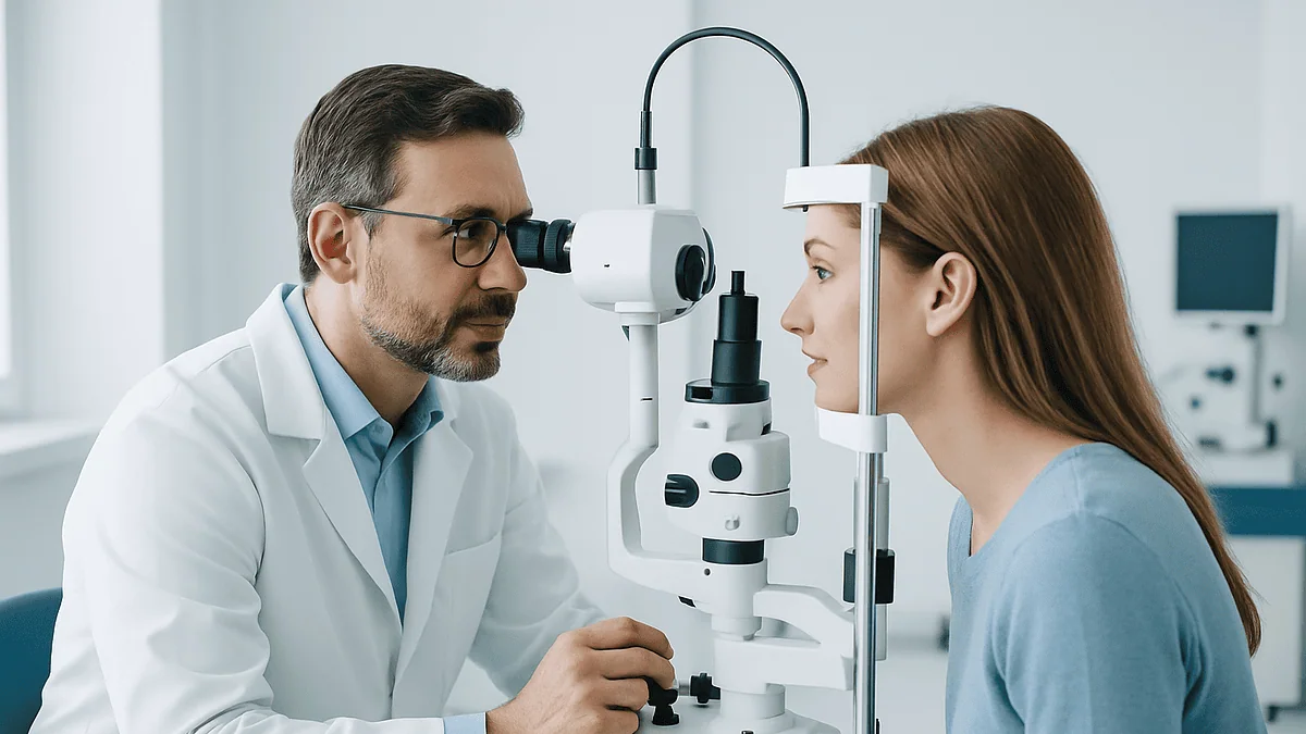
The third layer is the clinical examination. A slit lamp, essentially a microscope with a bright blade of light, lets the ophthalmologist inspect the cornea, lens and eyelids in fine detail. Dilating drops widen the pupil so the retina at the back can be examined properly. Mayo Clinic notes that the effects of dilation can last several hours, so most people are told not to drive afterward and to bring sunglasses.
None of these steps hurts, although the numbing drops sting for a moment and the dilated glare is tiresome. What surprises people is the redundancy. Several instruments measure overlapping things on purpose. When two independent machines agree, the surgeon can trust the number. When they disagree, that disagreement is itself useful information, and it often leads to a repeat visit rather than a surgery date.
Corneal thickness for LASIK: how pachymetry works and why it matters
Pachymetry is the measurement of corneal thickness, reported in micrometers, which are thousandths of a millimeter. A healthy cornea is thinner than a credit card, and laser reshaping works by vaporizing a precisely calculated sliver of tissue from within it. Every unit of prescription corrected costs a certain depth of tissue. The higher your prescription, the more the laser must remove.
There are two common ways to measure. An ultrasound probe touches the numbed cornea for a fraction of a second, sending a sound pulse through and timing its return, much like a depth sounder on a boat. Optical scanners do the same job without contact, building a thickness map of the whole cornea rather than a single point. Many clinics use both.
Why does the surgeon care so much? Because the cornea has to keep its job after surgery. Below the reshaped zone, enough untouched structural tissue, the stroma, must remain to hold the eye’s shape against the pressure inside it. If too much is removed, or if the cornea was already unusually thin or weak, the remaining tissue can gradually bulge forward over months or years. That complication is called ectasia, and it is one of the outcomes surgeons work hardest to avoid. Mayo Clinic lists thin corneas among the reasons a person may be advised against LASIK.
Thickness also quietly affects another test in the room. A thicker-than-average cornea resists the tonometer and can make eye pressure read higher than it really is; a thin cornea can make it read low. So the pachymetry number is not just a go or no-go gauge for the laser. It is the correction factor that helps the team interpret your pressure reading honestly.
Corneal topography: mapping the shape and screening for keratoconus
If pachymetry tells the surgeon how much cornea you have, topography tells them what shape it is in. Corneal topography projects rings of light onto the eye and analyzes the reflection to build a color-coded map of curvature, rather like an elevation map of a landscape. Warm colors mark steeper areas, cool colors flatter ones. Newer tomography systems go further, imaging the back surface of the cornea as well as the front.
A healthy cornea is a smooth, slightly flattened dome. Astigmatism, in which the cornea is more curved in one direction than another, shows up as a symmetrical bow-tie pattern that lasers handle routinely. The pattern the team is really hunting for is asymmetry: a localized steep zone, often below the center, that suggests the tissue is weaker there than elsewhere.
That pattern raises the question of keratoconus, a condition in which the cornea progressively thins and bulges into a cone shape. Mayo Clinic notes that it often begins in the late teens or twenties and can progress slowly for years. In its early form it may cause no symptoms at all, and glasses may still correct vision reasonably well, which is exactly why people do not know they have it. Laser reshaping removes tissue from a cornea that is already structurally compromised, and that is a combination surgeons decline.
Topography is also where recent contact lens wear causes the most trouble. Lenses can temporarily mold the cornea into a shape that mimics disease or hides it. This is the technical reason behind the request to switch to glasses before your scans, a request covered in its own section below. A single suspicious map is not a diagnosis. A repeat map, weeks later and lens-free, is what settles it.
What does the eye pressure test check before laser surgery?
Tonometry measures intraocular pressure, the pressure of the clear fluid that fills the front of the eye and keeps it firm. The familiar version blows a brief puff of air at the cornea and calculates pressure from how much the surface flattens. The more precise version uses a numbing drop and a small probe that gently touches the cornea. Neither takes more than a few seconds per eye.
MedlinePlus gives the usual reference range as roughly 10 to 21 millimeters of mercury. A reading above that range does not by itself mean glaucoma, the group of conditions in which raised pressure damages the optic nerve, but it does mean the team wants to look harder. Mayo Clinic lists glaucoma among the eye conditions that may make laser correction unsuitable, and a pressure reading is the fastest first screen.
There are two further reasons the number matters specifically before laser surgery. First, after the cornea has been thinned, tonometers tend to under-read pressure for the rest of your life. A reliable baseline recorded now becomes the reference point that every future optometrist will need, because an apparently “normal” post-surgery reading might be masking a real rise. Second, the anti-inflammatory steroid drops commonly prescribed in the weeks after surgery raise eye pressure in some people. Knowing your starting pressure, and whether you already sit near the top of the range, shapes how closely the team monitors you afterward. Any decision about those drops rests entirely with the prescribing clinician.
So the puff test is not the throwaway moment it feels like. It is a snapshot the team will still be consulting years from now.
Prescription stability: why surgeons want a year of steady numbers
Refraction is the measurement of the lens power your eye needs to focus clearly, expressed as the numbers on your glasses prescription. At a laser assessment it is usually done twice. The first is the standard “manifest” refraction with your eyes focusing naturally. The second, a cycloplegic refraction, uses drops that temporarily relax the focusing muscle inside the eye. That matters because people, especially younger ones, can unconsciously focus their way through a test and appear less far-sighted than they really are. The drops remove that effort and reveal the true number.
The laser corrects the prescription you have on the day of surgery. It cannot anticipate the prescription you will have in three years. If your eyes are still changing, the correction will drift out of date, and touch-up surgery removes more tissue from a cornea that has already given some up. That is why Mayo Clinic advises that a prescription should be stable for at least a year before LASIK is considered, and why the team will ask to see your old glasses or previous records rather than take your word for it.
Several things make a prescription wobble. The NHS notes that eyes can still be changing in the late teens and around the early twenties, which is why young adults are often asked to wait. Pregnancy and breastfeeding shift the cornea’s hydration and curvature through hormonal effects, and Mayo Clinic lists both as reasons to postpone. Poorly controlled blood sugar in diabetes can swing focus from week to week. Certain medicines alter vision or healing.
Stability is not a formality to hurry past. It is the difference between a correction that lasts and a correction you outgrow.
Pupils, tear film and the back of the eye: the rest of the workup
Three quieter tests round out the picture, and each one predicts a different kind of regret.
The first is pupil size in dim light. A laser treats a zone of a certain diameter. When the pupil widens at night beyond the edge of that treated zone, light entering through the untreated rim is focused differently, producing halos and starbursts around headlights. Mayo Clinic identifies large pupils as a factor that can increase this risk. Modern lasers treat wider zones than older ones, but a wider zone removes more tissue, so pupil size and corneal thickness are weighed together.
The second is the tear film. Laser surgery cuts some of the tiny corneal nerves that signal the eye to produce tears, and Mayo Clinic notes that eyes commonly feel unusually dry for the first six months or so afterward. Someone who already has dry eye, from screen work, contact lenses, certain medicines or an underlying condition, may find that temporary phase harder to tolerate. The team may place a yellow or green dye on the eye to see how quickly the tear layer breaks apart, or use a paper strip to gauge tear volume. Untreated dry eye is one of the most common reasons a person is asked to come back rather than proceed.
The third is the examination itself. Under the slit lamp the ophthalmologist checks the cornea for scars and the lens for early cataract, a clouding that laser correction cannot address and that might make a lens-based option more sensible. After dilating drops, a look at the retina rules out tears, thinning or other disease, because a nearsighted eye is often a longer eye, and a longer eye has a more stretched retina. None of this is about the laser directly. All of it is about whether your eye is a good place to use one.
LASIK eligibility criteria: who is usually suitable and who is asked to wait
Eligibility is a judgment, not a checklist, and the judgment belongs to the treating team. Still, the broad pattern is consistent across national guidance.
People who are generally considered suitable are adults whose prescription falls within the range the laser can correct, has been stable for at least a year according to Mayo Clinic’s guidance, and whose corneas are thick enough and regularly shaped to leave a safe structural reserve. They have no active eye disease, their tear film is adequate, and their expectations are realistic: sharper distance vision, not perfect vision at every distance for life.
People who are commonly asked to wait, rather than declined outright, include:
- Young adults whose prescription is still shifting; the NHS notes eyes can keep changing into the early twenties.
- Anyone pregnant or breastfeeding, because of temporary hormonal effects on the cornea.
- People with an active eye infection, inflammation or a recent injury.
- People with significant dry eye that has not yet been brought under control.
- People whose scans were taken too soon after removing contact lenses.
People who are more often advised against laser correction altogether include those with keratoconus or a suspicious topography map, corneas too thin for the tissue that would need removing, glaucoma or a cataract that is already affecting vision, and autoimmune or immune-suppressing conditions that impair corneal healing. Mayo Clinic groups these under conditions that increase risk or make outcomes less predictable.
The point worth holding onto is that a “not now” or a “not this procedure” is protective. The alternatives, discussed later, exist precisely for the eyes that fall outside this pattern.
Why stop wearing contacts before LASIK tests?
A contact lens sits on the cornea for twelve or more hours a day and, however gently, it presses. Soft lenses slightly flatten and dehydrate the surface. Rigid lenses reshape it more firmly. Either way, the cornea you present to the scanner immediately after years of lens wear is not quite the cornea you were born with. Surgeons call this corneal warpage.
The consequences run in both directions. Warpage can create an irregular map that looks like early keratoconus, leading to a needless refusal. It can also flatten a genuinely irregular cornea into a temporarily reassuring pattern, hiding a problem the laser would then make worse. And because the warped shape changes your refraction, the prescription programmed into the laser could be slightly wrong.
Mayo Clinic advises switching to glasses and stopping contact lens wear for at least a few weeks before the evaluation and the surgery itself. Rigid gas-permeable lenses need considerably longer than soft ones, and lenses worn for many years need longer still; the exact interval for your eyes is a decision for the surgical team, who may repeat the topography until two maps taken weeks apart match.
People find this the most inconvenient instruction of the whole process, particularly if their glasses are old or their prescription has drifted. Plan for it. Update your glasses if you need to, warn your workplace if you drive or use screens intensively, and treat the lens-free weeks as part of the assessment rather than an obstacle to it. Skipping the break does not save time. It usually means the scans have to be repeated.
Tests before eye laser surgery at a glance
Laid side by side, the individual tests stop feeling random. Each answers a distinct question the surgeon must settle before a laser is programmed.
| Test | What it measures | What it helps decide |
|---|---|---|
| Refraction (manifest and cycloplegic) | Your true prescription, with focusing effort removed | Whether the correction is within the treatable range and has been stable |
| Pachymetry | Corneal thickness in micrometers | Whether enough structural tissue will remain after treatment |
| Topography or tomography | Curvature of the front, and often back, corneal surface | Whether the shape is regular or shows signs of keratoconus or lens warpage |
| Tonometry | Pressure inside the eye | Screening for glaucoma and recording a baseline for life after surgery |
| Pupil measurement in dim light | How wide the pupil opens at night | Risk of halos and glare from light entering outside the treated zone |
| Tear-film assessment | Tear volume and how quickly the tear layer breaks up | How well the eye is likely to tolerate post-surgery dryness |
| Slit-lamp and dilated retinal examination | Cornea, lens and retina in magnified detail | Ruling out cataract, scarring, retinal thinning or other disease |
Notice how many of these tests interact. Thickness changes the meaning of the pressure reading. Contact lens history changes the trust placed in the topography. Pupil size changes how much thickness the laser will consume. A surgeon reading these results is not ticking boxes; they are building a single three-dimensional model of your eye and asking whether it can safely spare some tissue. That is why a good assessment ends with a conversation rather than a printout, and why the conversation sometimes ends with a recommendation you did not walk in hoping for.
What the results can lead to: LASIK, surface ablation, lenticule extraction or no laser at all
The assessment is not a pass-or-fail exam for one procedure. It is a sorting process, and it can point in several directions.
LASIK, the procedure most people picture, creates a thin hinged flap in the cornea, reshapes the tissue beneath it with an excimer laser, and lays the flap back. It requires enough thickness for both the flap and the reshaping, plus a structural reserve underneath. Cleveland Clinic describes it as the most commonly performed laser vision procedure, and it is the default when all the numbers line up.
Surface ablation, of which PRK is the original form, removes the cornea’s thin outer skin, the epithelium, and lasers the surface directly without a flap. Because no flap is made, it uses less depth and is sometimes considered when corneas are on the thinner side or when a person’s work or sport carries a risk of eye impact. The trade-off is a slower, more uncomfortable healing of the surface in the first days, described in NHS guidance on laser and lens surgery.
Lenticule extraction removes a lens-shaped disc of tissue through a small incision using a femtosecond laser, without a full flap. Its suitability depends on the prescription type and corneal findings.
For high prescriptions, thin corneas or early cataract, the team may raise lens-based options: an implantable lens placed inside the eye in front of the natural lens, or replacement of the natural lens itself. These are intraocular operations with their own risk profile, and the NHS outlines them alongside laser techniques as alternatives rather than upgrades.
And sometimes the right answer is glasses or contact lenses, unchanged. That is not a failure of the assessment. It is the assessment working.
What the days and weeks after the assessment usually look like
The first few hours are the least glamorous. Dilating drops leave near vision blurry and daylight harsh, and Mayo Clinic notes the effect can last several hours, so reading small print and driving are off the table until it wears off. If numbing drops were used for pachymetry or tonometry, the eye may feel gritty for a short while. Rubbing it is a bad idea, because the numbed surface will not warn you if you press too hard.
Results are often discussed the same day, but not always. If your topography was borderline, or you had removed contact lenses more recently than the team would like, expect an invitation to return in a few weeks for repeat scans. Two consistent maps taken weeks apart are worth far more than one perfect one. If dry eye was flagged, you may be asked to work on it first, which can mean weeks of lubricating drops, lid hygiene or other measures directed by the clinician before a decision is revisited.
If surgery is scheduled, the assessment shapes what follows. Mayo Clinic describes blurry, hazy vision immediately afterward, dryness that commonly persists for the first six months or so, and a period of roughly two to three months before healing settles and vision stabilizes. Follow-up visits during that window recheck pressure, particularly while anti-inflammatory drops are being used, and confirm the cornea is healing as the scans predicted. Every one of those timeframes is a typical range, not a promise, and your own course is something only your treating team can comment on.
Keep your pre-surgery records. The baseline pressure and thickness numbers will matter to eye care professionals you have not met yet.
What people often get wrong about pre-laser eye tests
“If I see 20/20 with glasses, I qualify.” Good corrected vision says nothing about corneal thickness, shape, pressure or tears. Plenty of people with crisp vision in glasses have corneas that should not be lasered, and early keratoconus is the classic example.
“A thick cornea means I am safe.” Thickness is one variable. A thick but irregularly shaped cornea can be a poorer candidate than a thinner, perfectly regular one. Surgeons read pachymetry and topography together, never alone.
“The puff test tells me whether I have glaucoma.” Pressure is a screen, not a diagnosis. MedlinePlus gives a reference range of about 10 to 21 mmHg, but glaucoma can occur with pressure inside that range and raised pressure can occur without glaucoma. Before laser surgery the reading is also a baseline for future comparison, which is arguably its more important job.
“Leaving contacts in until the day before is fine.” It is the single most common reason scans have to be repeated. Lens warpage can both mimic and mask disease, which is why Mayo Clinic advises a lens-free period of at least a few weeks before evaluation.
“Laser surgery means no glasses ever again.” Laser reshaping corrects distance focus. It does not stop the age-related stiffening of the natural lens that brings on reading difficulty in the forties and fifties. The NHS is explicit that many people will still need reading glasses later in life.
“If one team says no, another will say yes, so keep asking.” Second opinions are entirely reasonable. Hunting for a yes after several careful refusals is not. The tests are protective, and a decline grounded in your scans is a conclusion about your eyes, not about the surgeon’s mood.
Questions to ask your care team
The most useful thing you can bring to a results conversation is a short list of questions. Written down, so the dilating drops do not steal them from you.
- What was my central corneal thickness, and how much tissue would the planned correction remove?
- Did my topography show any asymmetry or anything you would call borderline? Would you like to repeat it?
- What was my eye pressure, and did you adjust it for my corneal thickness? Can I have the number for my records?
- How stable does my prescription look across the records I brought, and would you prefer to wait?
- How large were my pupils in dim light, and how does that relate to the treatment zone you would use?
- What did the tear-film tests show, and is there anything you want addressed before deciding?
- Given all of this, which procedure are you recommending, and which ones did you rule out for my eyes and why?
- What are the specific risks you would flag for me, as opposed to the general list?
- Will I be prescribed drops afterward, and how will you monitor my eye pressure while I use them?
- What would make you cancel or postpone on the day of surgery?
- If I decide not to go ahead, is there anything in today’s findings I should follow up on anyway?
That last question deserves emphasis. The assessment is a thorough eye examination in its own right. Early cataract, raised pressure, retinal thinning or a suspicious corneal map are worth knowing about whether or not you ever have laser surgery, and the team can advise on how those findings should be followed by your usual eye care provider.
Ask, too, for copies of the scans. They are yours, and they become more valuable with time.
When to call your doctor
Pre-operative testing is low-risk, but a few situations, both after the assessment and after any surgery that follows, deserve a same-day call to the treating team or, if you cannot reach them, urgent care.
After the assessment itself, seek care promptly for:
- Severe eye pain, especially with headache, nausea or a halo around lights, which can indicate a sudden rise in eye pressure after dilating drops
- A sudden loss or dimming of vision in either eye, or a curtain or shadow across part of your visual field
- A sudden shower of new floaters or flashes of light, which can signal a retinal tear
- Increasing redness with pain, light sensitivity or discharge, particularly if a numbed eye was rubbed or a contact lens was reinserted too soon
- Blur from dilation that has not begun to lift by the following morning
If you go on to have surgery, Mayo Clinic and the NHS both describe warning signs that should not wait for the next scheduled visit: pain that worsens rather than eases, vision that deteriorates after it had started to clear, a gritty sensation with heavy discharge, or any sense that the eye was struck or rubbed hard in the early days when a LASIK flap is still settling.
Between those extremes, many things are ordinary: mild stinging from drops, temporary light sensitivity, dryness, and fluctuating vision in the first weeks. Ordinary does not mean unmentionable. If something worries you, the team would rather hear about it early. Every decision about drops, follow-up timing, further tests or whether to proceed at all rests with the clinicians who examined your eyes, and this article is a guide to the conversation, not a substitute for it.
Frequently asked questions
LASIK consultation: what to expect at the first visit?
Expect a long appointment with several machines rather than a quick glasses check. You will discuss your medical history, have your prescription measured twice, have corneal thickness and shape scanned, have eye pressure checked, and have pupils and tears assessed. Dilating drops mean blurry near vision and glare for several hours afterward, so arrange transport and bring sunglasses and any old prescriptions.
How thick does a cornea need to be for laser eye surgery?
There is no single number that applies to everyone. The surgeon calculates how much tissue your particular prescription would remove and how much structural tissue would remain beneath, then weighs that against your corneal shape and pupil size. Thin corneas are a recognized reason to be steered toward surface ablation, a lens-based option or no surgery, and that judgment sits with the treating team.
What does the air puff test have to do with laser surgery?
The air puff measures pressure inside the eye, a screen for glaucoma and a baseline for the future. After laser surgery the thinner cornea makes pressure readings appear lower than they truly are, so the pre-surgery figure becomes the reference every later eye professional needs. It also helps the team plan monitoring if anti-inflammatory drops that can raise pressure are prescribed afterward.
Why does my prescription need to be stable before LASIK?
Because the laser corrects the prescription you have on the day, not the one you will have later. If your eyes are still changing, the result drifts out of date and any touch-up removes more tissue. Mayo Clinic advises stability for at least a year, and the team will usually want to see old records rather than rely on memory.
How long should I stop wearing contacts before the eye tests?
Mayo Clinic advises switching to glasses for at least a few weeks before both the evaluation and the surgery. Soft lenses need the shortest break; rigid gas-permeable lenses and decades of wear need considerably longer. The exact interval for you is set by the surgical team, who may repeat the corneal map until two scans weeks apart match.
Can the pre-laser tests detect keratoconus?
They are designed to. Corneal topography and tomography look for the asymmetric steepening and localized thinning that characterize keratoconus, sometimes before it causes any symptoms or shows on a routine eye test. A suspicious map is usually repeated after a contact lens break, because lens warpage can mimic the pattern. Confirmed keratoconus is a standard reason laser reshaping is not offered.
Are the tests before eye laser surgery painful?
No. The scans and photographs involve staring at lights, and the pressure test is either a brief puff of air or a gentle touch after a numbing drop that stings for a moment. Dilating drops cause hours of blurry near vision and light sensitivity, which is tiresome rather than painful. Most people find the length of the appointment the hardest part.
What happens if my pupils are large?
Large pupils in dim light are one of the factors Mayo Clinic links to a higher chance of night-time halos and glare, because light entering through the untreated rim of the cornea focuses differently. Surgeons may plan a wider treatment zone, which uses more corneal tissue, so pupil size is weighed together with thickness. It is rarely a sole reason to decline, but it shapes the plan.
Will these tests tell me whether I will still need reading glasses?
Not in the way many people hope. Laser reshaping corrects distance focus; it does not stop the natural lens stiffening with age, which is what brings on reading difficulty in the forties and fifties. The NHS notes many people who have laser surgery will still need reading glasses later. Ask the team how your age and current prescription fit into that picture.
What are the LASIK eligibility criteria if I have dry eyes?
Dry eye does not automatically rule you out, but it carries real weight. Laser surgery temporarily reduces tear production, with dryness commonly lasting around six months according to Mayo Clinic, so an eye that is already dry may struggle. Teams often ask people to bring dry eye under control first and then reassess. The decision, and any treatment used to get there, rests with your clinician.
References
This article is for general information only and is not a substitute for professional medical advice. Please consult a qualified doctor about your individual situation.

More from the Blog
Injections, Laser or Surgery for Macular Diseases: How the Retina Team Picks a Path
Macular disease treatment depends on what is happening in the macula. Leaking new blood vessels and swelling are usually treated with repeated anti-VEGF eye…

How Tear Duct Irrigation and Imaging Confirm Where the Blockage Sits
A blocked tear duct test locates where tears stop draining rather than simply confirming that they overflow. An eye specialist usually starts with a…
What Happens in the Operating Room During Cataract Surgery, Minute by Minute
During cataract surgery, a surgeon numbs the eye with drops or a small injection, makes an incision of a few millimeters at the edge…
Yellow Skin, Nausea and Blurry Hours After Eye Angiography: Expected Effects and Red Flags
After fluorescein eye angiography, a yellowish tint to the skin for a few hours, dark orange urine for about a day, brief nausea during…

Glasses, Contacts or Laser for Farsightedness: How the Decision Usually Unfolds
Farsightedness treatment options usually begin with glasses, which correct the focusing error with a convex lens and carry no medical risk. Contact lenses offer…

Massage, Probing, Stenting or DCR Surgery: How Blocked Tear Duct Treatment Is Chosen
Doctors choose blocked tear duct treatment by age, cause and where the blockage sits. In babies, most blockages open on their own, so gentle…