

Which Scans Show a Ureter Problem? Ultrasound, CT and Kidney Function Tests Explained

Key Takeaways
- Ultrasound reliably shows kidney swelling from a blockage but can miss stones in the middle of the ureter, where bowel gas blocks sound waves.
- Non-contrast CT is the most accurate test for finding a ureteral stone and measuring its size, which is the figure that guides whether to wait or treat.
- Hydronephrosis on a report means urine is pooling above a blockage or in a roomy system; it is a sign, not a diagnosis, and it does not by itself mean kidney damage.
- No scan measures kidney function; creatinine and eGFR from a blood test do, with an eGFR of 60 or higher in the normal range according to the NIH.
- The NHS describes a CT scan as equivalent to between a few months and a few years of natural background radiation, which is why ultrasound handles routine follow-up.
- Fever with flank pain, no urine output, or uncontrolled pain are red flags that need same-day care regardless of what any previous scan showed.
A non-contrast CT scan is the most accurate way to find a stone or blockage in a ureter, while ultrasound is often the first test because it uses no radiation and reliably shows swelling of the kidney (hydronephrosis) caused by a blockage. Neither scan measures how well the kidneys filter; blood and urine tests such as eGFR and creatinine do that. Your care team chooses based on your symptoms, age and kidney health.
The pain arrives on a Tuesday afternoon, low on one side of the back, then curls forward toward the groin. By evening there is a pink tinge in the toilet bowl. At the urgent care desk, the first question after the nurse checks a temperature is not about medicine. It is about pictures: “Do you want a ureter CT scan, ultrasound, or something else?” For most people that is the first time they have heard the word ureter said aloud.
The ureters are two narrow muscular tubes, each about as wide as a drinking straw, that carry urine from the kidneys down to the bladder. When one is blocked, kinked or narrowed, urine backs up and the kidney above it swells. That is what imaging is trying to catch.
The honest answer to “which scan” is that each test sees a different slice of the problem, and a blood test often matters as much as any picture. Here is how the pieces fit together.
What a ureter CT scan or ultrasound is actually looking for
Start with what can go wrong inside a tube the width of a straw. A stone that formed in the kidney can slip into the ureter and wedge at one of its naturally narrow points: where it leaves the kidney, where it crosses the pelvic blood vessels, or where it enters the bladder wall. Scar tissue can narrow the tube (a stricture). A kink at the junction with the kidney can slow drainage from birth. Less commonly, a growth inside the ureter or pressure from something outside it, such as an enlarged lymph node or a pregnant uterus, can squeeze it shut.
Every one of those problems leaves the same fingerprint: urine cannot get past, so it collects above the blockage. Radiologists (doctors who interpret scans) call that swelling hydronephrosis, which simply means water in the kidney. Imaging is designed to answer three questions in order. Is there swelling? If so, where does the tube change from wide to narrow? And what is sitting at that point?
Ultrasound is excellent at the first question and fair at the second. CT (computed tomography, a rotating X-ray that builds cross-sectional images) is strong at all three, which is why the Mayo Clinic describes CT as able to reveal even tiny stones that plain X-rays miss.
Then there is a fourth question that no picture can answer: is the kidney still filtering properly? A swollen kidney can look dramatic yet work almost normally for a while, and a quiet-looking kidney can be losing ground. That is why a blood sample for creatinine, a waste product the kidneys clear, is drawn alongside almost every ureter workup. Scans show shape; blood tests show function. You usually need both to make a safe decision.
How a kidney and ureter ultrasound works, step by step
An ultrasound scan bounces high-frequency sound waves off your organs and turns the returning echoes into a moving picture. MedlinePlus notes that it uses no ionizing radiation, which is why it is the default first look in pregnancy and childhood.

You may be asked to arrive with a comfortably full bladder. Fluid is a clear window for sound, and a filled bladder pushes bowel loops aside so the lower ends of the ureters and the bladder wall come into view. A sonographer (a technologist trained in ultrasound) spreads warm gel on your flank and abdomen and presses a hand-held probe against the skin. You will roll from your back to each side and hold your breath for a few seconds so the kidneys drop below the ribs. According to the NHS, most ultrasound scans last between 15 and 45 minutes.
What the operator is checking, in plain terms:
- Kidney length and the thickness of its outer filtering layer, the cortex.
- Whether the central collecting area is stretched with fluid, and how much.
- Bright spots with a dark shadow behind them, the classic look of a stone.
- Small puffs of urine squirting into the bladder from each ureter, called ureteral jets, which suggest the tube on that side is open.
- How much urine remains in the bladder after you empty it.
The middle third of each ureter usually hides behind gas-filled bowel, and sound waves cannot pass through gas. That single anatomical fact explains most of ultrasound’s blind spot for ureteral stones, and it is why a clear ultrasound never fully closes the question if pain persists.
Can ultrasound detect kidney stones in the ureter?
Sometimes, and the qualifier matters. Ultrasound sees stones best at the two ends of the ureter: just below the kidney and just above the bladder, where a full bladder provides a clear acoustic window. In the long middle stretch behind the bowel, a stone can sit undetected while the scan looks reassuring.
Because of that, sonographers lean on indirect evidence. A swollen kidney on one side, a missing ureteral jet on the same side, or a glittering “twinkle” behind a bright spot on color Doppler (a setting that shows movement) all raise the likelihood of a stone even when the stone itself is not visible. Interpreting those clues takes experience, and results vary with the operator, the patient’s body shape and how much bowel gas is present that day.
Non-contrast CT is the reference standard for locating a ureteral stone and measuring its size, which is the number that drives decisions about whether it is likely to pass on its own or needs a procedure. The Mayo Clinic describes CT as able to show even tiny stones, and it also measures stone density, a hint about composition.
Why, then, do so many emergency departments start with ultrasound? Radiation, mostly. A strategy of ultrasound first, with CT reserved for people whose picture is unclear or whose symptoms do not settle, spares a large share of patients a CT while catching the ones who need one. It is a triage tool rather than a final verdict.
The practical translation: if an ultrasound finds no stone but your kidney is swollen, or your pain continues, expect your team to move to CT rather than stop there. A normal ultrasound in a person whose pain has vanished is a different, more reassuring situation.
What hydronephrosis on ultrasound means, and what it does not
Hydronephrosis on ultrasound is the finding most likely to appear on a report you read before anyone explains it. It describes a kidney whose inner collecting system, normally a tight set of funnels, has ballooned with urine. The Cleveland Clinic describes it as swelling caused by a build-up of urine, and stresses that it is a sign of an underlying problem rather than a disease in itself.

Radiologists usually grade it. Mild means the funnels are slightly widened. Moderate means the central area is clearly distended. Severe means the funnels are so stretched that the filtering rim of kidney tissue looks thinned. The grade helps, but on its own it does not tell anyone how long the blockage has been there or how well the kidney is working.
The list of causes is longer than most people expect:
- A stone or stricture in the ureter, the classic culprit.
- Urine flowing backward from the bladder toward the kidney (vesicoureteral reflux), most often found in children.
- An overfull bladder at the time of the scan, which can temporarily widen both sides.
- Pregnancy, in which the uterus presses on the ureters and hormones relax them, more often on the right.
- A normal anatomical variant in which the kidney’s funnel sits outside the kidney and always looks a little roomy.
Two patterns carry different meanings. Swelling on one side points to that side’s ureter. Swelling on both sides points lower, to the bladder outlet or the prostate in men. A repeat scan after you empty your bladder, or a nuclear medicine drainage study, often settles whether the widening is a true obstruction or simply a roomy but freely draining system.
Why would a urologist order an ultrasound first?
A urologist (a surgeon who specializes in the urinary tract) reaches for ultrasound for reasons that have little to do with it being the most detailed picture. It is not. Its strengths are practical.
It can be repeated without limit. Someone who has already passed three stones does not need a fresh CT for every episode; a quick ultrasound showing whether the kidney is swollen answers the urgent question and the person’s own history fills in the rest. For children and pregnant women, it is the standard first test because it avoids radiation entirely.
It answers a specific set of questions well. Has a known stone moved or passed? Has swelling settled after a stent (a thin plastic tube placed inside the ureter to keep it open) was put in? Is the bladder emptying completely? How large are the kidneys in someone with long-standing kidney disease, where shrunken, scarred kidneys tell a story? Ultrasound handles all of these in a single unhurried visit.
It is also the tool for following things that may never need treatment. A mild widening found by accident on a scan done for another reason is often re-imaged with ultrasound to see whether it changes, rather than treated on the strength of a single picture.
There is a logic to the order of tests, and ultrasound sits at the front of it. Think of it as the general practitioner of imaging: broad, safe, quick, and very good at deciding who needs the specialist test. When the urologist orders it, the message is usually “let us look without exposing you to anything,” not “we are not taking this seriously.” If the ultrasound raises a question it cannot answer, the next step follows quickly.
CT urogram for ureter problems: how a ureter CT scan works
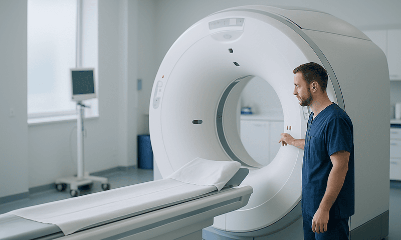
A CT scanner is a doughnut-shaped machine that rotates a thin X-ray beam around you and stitches the results into slices, then into a three-dimensional map. You lie on a table that glides through the ring; the NHS notes the scan itself usually takes around 10 to 20 minutes, most of it positioning.
Two versions matter for the ureter. A non-contrast CT is the stone-hunting scan. Nothing is injected, and the slices are read for the bright dot of a stone, the exact level where the ureter narrows, and the fat around the kidney, which turns hazy when urine is backing up under pressure. It also catches look-alikes that can mimic a stone: appendicitis, an inflamed ovary, a leaking aortic aneurysm.
A CT urogram adds contrast, an iodine-based liquid injected into a vein that the kidneys filter and pass into the urine. Images are taken in phases. Early scans show blood flow to the kidneys; a delayed set, often ten minutes or more after injection, catches the contrast filling the ureters and bladder so the tubes light up from end to end. That outline is what reveals a stricture, a growth pushing into the tube, or a filling defect where something is blocking flow. Because contrast can be hard on kidneys that are already struggling, a recent creatinine result is usually required before it is given, and the radiology team will ask about allergies and any medicines that need adjusting around the injection.
Low-dose stone protocols now use a fraction of the radiation older scans did, and many centers use them routinely. Even so, CT remains the test your team will think twice about ordering repeatedly in a young stone-former, which brings the conversation back to ultrasound for follow-up.
Ureter CT scan vs ultrasound: a side-by-side table
People often ask for the “best” scan. The more useful question is which test answers the question in front of you. This table lines them up, with kidney function tests included because they belong in the same conversation.
| Test | Best at showing | Weak spot | Radiation | Typical role |
|---|---|---|---|---|
| Ultrasound | Kidney swelling, kidney size and scarring, bladder emptying, stones at either end of the ureter | Mid-ureter stones hidden by bowel gas; operator-dependent | None | First look, pregnancy, children, follow-up |
| Non-contrast CT | Exact stone location, size and density; causes of pain outside the urinary tract | Shows shape only, not function; radiation | Yes | Suspected stone in adults when the answer changes treatment |
| CT urogram (with contrast) | Strictures, growths inside the ureter, blood in the urine without a clear cause | Needs adequate kidney function; contrast reactions possible | Yes | Painless visible blood in urine, complex anatomy |
| Blood tests (creatinine, eGFR) | How well both kidneys filter, whether a blockage is affecting function | Cannot say which side or why | None | Alongside any imaging; before contrast |
| Nuclear drainage scan (renogram) | How much each kidney contributes and whether urine truly drains | Little anatomical detail | Small | Sorting true obstruction from a roomy system |
Read across the rows and a pattern emerges. Ultrasound and CT compete only for one job, finding a stone, and even there they cooperate more than they compete. For everything else they occupy different lanes. The NHS describes CT as exposing a person to more radiation than a plain X-ray, roughly the equivalent of between a few months and a few years of natural background radiation depending on the scan, which is precisely why the radiation-free tests carry the routine work and CT is saved for the moments when its precision changes what happens next.
Can an ultrasound tell if your kidneys are failing? Where kidney function tests come in
Not on its own. Ultrasound shows structure, and structure is only a rough proxy for function. It can reveal kidneys that have shrunk and scarred over years, a thinned filtering rim, cysts, or the swelling of a blockage. It can suggest that a kidney has been under strain. What it cannot do is put a number on how well blood is being cleaned today.
That number comes from a blood test. Creatinine is a waste product of normal muscle turnover that healthy kidneys remove steadily. Laboratories feed the result, with age and sex, into a formula that produces the estimated glomerular filtration rate, or eGFR, a measure of how much blood the kidneys filter per minute. The NIH’s kidney institute explains that an eGFR of 60 or higher is in the normal range, and that a result below 60 persisting for three months or more indicates chronic kidney disease. A urine test for albumin, a protein that should stay in the blood, adds a second dimension; the same source notes that an albumin-to-creatinine ratio above 30 signals kidney damage.
In a ureter problem these tests do two jobs. First, they show whether a blockage is hurting function right now. A creatinine that climbs over a day or two in someone with a swollen kidney turns a wait-and-see plan into an urgent one, especially if the other kidney is absent or weak. Second, they gate the use of contrast, because iodine dye is harder on kidneys that are already compromised.
When the question is not “how well do the kidneys work overall” but “how well does this one kidney work,” a nuclear medicine renogram fills the gap. A small tracer dose is injected, and a camera tracks how each kidney takes it up and drains it, giving a percentage split between the two sides and a drainage curve. Scans, blood and tracer together give the picture no single test can.
Who usually gets which scan, and who is asked to wait
Guideline pathways are less mysterious than they seem from the waiting room. A few typical situations show the logic.
An adult with a first episode of sudden one-sided flank pain and blood in the urine is usually offered a non-contrast CT, because a precise stone size and location decides whether to wait, prescribe medicine to help passage, or plan a procedure. If that person is pregnant, or is a child or teenager, ultrasound comes first and CT is held back unless the picture stays unclear and the result would change management.
Someone with a long history of stones and a familiar pain often gets ultrasound alone, plus blood and urine tests, with CT reserved for a change in pattern, fever, or pain that will not settle. Painless visible blood in the urine, particularly in older adults or smokers, points toward a CT urogram and a look inside the bladder with a camera, because the concern shifts from stones to growths.
Who is asked to wait, and why:
- A person with mild swelling found by chance, no symptoms and normal kidney function, is commonly re-scanned with ultrasound over months rather than sent straight to CT.
- Someone with a low eGFR may have contrast postponed or replaced with a non-contrast CT, MRI or nuclear scan until the kidneys are protected.
- A person with a previous serious contrast reaction will have the plan reworked rather than rushed.
- During pregnancy, CT is deferred whenever ultrasound and, if needed, MRI can answer the question.
None of these are rules that a patient can apply to themselves from a chair. Age, the other kidney, fever, pain control, and how quickly the creatinine is moving all shift the balance. The treating team weighs them together, and the right answer for a neighbor with the same symptom may be different from yours.
What the days and weeks after the scan usually look like
The scan is the short part. What follows depends on what it found.
Results from an ultrasound are often discussed the same day, because the sonographer or radiologist can see the key findings as they scan. CT images are read by a radiologist who writes a formal report; in an emergency setting that can be fast, while a scheduled scan is usually reviewed at a follow-up appointment. Ask when and how you will hear back before you leave.
If a small stone was found and your team judges it likely to pass, the coming weeks involve drinking enough fluid to keep urine pale, pain control agreed with your clinician, and often straining urine through a fine mesh so the stone can be caught and sent for analysis. Some clinicians prescribe a medicine from the alpha-blocker class, which relaxes smooth muscle in the lower ureter and may ease passage; whether it suits you, and for how long, is a decision for the prescriber. A repeat ultrasound or plain X-ray is commonly arranged to confirm the stone has gone, because absence of pain does not always mean absence of stone.
If the scan showed a blockage with fever or a rising creatinine, the timeline collapses. Infected urine trapped above a blockage needs draining, usually with a stent placed through the bladder or a small tube through the skin into the kidney (a nephrostomy), followed later by definitive treatment once the infection has settled. If a stricture or growth was seen, expect further tests, often a camera study of the ureter itself, before any decision.
Across all of these paths, one thread runs: kidney function is rechecked. A creatinine that returns to baseline after drainage is one of the most reassuring numbers in the whole process, and one no scan can provide.
Radiation, contrast and other risks, in neutral terms
Every test carries a trade. Naming the trades plainly helps the conversation with your team.
Ultrasound has no known harmful effects at diagnostic settings, according to MedlinePlus, and can be repeated freely. Its risks are risks of information: a missed mid-ureter stone, or a widened system reported as obstruction when it is a harmless variant, either of which can steer a plan in the wrong direction until another test corrects it.
CT involves ionizing radiation. The NHS frames a single CT as equivalent to between a few months and a few years of natural background radiation, depending on the body part and technique. For most adults facing a real question about a blocked kidney, that exposure is justified; the concern is cumulative dose in people who need many scans over a lifetime, which is why low-dose protocols and ultrasound follow-up exist. The NHS also notes CT is generally avoided in pregnancy unless there is a strong reason.
Contrast adds two further considerations. Iodine-based dye can cause allergic-type reactions, most mild and brief, rarely serious. It can also transiently affect kidney function, chiefly in people whose kidneys are already impaired or who are dehydrated, which is why creatinine is checked beforehand and fluids are encouraged around the scan.
Incidental findings deserve a mention. A CT of the abdomen images everything from the lung bases to the pelvis, and it is common for a report to describe something unrelated, a liver cyst or a small adrenal nodule, that then needs its own reassurance or follow-up. This is not a reason to avoid the scan, but it is a reason not to be alarmed by a long report.
Alternatives exist. MRI can outline the urinary tract without radiation, though it is slower, less available and poor at showing stones. A plain X-ray of the kidneys, ureters and bladder is cheap in radiation terms and useful for tracking a stone already known to be visible. Your team will choose from the whole menu, not just the two headline options.
What people often get wrong about ureter imaging
Some misunderstandings show up in almost every clinic. Correcting them saves worry and, occasionally, kidneys.
“Ultrasound is safer, so it must be the better test.” Safer in one dimension, radiation, is not the same as better. A missed stone that blocks a kidney for weeks carries its own harm. The right test is the one that answers the question with the least exposure, which is sometimes CT.
“My ultrasound was clear, so there is no stone.” A clear ultrasound with no kidney swelling and vanished pain is reassuring. A clear ultrasound with ongoing pain is not a verdict; the middle ureter is simply hard to see.
“Hydronephrosis means my kidney is damaged.” It means urine is pooling. Kidneys tolerate short periods of swelling well, and a blockage relieved in time typically leaves function intact. Duration and infection are what turn swelling into damage, which is why the timing of treatment matters more than the word on the report.
“The CT will tell me how well my kidneys work.” CT shows anatomy in fine detail and function only by inference. The blood test drawn the same day is the functional measurement.
“No pain means no blockage.” Pain comes from sudden stretching. A slowly developing narrowing can obstruct a kidney silently for months, which is exactly why an incidental finding of swelling gets followed even in someone who feels fine.
“The stone measured on ultrasound is the exact size.” Ultrasound tends to overestimate stone size compared with CT, because the bright echo blurs the edges. Decisions that hinge on a millimeter or two are usually made on CT measurements.
“One CT scan will give me cancer.” The added lifetime risk from a single medically justified CT is small and, for most people, far outweighed by the value of the answer. The sensible caution is about avoiding scans that do not change management, not about refusing one that does.
Questions to ask your care team
A scan appointment moves quickly. Arriving with a handful of questions turns it from something done to you into something done with you. Choose the ones that fit your situation.
- What specific question is this scan meant to answer, and what will we do differently depending on the result?
- Why ultrasound rather than CT for me, or the reverse? Is my age, kidney function or pregnancy part of that reasoning?
- Will contrast be used? Has my creatinine been checked recently enough, and is there anything I take that needs adjusting around the injection?
- If the ultrasound does not show a stone but my pain continues, what is the next step and how soon?
- Is this a low-dose CT protocol? Roughly how does the radiation compare with what I have already had this year?
- If swelling is found, how will you tell whether it is a true blockage or a system that simply looks roomy?
- What is the plan for rechecking my kidney function, and what result would change the plan?
- Do I need to do anything before the scan, such as arriving with a full bladder, fasting or bringing previous images?
- How and when will I get the results, and who will explain them?
- If a stone is found, what size or location would make you recommend a procedure rather than waiting?
- Are there findings on the report that are unrelated to my ureter, and do any of them need follow-up?
- Should I keep a record of this scan for future comparisons?
Write the answers down or ask for them in the after-visit summary. Imaging decisions in the urinary tract are often revisited, and the person best placed to spot a change on the next scan is the one who understood the first.
When to call your doctor: red-flag signs with a ureter problem
Most ureter problems are uncomfortable rather than dangerous. A few situations are different, and imaging results should never delay a call if these appear.
Seek urgent care the same day, or emergency care if it is out of hours, for any of the following:
- Fever, shaking chills or feeling generally unwell alongside flank pain, because infection trapped above a blockage can become serious within hours.
- Pain that cannot be controlled with the measures your clinician agreed, or that is accompanied by repeated vomiting so you cannot keep fluids down.
- Passing very little or no urine over several hours, which can signal a blockage on both sides or in a single working kidney.
- Heavy bleeding in the urine, with clots or urine that is frankly red rather than pink-tinged.
- Confusion, a racing heartbeat, or feeling faint, which can be early signs of a spreading infection.
Contact your doctor or the clinic that arranged your scan within a day or two if pain persists after a reassuring ultrasound, if a known stone has not appeared after the window your team described, if you develop new burning or urgency when passing urine, or if you are pregnant and have any flank pain at all. Anyone with one kidney, a transplant, or a known low eGFR should have a lower threshold for calling than the general public.
A note on incidental findings: a report that mentions mild swelling or a small stone in someone who feels well is a reason for a planned follow-up conversation, not an emergency. Your treating team holds the full picture, including your blood results and history, and every decision about further scans or treatment sits with them.
Frequently asked questions
Kidney ultrasound vs CT scan: which one should I ask for?
Neither is universally better; they answer different questions. Ultrasound is the radiation-free first look that shows kidney swelling and bladder emptying, while CT pinpoints a stone’s exact size and location and finds causes of pain outside the urinary tract. Your team chooses based on your age, symptoms, kidney function and whether the result would change treatment. Asking why a particular test was chosen is more useful than asking for one by name.
Can ultrasound detect ureteral stones?
Sometimes. Ultrasound sees stones near the kidney and near the bladder reasonably well, but the middle ureter is usually hidden behind bowel gas, so stones there are often missed. Sonographers rely on indirect clues such as kidney swelling or an absent urine jet on one side. A non-contrast CT is the more reliable test for locating and measuring a ureteral stone, which is why ongoing pain after a clear ultrasound usually leads to CT.
What is an ultrasound of the ureter used for?
It is used to check whether urine is backing up into the kidney, to look for stones at either end of the ureter, to see whether a known stone has passed, to confirm that swelling has settled after a stent, and to measure how much urine stays in the bladder after voiding. It is also the standard first test in children and during pregnancy because it uses no radiation and can be repeated freely.
Can an ultrasound tell if your kidneys are failing?
Not directly. Ultrasound shows kidney size, scarring, cysts and swelling, which can hint at long-standing damage, but it cannot measure how well the kidneys filter blood. That comes from a blood test for creatinine and the calculated eGFR, plus a urine test for albumin. The NIH notes an eGFR below 60 persisting for three months indicates chronic kidney disease. Scans and blood tests are read together.
Why would a urologist order an ultrasound instead of a CT?
Usually to avoid radiation when a detailed picture is not needed. Ultrasound answers the urgent question of whether a kidney is swollen, tracks known stones, follows incidental findings over time and checks bladder emptying, all without exposure. It is the first-line test in pregnancy and childhood. If the ultrasound raises a question it cannot resolve, the urologist moves to CT or another test rather than stopping there.
What does mild hydronephrosis on ultrasound mean?
It means the kidney’s inner collecting system is slightly wider than usual, which can result from a partial blockage, urine flowing backward from the bladder, a very full bladder at the time of the scan, pregnancy, or a harmless anatomical variant. Mild swelling in someone with no symptoms and normal kidney function is commonly re-scanned rather than treated. The finding needs context from your history and blood tests, not a diagnosis on its own.
Do I need a full bladder for a kidney and ureter ultrasound?
Often yes, and the appointment letter will say so. A comfortably full bladder gives sound waves a clear window to the lower ureters and bladder wall, and it pushes bowel out of the way. Some scans are also repeated after you empty your bladder to see whether swelling settles. If you are unsure about the instructions, or have a condition that makes holding urine difficult, call the department beforehand.
Is contrast dye needed for a ureter CT scan?
Not for a stone search. A non-contrast CT shows stones clearly without any injection. Contrast is added for a CT urogram, which outlines the ureters and bladder to look for narrowings or growths, most often when blood in the urine has no obvious cause. Because contrast can affect kidneys that are already impaired, a recent creatinine result and an allergy check are needed before it is given.
How long do the scans take?
According to the NHS, most ultrasound scans last between 15 and 45 minutes, and a CT scan itself usually takes around 10 to 20 minutes, with much of that spent positioning you on the table. A CT urogram takes longer because delayed images are captured after the contrast has reached the ureters. Waiting for a full radiology report can take longer than the scan, so ask when to expect results.
Can I have a CT scan for a ureter problem if I am pregnant?
It is generally avoided. Ultrasound is the first test in pregnancy, and MRI can be used if more detail is needed, both without ionizing radiation. Pregnancy itself often causes mild widening of the ureters, especially on the right, which makes interpretation trickier. If your team believes CT is the only way to answer a question that affects your safety, they will discuss the reasoning and the precautions with you.
References
- CT scan (NHS)
- Ultrasound scan (NHS)
- Hydronephrosis (Cleveland Clinic)
- Chronic Kidney Disease Tests and Diagnosis (NIH NIDDK)
This article is for general information only and is not a substitute for professional medical advice. Please consult a qualified doctor about your individual situation.

More from the Blog

Passing Stone Fragments After ESWL: What the First Days Feel Like and How Fluids Help
After ESWL, the crushed stone leaves the body as sand-like grit and small fragments in the urine, usually over the following days to weeks.…

How Hemolytic Uremic Syndrome Is Treated in Hospital: Fluids, Transfusion and Dialysis Support
Hemolytic uremic syndrome is treated in hospital with supportive care rather than a single drug: carefully balanced intravenous fluids, close monitoring of kidney function…

What Do Tests for Urethra Diseases Involve? Cystoscopy, Uroflowmetry and Ultrasound Explained
Tests for urethra diseases usually combine a urine sample, uroflowmetry (urinating into a machine that measures flow rate), an ultrasound of the bladder and…

What Makes a UTI Complicated? Obstruction, Catheters, Pregnancy and Kidney Involvement
A UTI is called complicated when something about the person or their urinary tract raises the risk that standard treatment will fail or the…

Renal Replacement Therapy Options: Hemodialysis, Peritoneal Dialysis and Transplant Compared
Hemodialysis filters blood through a machine, usually three times a week; peritoneal dialysis uses the lining of the abdomen as a filter at home,…
How Do Urinary Biomarkers Fit Into Bladder Cancer Surveillance Between Cystoscopy Visits?
Urinary biomarkers are laboratory tests on a urine sample that look for cancer-related cells, proteins or DNA changes. In bladder cancer surveillance they are…