

Which Spine Operation Fits Which Problem? Decompression, Fusion and Disc Surgery Compared

Key Takeaways
- Decompression, discectomy and fusion solve three different mechanical problems: a narrowed canal, a herniated disc fragment, and an unstable or deformed segment.
- Decompression and disc surgery target leg symptoms such as sciatica and walking-induced heaviness; back ache alone is a weaker indication for any of the three.
- The NHS describes a typical 1–4 day hospital stay and roughly 4–6 weeks to regain expected mobility after lumbar decompression, with nerve recovery continuing beyond that.
- Mayo Clinic describes a return to desk-type work in about 2–6 weeks after microdiscectomy, while fusion takes several months for bone to knit solidly.
- Each operation has a signature risk: recurrent herniation after discectomy, instability after extensive decompression, and non-union or adjacent-segment wear after fusion.
- No reliable single percentage exists for spinal fusion regret or for L4-L5 decompression success; outcomes depend on how well symptoms match imaging and why the operation was done.
Decompression, fusion and disc surgery treat different mechanical problems. Decompression removes bone or ligament that is squeezing nerves, usually for spinal stenosis. Disc surgery (discectomy) removes a fragment of herniated disc pressing on a nerve root. Fusion joins two or more vertebrae when instability or deformity, not simple nerve pressure, drives the symptoms. Surgeons match the operation to the imaging finding and the pattern of pain, and the choice always rests with the treating team.
A retired carpenter sits in a clinic corridor with an MRI report folded into quarters. One line says “disc protrusion L4-L5.” Another says “moderate central canal stenosis.” A friend had a fusion and swears by it; a neighbor had a quick disc operation and was walking the dog in a fortnight. He wants to know which story is his.
That confusion is understandable, because the question of decompression vs fusion vs disc surgery is really three questions wearing one coat. Each operation solves a specific mechanical problem, and the right one depends less on how bad the pain feels than on what is actually pressing on what, and whether the spine itself has become unstable.
This explainer walks through what each operation does, who is usually offered which, what the first weeks tend to look like, and where the popular wisdom goes wrong. It will not tell you which surgery to have. It should help you understand the conversation when your surgeon does.
Decompression vs fusion vs disc surgery: what the three names actually mean
Start with the anatomy, because the names describe what the surgeon does to it. Your spine is a stack of bones called vertebrae, separated by discs that act as cushions. Behind each disc runs the spinal canal, a bony tunnel carrying the spinal cord and, lower down, a bundle of nerve roots. Those roots leave the canal through side openings called foramina. Anything that narrows the tunnel or the side openings can squeeze a nerve, and squeezed nerves hurt, tingle or weaken the muscles they supply.
Decompression means making room. The surgeon removes whatever is crowding the nerve: a slice of the bony roof (the lamina), thickened ligament, or bone spurs around a foramen. The disc may be left entirely alone. Mayo Clinic describes laminectomy, the commonest form, as creating space by removing the back part of a vertebra that covers the spinal canal.
Disc surgery, usually called discectomy or microdiscectomy, is decompression with a narrower target. The surgeon removes the piece of disc that has bulged or broken off and is pressing on a nerve root. The rest of the disc stays in place.
Fusion is a different idea altogether. Instead of removing something, the surgeon joins two or more vertebrae so they heal into a single bone. Mayo Clinic explains that fusion is used when movement between vertebrae is itself the problem, as in instability, slippage or deformity. Screws and rods hold things still while bone graft does the actual joining.
The opinion that matters most here, grounded in guideline-level evidence: these operations are not a ladder from mild to severe. A person with severe leg pain may need only a small disc operation. A person with modest symptoms but a slipping vertebra may be a candidate for fusion. The imaging finding and the symptom pattern, not the intensity of pain, usually decide.
How does lumbar decompression surgery work, step by step?
Picture a hallway that has grown narrower over decades. The walls (facet joints) have thickened with arthritis, the ceiling (ligamentum flavum, the yellow ligament lining the canal) has bulged inward, and the floor (the disc) has sagged. The hallway is the spinal canal, and the narrowing is spinal stenosis. Cleveland Clinic describes stenosis as this gradual loss of space, most often from wear-related changes in people over 50.

Decompression widens the hallway. Under general anesthesia, the surgeon makes an incision over the affected level and gently moves the back muscles aside. In a laminectomy the lamina is removed; in a laminotomy only a window is cut in it. A foraminotomy enlarges the side opening where a single nerve root exits. Thickened ligament and bone spurs are trimmed until the nerves lie free. The NHS lists these three techniques as the standard components of lumbar decompression surgery and notes that they are often combined in one operation.
Several points matter for expectations. Decompression is designed to relieve pressure on nerves, so the symptoms most likely to respond are the ones that nerves cause: leg pain, numbness, the heavy-legged tiredness that forces people to sit after walking a block. Low back ache from arthritic joints is a different mechanism and often responds less, a distinction the NHS makes plainly.
Nerves recover slowly. Pain often eases quickly once pressure is off, but numbness and weakness that were present for months may take weeks or longer to improve, and some long-standing changes do not fully reverse. Surgeons tend to frame decompression as stopping further damage and giving nerves the chance to heal, rather than guaranteeing a particular result.
Minimally invasive versions use smaller incisions and tubular retractors to reach the same target. The goal and the tissue removed are the same; the route in is narrower.
What happens in disc surgery (discectomy and microdiscectomy)?
A disc has a tough outer ring and a soft center, a little like a jam doughnut. When the ring tears, jam escapes. If that escaped material presses on a nerve root, the result is sciatica: pain running from the buttock down the leg, often with tingling or weakness in the foot. MedlinePlus notes that most herniated discs settle without surgery, and that operation is generally reserved for people whose pain or weakness persists or progresses despite a reasonable period of non-surgical care.
Discectomy removes the escaped material. The surgeon reaches the disc from the back, usually through a small incision, and often lifts a tiny window of bone or ligament to see the nerve. The nerve is moved aside with care and the herniated fragment is pulled out. Loose pieces inside the disc space may also be removed so they cannot escape later. Mayo Clinic describes the microdiscectomy variant, in which a microscope or magnifying loupe allows the same work through a smaller opening.
Two things surprise patients. First, most of the disc stays. The operation does not replace or rebuild the cushion; it removes the part that is causing trouble. Second, the disc wall that tore is not sewn shut in any durable way, which is why a herniation can recur through the same weak spot. Mayo Clinic lists recurrent herniation among the recognized risks of diskectomy, alongside bleeding, infection, spinal fluid leak and nerve injury.
What discectomy does well is leg pain. Because it removes a discrete compressor from a single nerve, relief of sciatica can be noticeable within days. Back pain alone is a weaker reason to operate, and most spine guidelines treat disc surgery as a leg-symptom operation first.
Variants include endoscopic discectomy, performed through a camera-equipped tube, and cervical discectomy in the neck, which is usually combined with fusion or an artificial disc because of the neck’s anatomy.
What spinal fusion does, and why the screws are not the point
People often picture fusion as hardware holding the spine together. The hardware is scaffolding. The real operation is biological: persuading two vertebrae to grow into one solid bone, the way a broken forearm knits. Mayo Clinic describes fusion as placing bone or bone-like material between vertebrae, with metal plates, screws and rods holding them still so the graft can heal over months.
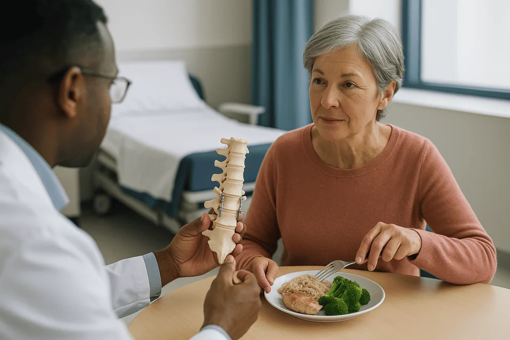
Why would anyone want less movement in their spine? Because at some levels, movement has become the disease. A vertebra may have slipped forward on the one below (spondylolisthesis). A curve may be progressing (scoliosis or kyphosis). A fracture or tumor may have weakened a segment. Or decompression may require removing so much bone that the segment would be unstable afterward. In each case, the painful or dangerous thing is motion at a damaged joint, and fusion is designed to eliminate that motion.
Fusion comes in several routes. Posterior fusion goes through the back. Interbody fusion places a spacer cage into the disc space after removing the disc, which also restores height and widens the nerve openings; the acronyms TLIF, PLIF, ALIF and LLIF describe the direction of approach. In the neck, anterior cervical discectomy and fusion (ACDF) removes a disc from the front and fuses the two vertebrae.
The trade-offs are real. A fused segment transfers its share of bending to the levels above and below, which is one reason surgeons prefer to fuse as few levels as possible. Bone may fail to knit (a non-union, sometimes called pseudarthrosis), which can mean persistent pain and occasionally revision surgery. Smoking, poor bone density and some medical conditions reduce the chance of solid fusion, and surgeons routinely address these before operating.
Fusion is often combined with decompression. Many operations billed as “fusion” include freeing the nerves first; the fusion stabilizes what the decompression loosened.
Who is usually offered which operation, and who is asked to wait?
Spine surgeons tend to sort people into a small number of patterns, and matching the pattern matters more than the label on the scan.
Leg pain from a single herniated disc, persisting after several weeks of non-surgical care, points toward discectomy. MedlinePlus and the NHS both frame surgery for a slipped disc as an option when symptoms have not settled with time, activity modification and medicines, or when weakness is worsening.
Leg heaviness, pain or numbness brought on by walking and relieved by sitting or leaning on a shopping cart is the classic story of lumbar stenosis, and it points toward decompression. Cleveland Clinic lists this posture-dependent pattern, called neurogenic claudication, as the hallmark symptom.
Back pain with a vertebra that slips or shifts on bending X-rays, a progressing curve, a fracture or a segment destabilized by prior surgery points toward fusion, often combined with decompression. Mayo Clinic lists deformity, instability and herniated disc with instability among the reasons fusion is considered.
Who is usually asked to wait? Several groups, and the waiting is clinical judgment rather than a brush-off:
- People in the first weeks of a disc herniation without weakness, because most improve without surgery.
- People whose main complaint is back ache without leg symptoms and without instability on imaging, where surgery has a weaker evidence base.
- People whose scan shows age-related changes that do not match their symptoms; many pain-free adults have bulging discs on MRI.
- People with untreated factors that raise surgical risk, such as active smoking, uncontrolled diabetes or very low bone density, who are usually asked to work on these first.
The one group never asked to wait is anyone with signs of cauda equina syndrome, covered in the final section. That is an emergency, not a scheduling question.
Laminectomy vs discectomy vs fusion: a side-by-side table
Tables flatten nuance, but a side-by-side view helps people see why their surgeon chose one door and not another. Timeframes below come from the NHS lumbar decompression guidance and Mayo Clinic procedure pages, and they are typical ranges, not promises.
| Feature | Decompression (laminectomy) | Disc surgery (discectomy) | Spinal fusion |
|---|---|---|---|
| Core problem treated | Narrowed canal or foramen (stenosis) | Herniated disc fragment on a nerve root | Instability, slippage, deformity, fracture |
| What is removed | Bone (lamina), ligament, spurs | Escaped disc material only | Often disc, then bone graft added |
| Hardware | Usually none | None | Screws, rods, often a cage |
| Motion at the level | Preserved | Preserved | Deliberately eliminated |
| Symptom most likely to improve | Leg pain, walking distance | Leg pain (sciatica) | Instability-related back pain, nerve symptoms if decompression included |
| Typical hospital stay | 1–4 days (NHS) | Often same day or overnight (Mayo Clinic) | Commonly 2–3 days (Mayo Clinic) |
| Typical return to light activity | About 4–6 weeks to expected mobility (NHS) | Desk-type work in about 2–6 weeks (Mayo Clinic) | Several months for bone to heal (Mayo Clinic) |
| Distinctive risk | Instability if much bone removed | Recurrent herniation | Non-union, adjacent segment wear |
Two lines deserve a second look. “Motion preserved” versus “motion eliminated” is the philosophical divide in spine surgery: decompression and discectomy keep the joint working, fusion retires it. And “symptom most likely to improve” explains why a surgeon may decline to operate on someone with severe back ache and a clean-looking canal. The operation has to have a target.
Remember that combinations are common. A laminectomy plus fusion is a single operation with two purposes; its recovery follows the fusion column.
Does the level matter? What L4-L5 decompression surgery involves
Search engines fill with questions about L4-L5 and L5-S1 because those two levels carry the most load and wear fastest. Lumbar vertebrae are numbered L1 to L5 from the waist down; S1 is the top of the sacrum, the triangular bone at the base of the spine. L4-L5 is the disc between the fourth and fifth lumbar vertebrae, roughly level with the top of the hip bones.
The level matters because each nerve root supplies a map of skin and muscle. Pressure at L4-L5 typically affects the L5 root, which supplies the outer calf, the top of the foot and the muscles that lift the foot and big toe. Pressure at L5-S1 usually affects the S1 root, which runs to the back of the calf, the sole and the muscles that push the foot down. Surgeons use this map to check that the scan finding matches the symptoms before they operate on it.
L4-L5 decompression surgery, whether a laminectomy for stenosis or a microdiscectomy for a herniation, follows the same steps as at any other level. The surgeon confirms the level with an X-ray during the operation, since counting vertebrae by feel is unreliable, then clears the nerve.
People ask for “the success rate of L4-L5 decompression” and deserve an honest answer: no single figure exists, because studies define success differently, mix stenosis with herniation, and follow patients for different lengths of time. The NHS states that lumbar decompression is often effective for leg symptoms such as pain and numbness, while being more variable for back pain and less predictable for weakness that has been present a long time. That qualitative picture is more reliable than any one percentage lifted from a single trial.
What shifts the odds in the individual is well established in guideline language: a clear match between symptoms and imaging, leg-dominant pain, shorter duration of nerve symptoms, and good general health. Your surgeon’s estimate for you is worth more than a population number.
Which back surgery is hardest to recover from?
Ask a ward physiotherapist and the answer is immediate: multi-level fusion, especially when it includes correction of a curve. The reason is not mysterious. Fusion involves more dissection of muscle, more bone work, hardware, and a biological healing process that takes months rather than weeks. Mayo Clinic notes that it can take several months for the vertebrae to fuse, and that activity is restricted during that period to protect the healing bone.
At the other end sits single-level microdiscectomy. The incision is small, muscles are moved rather than cut, no bone needs to heal, and Mayo Clinic describes a return to desk-type work for many people within about 2–6 weeks, with longer, around 6–8 weeks, for jobs involving heavy lifting or driving.
Decompression without fusion sits between the two. The NHS describes a hospital stay of 1–4 days and around 4–6 weeks to reach the expected level of mobility, with improvement continuing beyond that as nerves recover.
Several factors matter as much as the operation’s name:
- Number of levels: a three-level fusion is a different recovery from a one-level fusion.
- Approach: front, side and back approaches disturb different muscles and have different early discomforts.
- Age and fitness: people who were walking regularly before surgery tend to walk sooner after it.
- Duration of nerve compression: long-standing numbness and weakness improve more slowly, if at all.
- Smoking and bone density, which affect fusion specifically.
“Hardest” also has a psychological dimension. Fusion patients often describe the frustration of feeling well before the bone is actually healed and being asked to hold back anyway. Having that expectation set in advance, by the surgical team rather than by a search engine, makes the restriction easier to live with.
Microdiscectomy recovery time and the first weeks after each operation
The first morning after any of these operations looks more alike than people expect. Nurses encourage sitting up and walking within hours, because early movement reduces clot and lung risks. The NHS notes that most people are up and walking the day after lumbar decompression. Pain at the incision is normal; the leg pain that brought you to surgery is often already different, sometimes dramatically so after discectomy, though soreness and intermittent nerve twinges are common while the irritated root settles.
Discharge follows the table above: often the same or next day for microdiscectomy, 1–4 days for decompression per the NHS, commonly 2–3 days for fusion per Mayo Clinic. Everyone leaves with instructions that boil down to the same four verbs: walk, avoid bending and twisting under load, keep the wound dry and clean, and report anything that worsens.
Microdiscectomy recovery time is the most searched of the three, so here is the honest version. Mayo Clinic describes return to work in roughly 2–6 weeks depending on job demands. Walking should increase daily. Lifting limits are usually eased over the first several weeks on the surgeon’s advice. Residual numbness may outlast pain by weeks or months.
Decompression follows a similar arc on a slower clock. The NHS suggests around 4–6 weeks to regain expected mobility, with physiotherapy often starting in those weeks to rebuild trunk strength. Driving is generally discouraged until you can perform an emergency stop comfortably and are no longer taking medicines that impair alertness; the exact timing is a conversation with your team.
Fusion patients live in two timelines. Pain and walking improve over weeks; bone healing takes months, and X-rays at follow-up visits track it. Some surgeons use a brace during early healing, others do not; neither practice is universally preferred in guidelines. Restrictions on bending, lifting and twisting typically last until imaging shows solid fusion, and that date is set by the surgeon, not the calendar.
Risks and alternatives, described in plain clinical language
Every spine operation carries a shared set of risks and each has its own signature complication. Shared risks, listed by Mayo Clinic and the NHS across these procedures, include infection, bleeding, blood clots in the legs or lungs, a tear in the membrane around the nerves (dural tear) causing a spinal fluid leak, nerve injury producing new numbness or weakness, and the general risks of anesthesia. None is common; all are discussed during consent because they are serious when they occur.
The signature risks are the ones worth understanding before choosing a path. For discectomy, it is recurrence: disc material escaping again through the same tear, which Mayo Clinic lists specifically. For decompression, it is instability if enough bone and joint has to be removed that the segment becomes loose, sometimes requiring a later fusion. For fusion, it is non-union, in which the bone fails to knit and pain persists, and adjacent segment degeneration, in which the levels above and below wear faster because they absorb the fused segment’s share of movement. Hardware can also loosen or, rarely, break.
Alternatives exist at every stage, and surgeons are expected to discuss them. For herniated discs, MedlinePlus describes time, activity modification, physical therapy, anti-inflammatory medicines and steroid injections near the nerve root as the usual sequence, with most people improving within weeks. For stenosis, walking programs, flexion-based exercises that open the canal, and epidural injections may manage symptoms for long periods. For instability, bracing and targeted strengthening are sometimes tried first, though they do not fix a slipped vertebra.
Medicines in these pathways work by reducing inflammation around the nerve or dampening nerve pain signaling. They do not shrink a herniation or widen a canal, and decisions about starting, continuing or stopping any of them belong with the prescribing clinician.
One alternative deserves a caution. Non-surgical “spinal decompression therapy” using motorized traction tables is heavily marketed. The evidence that it changes disc anatomy or outperforms ordinary physical therapy is weak, and mainstream guidelines do not endorse it as a treatment for stenosis or herniation.
Spinal fusion regret rate: what the evidence actually shows
“What percentage of people regret spinal fusion?” is one of the most typed spine questions, and the honest answer is that no trustworthy single number exists. Regret is not a standard outcome measure in surgical trials; studies report pain scores, function scales and whether a person would choose the same surgery again, and they do so in populations that differ enormously in why they were fused.
What the research pattern does show is more useful than a percentage. Fusion performed for a clear structural problem, such as a slipping vertebra with matching nerve symptoms, tends to produce more consistent satisfaction than fusion performed for back pain alone with only degenerative changes on MRI. Mayo Clinic puts this plainly: fusion is most likely to help when the source of pain has been identified, and back pain without a clear cause is a weaker indication.
Several themes recur among people who report disappointment:
- Expecting fusion to remove all back pain, when its realistic goal is to eliminate pain from an unstable segment.
- Being fused for pain that was not clearly coming from the fused level.
- Non-union, which leaves pain in place and sometimes requires a second operation.
- Adjacent segment problems years later, which can feel like the operation failing even though the fused level is solid.
- Recovery that took longer than the person had mentally budgeted for.
The reverse is also true. People who went in understanding that fusion stabilizes rather than rejuvenates, who had leg symptoms that matched the operated level, and who completed rehabilitation tend to report that they would choose it again.
If a percentage is quoted to you, ask what it measured, in whom, and how long after surgery. A satisfaction figure from a trial of young adults with a single slipped vertebra tells you little about a three-level fusion for degenerative scoliosis. Your surgeon’s experience with people like you is the number that counts.
What can you never do again after spinal fusion? Separating rules from myths
This question carries a lot of quiet fear, so it deserves a careful answer. There is a short list of true permanent changes, a longer list of temporary restrictions that people mistake for permanent ones, and a great deal of individual variation that only your surgeon can speak to.
The permanent change is the obvious one: the fused segment will not move again. For a single lumbar level, most people do not notice the loss in daily life, because the hips and the other four lumbar levels absorb bending. Multi-level fusions, and fusions that include the lower lumbar levels or the sacrum, reduce flexibility more noticeably; touching your toes may become a different movement, led by the hips.
Temporary restrictions, usually lasting until imaging confirms solid bone, typically include heavy lifting, repetitive bending and twisting, high-impact sport and prolonged sitting without breaks. Mayo Clinic frames these as protecting the graft during the months it takes to fuse. They are lifted progressively, on the surgeon’s schedule, not abandoned forever.
Activities people assume are banned but which many fused patients return to, with clearance and often with modified technique, include swimming, cycling, walking long distances, golf, gardening, and most forms of work. Contact sports and activities with high fall risk are judged case by case, weighing the number of levels fused and the hardware present.
Two genuine long-term considerations are worth raising with your team. First, fused levels pass more stress to their neighbors, so lifelong attention to trunk strength, weight and posture is sensible. Second, airport scanners and MRI machines: modern spinal implants are generally compatible with MRI and do not usually trigger security alarms, but you should carry your implant information and tell any future imaging department.
The honest bottom line: fusion changes how one segment of your spine behaves. It does not, for most people, define what their life can contain.
What people often get wrong about decompression, fusion and disc surgery
Spine surgery attracts myths the way a magnet attracts filings. A few of the most persistent, corrected against mainstream evidence:
“A bad MRI means I need surgery.” It does not. Disc bulges, dehydration and mild stenosis are common in adults with no pain at all. Surgeons operate on symptoms that match imaging, not on images alone. The NHS and MedlinePlus both describe surgery for disc problems as a step taken after non-surgical care has been tried, except where weakness is progressing or there are emergency signs.
“Fusion is the strongest fix, so it is the best.” Fusion is the right tool for instability and the wrong tool for a simple herniation. More operation is not more benefit; it is more risk and longer recovery aimed at a problem you may not have.
“Disc surgery replaces the disc.” Discectomy removes a fragment and leaves most of the disc. Artificial disc replacement is a separate operation with its own narrow indications.
“Once a disc is operated on, it cannot herniate again.” It can, through the same weakened wall. Mayo Clinic lists recurrent herniation among the risks of diskectomy.
“Surgery will fix my back pain.” Decompression and discectomy target leg symptoms. Back ache from arthritic joints or muscles usually responds less, and surgeons are expected to say so beforehand.
“Traction tables decompress discs non-surgically.” Marketed devices do not reliably change disc anatomy, and mainstream guidelines do not endorse them as treatment for herniation or stenosis.
“Numbness should vanish immediately.” Pain often eases quickly; numbness and weakness follow nerve-healing time, measured in weeks to months, and may not fully resolve if compression was prolonged.
“Bed rest helps recovery.” Early walking is standard after all three operations. Prolonged rest raises clot risk and slows return to function.
Questions to ask your care team before deciding
A good consultation leaves you able to explain, in your own words, what is pressing on what and what the operation will do about it. These questions tend to get there. Bring someone to take notes; it is hard to listen and write at the same time when the subject is your own spine.
- Which exact structure is causing my symptoms, and does my scan match the pattern of my pain, numbness or weakness?
- Are you proposing decompression, disc removal, fusion, or a combination, and why that one rather than the others?
- Which of my symptoms is this operation designed to improve, and which may not change?
- What happens if I choose to wait, and are there non-surgical options I have not yet tried?
- How many levels are involved, and does that change the recovery you expect for me?
- Will I have hardware, and what are the implications for future imaging and for the levels above and below?
- What is the specific complication you worry about most in my case, and how often have you seen it?
- What would make you stop and change plan during the operation?
- How long will I be in hospital, when can I shower, drive, sit at a desk, lift my grandchild?
- What rehabilitation will I need, and who arranges it?
- Is there anything I should change beforehand, such as smoking, weight, blood sugar control or bone health, that would improve the outcome?
- Who do I call, and at what number, if something worries me after discharge?
One more, which patients rarely ask and surgeons respect: “If this were your own spine, with my scan and my symptoms, what would make you hesitate?” The answer often reveals more than any statistic.
When to call your doctor: red-flag signs before and after spine surgery
Most back and leg pain, even severe pain, is not an emergency. A small set of signs is, and knowing them is more valuable than any surgical comparison. They apply whether you are waiting for a decision, recovering from an operation, or years down the line.
Seek emergency care immediately, without waiting for a routine appointment, if you notice:
- New difficulty passing urine, loss of bladder control, or loss of bowel control.
- Numbness or tingling around the genitals, inner thighs or anus (the “saddle” area).
- Weakness spreading rapidly in one or both legs, or a foot that suddenly drags or cannot be lifted.
- Severe back pain with fever, unexplained weight loss, or a history of cancer.
- Back pain after a significant fall or accident.
The first three together form cauda equina syndrome, compression of the nerve bundle at the base of the spine. The NHS describes it as a surgical emergency in which delay risks permanent bladder, bowel and leg damage. It can arise from a large disc herniation before any surgery and, rarely, from bleeding or swelling after one.
After surgery, call your surgical team the same day if you have:
- Increasing redness, warmth, swelling or discharge from the wound, or the wound edges opening.
- Fever or chills.
- Clear fluid leaking from the incision, or a severe headache that is worse on standing, which can signal a spinal fluid leak.
- New or worsening numbness or weakness compared with the day you went home.
- Calf pain or swelling in one leg, or sudden chest pain or breathlessness, which can indicate a blood clot and need emergency assessment.
- Leg pain that returns sharply after a period of relief.
None of these lists is a diagnosis. They are reasons to pick up the phone. Your treating team knows your operation and your anatomy; they would always rather hear from you about a false alarm than about a true one a day late.
Frequently asked questions
What is the difference between laminectomy vs discectomy?
A laminectomy removes part of the bony roof of the spinal canal to relieve pressure from stenosis, while a discectomy removes a fragment of herniated disc pressing on a nerve root. Both are forms of decompression and both preserve motion at the level. Laminectomy is typically chosen for age-related canal narrowing; discectomy for a discrete disc herniation causing sciatica. Surgeons sometimes combine a small laminotomy with discectomy to reach the disc.
What is the hardest back surgery to recover from?
Multi-level spinal fusion, particularly when it corrects a curve, is generally the most demanding recovery because muscle dissection is greater, hardware is placed, and bone must heal over several months according to Mayo Clinic. Single-level microdiscectomy is usually the least demanding, with Mayo Clinic describing return to desk work in roughly 2–6 weeks. Individual factors such as age, fitness, smoking and how long nerves were compressed matter as much as the operation’s name.
What percentage of people regret spinal fusion?
There is no reliable single figure, because regret is not a standard research outcome and studies mix very different reasons for fusion. The consistent pattern is that fusion for a clear structural problem, such as a slipping vertebra with matching nerve symptoms, produces more consistent satisfaction than fusion for back pain alone with only degenerative changes on MRI. Ask your surgeon how people with your specific condition have fared.
What is the success rate of L4-L5 decompression surgery?
No single success rate applies, since studies define success differently and combine stenosis with disc herniation. The NHS describes lumbar decompression as often effective for leg pain and numbness, more variable for back pain, and less predictable for long-standing weakness. Factors that favor a good result include leg-dominant symptoms that match the imaging, shorter duration of nerve compression and good general health. Your surgeon’s estimate for your case is more meaningful than a population number.
What can you never do again after spinal fusion?
The only permanent change is that the fused segment no longer moves; for a single level most people barely notice this in daily life. Restrictions on heavy lifting, twisting and high-impact activity are usually temporary, lasting until imaging confirms solid bone, which Mayo Clinic notes can take several months. Many people return to swimming, cycling, golf, gardening and most jobs with clearance. Contact sports are judged individually by the surgical team.
How long is microdiscectomy recovery time?
Mayo Clinic describes return to work in about 2–6 weeks after diskectomy, with longer, around 6–8 weeks, for jobs involving heavy lifting or long driving. Walking is encouraged from the first day, and leg pain often eases quickly, though numbness can take weeks or months to settle. Lifting and bending limits are relaxed progressively on the surgeon’s advice. These are typical ranges, not guarantees, and vary with age, fitness and how long the nerve was compressed.
Is spinal fusion better than decompression?
Neither is better; they treat different problems. Decompression relieves nerve pressure while keeping the segment mobile and is the standard operation for stenosis without instability. Fusion eliminates motion and is reserved for instability, slippage, deformity or segments left unstable by extensive decompression. Adding fusion where no instability exists increases risk and recovery time without a clear target. The imaging findings and symptom pattern, reviewed by the treating team, determine which fits.
Does non-surgical spinal decompression therapy work for L4-L5 discs?
The evidence is weak. Motorized traction tables marketed as spinal decompression therapy have not been shown to change disc anatomy or to outperform standard physical therapy in good-quality trials, and mainstream guidelines do not endorse them for herniation or stenosis. Most herniated discs improve over weeks with time, activity modification, physiotherapy and medicines, as MedlinePlus describes. Discuss any proposed therapy with your treating clinician before committing to a course.
Can a disc herniate again after discectomy?
Yes. Discectomy removes the escaped fragment but leaves most of the disc, and the torn outer wall is not durably repaired, so material can herniate again through the same weak spot. Mayo Clinic lists recurrent herniation among the recognized risks of diskectomy. Recurrence does not always need another operation; many are managed non-surgically. Maintaining trunk strength, avoiding prolonged heavy lifting early on, and following your surgeon’s activity plan are the usual advice.
When is spine surgery an emergency?
Immediately, when there are signs of cauda equina syndrome: new bladder or bowel dysfunction, numbness around the genitals or inner thighs, or rapidly spreading leg weakness. The NHS describes this as a surgical emergency where delay risks permanent damage. Severe back pain with fever, unexplained weight loss, a cancer history or a significant injury also needs urgent assessment. Everything else, including severe but stable sciatica, is usually a planned decision made with the treating team.
References
This article is for general information only and is not a substitute for professional medical advice. Please consult a qualified doctor about your individual situation.

More from the Blog

Which Symptoms After Spine Surgery Need a Same-Day Call? Wound, Bladder and Leg Changes
Call your surgical team the same day if you notice new or worsening leg weakness, numbness spreading between the legs or around the genitals,…

Who Benefits From the Schroth Method? Curve Size, Age and Growth Stage Considerations
The Schroth method is usually considered for people with idiopathic scoliosis and mild to moderate curves, most often growing adolescents with Cobb angles roughly…

Who Is Suitable for Endoscopic Disc Surgery? Herniation Type, Location and Prior Surgery
An endoscopic disc surgery candidate is typically an adult with a single, imaging-confirmed lumbar disc herniation that presses on one nerve root, causing leg…
How Long Does an Artificial Disc Last? Wear, Motion Over Time and Realistic Expectations
Nobody can promise a fixed number of years for an artificial disc. Published follow-up now stretches to roughly seven to ten years for several…

Laminectomy Recovery: Healing After Spinal Decompression
Most people recovering from a laminectomy walk within a day of surgery, return to light activity in a few weeks, and resume most normal…

When Is Spinal Fusion Recommended? Instability, Deformity and Failed Non-Surgical Care
Spinal fusion is usually recommended when a segment of the spine is unstable or misaligned, when a deformity such as severe scoliosis or a…