

Which Tests Confirm Emphysema? Spirometry, Chest CT and Oxygen Checks Explained

Key Takeaways
- Spirometry confirms airflow obstruction, but only the gas transfer test and chest CT directly show the alveolar damage that defines emphysema.
- A post-bronchodilator FEV1/FVC ratio below 0.70 is the usual threshold for COPD in guidance summarised by the NHS and NHLBI, though age-adjusted limits are increasingly used.
- A normal chest X-ray does not exclude emphysema; plain films are insensitive and serve mainly to rule out other causes.
- Resting pulse oximetry can stay normal for years, so oxygen drops are often detected only during a six-minute walk test or overnight monitoring.
- Guidelines encourage one-time alpha-1 antitrypsin testing for everyone diagnosed with COPD, because the inherited deficiency can cause emphysema in never-smokers and affects relatives.
- Emphysema damage does not regrow, but the Mayo Clinic notes that stopping smoking slows the rate of lung function decline toward that of a non-smoker.
Emphysema is usually confirmed with a combination of tests rather than one. Spirometry, a breathing test, shows whether airflow is obstructed; a gas transfer test and chest CT scan show whether the air sacs themselves are damaged; and oxygen checks such as pulse oximetry or arterial blood gas measure how well the lungs are moving oxygen into the blood. Doctors combine these results with history and examination before naming the condition.
The breathing booth is smaller than people expect. You sit on a stool, a soft clip pinches your nose shut, and a technician asks you to blow into a tube as if you were trying to put out every candle on a cake from across the room. Then she asks you to do it again. And again. By the third attempt your cheeks ache and you wonder how a plastic tube could possibly say anything meaningful about the inside of your chest.
Yet that tube, plus a scan and a small finger probe, is how most people learn whether the breathlessness that crept up on the stairs has a name. Emphysema diagnosis tests are not dramatic. They are quiet, repeatable measurements, and that is exactly their strength.
This explainer walks through what each test does, what it cannot do, how the results fit together, and which questions are worth bringing to the appointment where the pieces are put in place.
How emphysema diagnosis tests fit together
Emphysema is a form of chronic obstructive pulmonary disease, or COPD, in which the tiny air sacs at the ends of the airways, the alveoli, are damaged and merge into larger, floppier spaces. Fewer, bigger sacs means less surface area for oxygen to cross into the blood, and airways that collapse more easily when you breathe out. The lungs trap air. Breathing out gets slow, and breathing in feels crowded.
No single measurement captures all of that. So clinicians build the diagnosis in layers. A detailed history comes first: how long breathlessness has been present, what brings it on, whether there is a long-term cough, and crucially, exposure to tobacco smoke, workplace dust, fumes, or indoor smoke from solid fuels, which the Mayo Clinic lists as the main causes.
Spirometry comes next, because it answers the single most important question: is airflow out of the lungs obstructed, and does that obstruction persist after an inhaled bronchodilator? Obstruction that does not fully reverse is the defining feature of COPD, and emphysema is one of the patterns within it.
From there, the picture is refined. A gas transfer test asks whether the alveolar wall is still doing its job. A chest CT scan, a cross-sectional X-ray that builds detailed slices of the chest, shows the actual holes and thinning that spirometry can only infer. Oxygen checks measure the downstream consequence: whether blood leaving the lungs is carrying enough oxygen at rest, on exertion, or overnight.
Think of it as three questions in sequence. Is air flowing out normally? Is the lung tissue structurally intact? Is the body getting the oxygen it needs? Emphysema diagnosis tests are simply the tools that answer each one, and the diagnosis sits in the overlap of the answers rather than in any single number.
Who is usually tested, and who is usually asked to wait
Guidelines are fairly consistent about who should have spirometry. The NHS advises assessment for people aged over 35 who have a relevant risk factor, most often smoking, together with symptoms such as breathlessness on exertion, a persistent cough, frequent winter chest infections, or wheeze. The test is also offered to people with a family history of early lung disease or a known alpha-1 antitrypsin deficiency, a genetic condition described below.
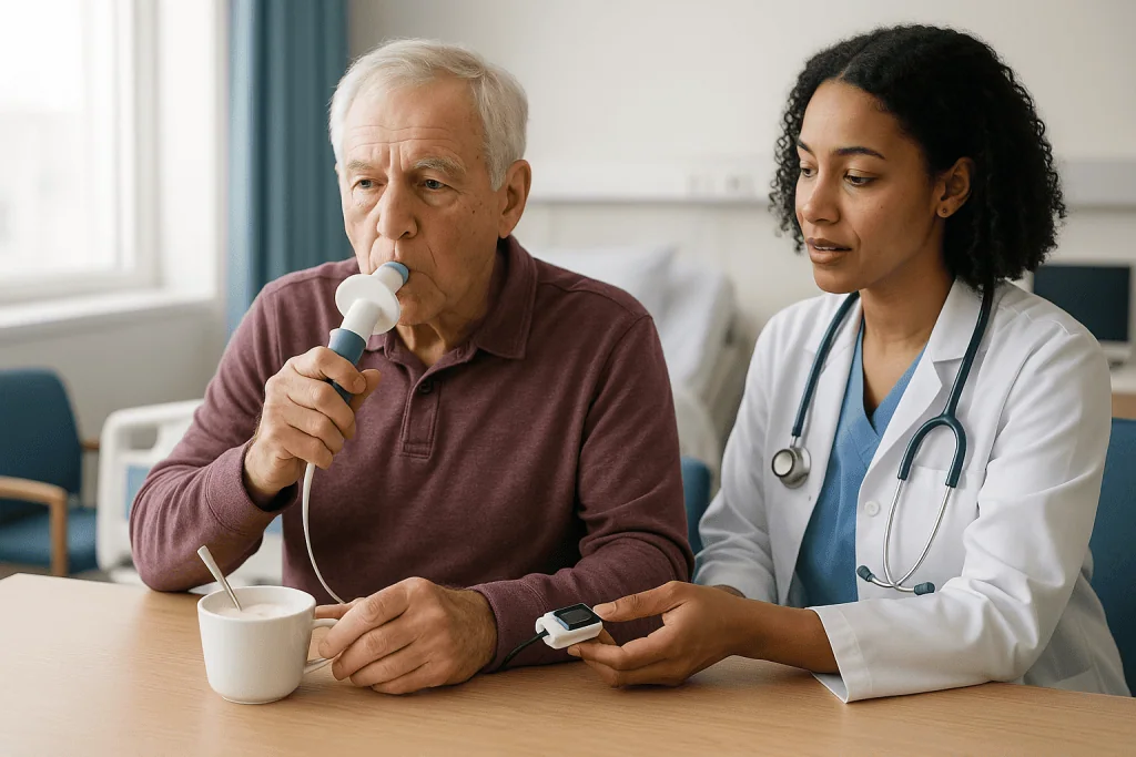
Who waits? Someone in the middle of an acute chest infection or a flare of wheeze is usually asked to come back once the episode has settled, because inflamed airways can exaggerate obstruction and make a one-off result unreliable. People who have recently had chest or abdominal surgery, a heart attack, an unstable aneurysm, or a collapsed lung are typically deferred as well, since the forced blowing of spirometry briefly raises pressure inside the chest. Johns Hopkins Medicine lists these among the reasons a pulmonary function test may be postponed.
Screening people who have no symptoms is a different matter. Major guidelines, including those summarised by the NHLBI, do not recommend spirometry for symptom-free adults, even smokers, because finding mild obstruction in someone who feels well has not been shown to change outcomes on its own. The test is for people with a reason to be tested.
Chest CT is used more selectively. It is not a routine first step for everyone with suspected COPD. It tends to be requested when spirometry and symptoms do not match, when another diagnosis such as lung fibrosis or a tumour needs excluding, when a procedure such as lung volume reduction is being considered, or when a person qualifies for lung cancer screening for other reasons.
The treating team decides the sequence. If you are asked to wait, it is usually so that the result you eventually get is one that can be trusted.
Spirometry for emphysema: what actually happens in the test
Spirometry measures how much air you can blow out and how fast. A spirometer is the device that records it, either a desktop unit with a mouthpiece or a small handheld sensor connected to a computer.
The routine is simple but demanding. You take the deepest breath you can, seal your lips around the mouthpiece, then blast the air out as hard and as long as possible, often for six seconds or more, until the technician tells you to stop. Most people need at least three good attempts so that the best two agree closely; the machine flags blows that start late, end early, or include a cough. The whole session, including rest between efforts, usually takes well under an hour according to Johns Hopkins Medicine.
Then comes the part specific to COPD assessment. You inhale a short-acting bronchodilator, a medicine that relaxes the muscle around the airways, wait roughly a quarter of an hour, and repeat the blows. This is not treatment. It is a diagnostic step. If the obstruction largely disappears, the pattern points more toward asthma. If it persists, that persistence is the hallmark of COPD, of which emphysema is one form.
Preparation matters more than people realise. You may be asked to avoid your usual inhaler for a set number of hours beforehand, to skip a heavy meal, to wear loose clothing, and not to smoke or exercise hard immediately before. Follow those instructions exactly as given by the testing centre, because a result distorted by a recent inhaler puff may have to be repeated.
Spirometry is safe for most people. Light-headedness, a brief cough, or tiredness are the usual effects. The nose clip and the forceful blowing feel odd, not painful. The technician’s job is partly coaching; a good one will cheer you through the last seconds of each exhale.
Reading the numbers: FEV1, FVC and the 0.70 ratio
Two figures carry most of the weight. FVC, forced vital capacity, is the total volume of air you can push out after a full breath in. FEV1, forced expiratory volume in one second, is how much of that air comes out in the first second. Healthy lungs empty quickly, so FEV1 is normally a large share of FVC.

In emphysema the airways narrow and collapse as you exhale, so air leaks out slowly. FEV1 falls much more than FVC, and the ratio between them drops. The NHLBI and NHS summaries of international guidance describe a post-bronchodilator FEV1/FVC ratio below 0.70 as the usual threshold for airflow obstruction. Some laboratories use an age-adjusted lower limit of normal instead, which matters at the extremes of age: a fixed 0.70 can over-label healthy older adults and miss younger people with early disease.
Severity is then graded by FEV1 as a percentage of what is predicted for someone of your age, sex, height and ancestry. The NHLBI describes four stages from mild to very severe based on that percentage. Two people with the same stage can feel very different, which is why symptom scores and flare history sit alongside the numbers.
What spirometry cannot say is why the airflow is obstructed. Chronic bronchitis, long-standing asthma that has become fixed, bronchiectasis and emphysema can all produce an obstructive pattern. The ratio tells you the door is sticking; it does not tell you whether the hinge or the frame is at fault.
Patients often ask for their numbers, and that is reasonable. Ask what your FEV1 is as a percentage of predicted, what the ratio was before and after the bronchodilator, and whether the technician rated the test quality as good. Those three answers are the foundation that everything else is built on.
Gas transfer and lung volumes: what spirometry cannot see
Here is where emphysema separates itself from its COPD cousins. The damage is in the alveolar walls, where oxygen crosses into capillaries. Spirometry never measures that surface. A gas transfer test does.
The test goes by several names: DLCO, TLCO, or diffusing capacity. You breathe in a single breath of air containing a tiny, harmless trace of carbon monoxide plus a tracer gas, hold it for about ten seconds, and breathe out. The analyser measures how much carbon monoxide was absorbed across the lung surface during the hold. Less absorbed means less working alveolar membrane. In emphysema the gas transfer value is typically reduced, often out of proportion to the spirometry result, whereas in asthma or pure chronic bronchitis it is usually preserved. Johns Hopkins Medicine describes diffusing capacity as a standard part of a full pulmonary function test battery.
Lung volume measurement completes the set. You sit inside a clear booth called a body plethysmograph, sometimes nicknamed the phone box, and breathe against a shutter that briefly closes. Pressure changes inside the sealed booth let the computer calculate how much air stays in your lungs even after you have exhaled fully. Emphysema traps air, so residual volume and total lung capacity are frequently enlarged. The lungs are big but not useful, like an overinflated balloon that has lost its spring.
These two tests explain a common puzzle: people whose spirometry looks only mildly abnormal yet who are strikingly breathless. Their gas exchange surface may be far more damaged than their airflow suggests.
Not every clinic has a full laboratory, so you may be referred to a hospital-based department for these measurements. They are painless, take longer than spirometry, and give the diagnosis a physiological backbone that a chest scan alone cannot.
Chest CT for emphysema: what the scan shows and when it is ordered
A plain chest X-ray is often done early, mostly to rule out other causes such as pneumonia, heart enlargement, or a mass. It can hint at emphysema through flattened diaphragms and dark, overexpanded lungs, but the Mayo Clinic notes it is not sensitive enough to confirm or exclude the condition.
Computed tomography is different. A CT scanner rotates an X-ray beam around the chest and reconstructs it into thin slices, so radiologists can see individual lung regions. Emphysema appears as areas of abnormally low density: dark pockets where tissue has been destroyed. The pattern matters. Centrilobular emphysema, clustered around the small airways in the upper lungs, is classically linked with smoking. Panlobular emphysema, spread evenly through whole lobules and often worse at the lung bases, is the pattern associated with alpha-1 antitrypsin deficiency. Paraseptal emphysema sits just beneath the lung lining and is sometimes found incidentally.
Modern software can quantify the proportion of lung below a density threshold, giving an emphysema score, and can map which lobes are most affected. That mapping becomes important if a procedure to deflate the most damaged regions is ever considered.
The scan itself is quick. You lie on a table that slides through a ring, hold your breath for a few seconds, and it is done. Contrast dye is usually not needed for emphysema assessment. Radiation exposure is higher than a plain X-ray, which is one reason CT is used thoughtfully rather than routinely.
A word of balance: CT can show emphysema in people whose spirometry is still normal, and it can show modest emphysema in people who have no symptoms. A scan finding is not by itself a clinical diagnosis of COPD. The treating team weighs what the image shows against how your lungs actually function and how you actually feel.
Oxygen checks: pulse oximetry, arterial blood gas and the walk test
Damaged alveoli eventually struggle to load enough oxygen into the blood. Oxygen checks measure that consequence, and they also guide decisions about supplemental oxygen later on.
Pulse oximetry is the familiar finger clip. It shines two wavelengths of light through the fingertip and estimates the percentage of haemoglobin carrying oxygen, the saturation. It is painless and instant, and it is often checked at rest, during exertion, and sometimes overnight. Readings can be thrown off by cold hands, nail polish, poor circulation, or darker skin pigmentation, which several studies have shown can lead oximeters to overestimate saturation. A single low or high number is a prompt for a better measurement, not a verdict.
That better measurement is the arterial blood gas, or ABG. A small sample is drawn from an artery, usually at the wrist, and analysed for oxygen, carbon dioxide and acidity. It is more uncomfortable than a vein draw, often described as a sharp pinch, and the puncture site may ache briefly. The ABG shows not only whether oxygen is low but whether carbon dioxide is building up, which happens when the lungs can no longer blow it off efficiently. The Mayo Clinic lists it among the tests used to assess how well the lungs are exchanging gases.
The six-minute walk test adds a real-world dimension. You walk back and forth along a measured corridor at your own pace for six minutes while saturation, pulse and breathlessness are recorded. The distance covered and whether saturation drops during exertion say a great deal about how emphysema affects daily life. It is a test some people find reassuring and others find revealing, because the drop in oxygen on walking can be invisible at rest.
None of these checks confirms emphysema alone. They describe how hard the condition is biting.
Blood tests, including alpha-1 antitrypsin
Blood tests do not diagnose emphysema, but one of them can explain it. Alpha-1 antitrypsin is a protein made in the liver that protects lung tissue from an enzyme released by white cells during inflammation. In alpha-1 antitrypsin deficiency, an inherited condition, low levels of the protein leave the alveoli unprotected, and emphysema can develop early, sometimes in people who have never smoked.
The test is a simple blood sample measuring the protein level, often followed by genetic typing if the level is low. The Mayo Clinic and NHLBI both describe this test as part of the diagnostic work-up, and international guidance has long encouraged testing anyone diagnosed with COPD at least once, because the result changes the advice given to relatives and can alter long-term management options. The deficiency is under-recognised; many people live for years with a COPD label before anyone checks.
Other blood tests serve supporting roles. A full blood count can reveal polycythaemia, an excess of red cells that the body produces in response to chronically low oxygen, and it can pick up anaemia, which worsens breathlessness for a completely different reason. A blood eosinophil count, measuring a type of white cell linked to allergic inflammation, is sometimes used by specialists when weighing which classes of inhaled medicine are most likely to help; that decision rests with the prescribing clinician. Blood tests may also check heart strain markers if the team suspects the heart is contributing to the breathlessness.
A sputum sample is occasionally requested during a flare to identify infection, and tests for tuberculosis may be considered in people with relevant exposure. These are context-dependent, not standard for everyone.
If no one has mentioned alpha-1 testing and you have been diagnosed with emphysema, it is a fair question to raise.
Emphysema diagnosis tests compared: what each one answers
Laying the tests side by side makes the logic visible. Each answers a different question, and a thorough work-up uses several.
| Test | What it measures | What it can confirm | Main limitation |
|---|---|---|---|
| Spirometry | Volume and speed of exhaled air, before and after bronchodilator | Persistent airflow obstruction, the defining feature of COPD | Cannot show whether air sacs are damaged or why airflow is obstructed |
| Gas transfer (DLCO) | How well gas crosses the alveolar wall | Loss of working lung surface, typical of emphysema | Also reduced in anaemia and some other lung diseases |
| Lung volumes | Total capacity and trapped air after full exhale | Hyperinflation and air trapping | Requires specialist equipment |
| Chest X-ray | Two-dimensional image of chest | Mainly excludes other causes | Insensitive for early or moderate emphysema |
| Chest CT | Detailed cross-sectional images | Visible tissue destruction, pattern and distribution | Radiation exposure; findings may not match symptoms |
| Pulse oximetry | Estimated blood oxygen saturation | Low oxygen at rest, on exertion or overnight | Affected by circulation, skin pigmentation, movement |
| Arterial blood gas | Oxygen, carbon dioxide and acidity in arterial blood | Degree of gas exchange failure | Uncomfortable; snapshot only |
| Alpha-1 antitrypsin | Protective protein level and genotype | Inherited cause of emphysema | Does not measure lung damage itself |
Notice that only the gas transfer test and CT scan speak directly to emphysema rather than COPD in general. Spirometry establishes that obstruction exists; the physiology and imaging tests say what kind. Oxygen checks measure impact. The Cleveland Clinic and Mayo Clinic both describe this layered approach in their patient guidance.
The order is not fixed. A person who arrives with a CT done for another reason may work backwards to spirometry; someone with a strong family history may have alpha-1 testing early. The sequence bends to the individual, which is one more reason the final interpretation belongs with a clinician who has all the pieces in front of them.
What the following days and weeks usually look like
Diagnosis rarely happens in one visit. A typical path begins in primary care with a history, an examination with a stethoscope, perhaps a chest X-ray, and either spirometry on site or a referral to a testing service. Basic spirometry results are often available the same day because the machine calculates them as you blow; the technician or clinician then interprets quality and compares against predicted values.
If the result shows obstruction, a referral to a respiratory clinic or lung function laboratory for gas transfer and lung volumes may follow. Waiting times vary by health system and are not something any article can promise. The tests themselves are a single appointment, typically lasting around an hour to ninety minutes for a full battery according to Johns Hopkins Medicine. Results usually go to the referring clinician within a few days to a couple of weeks for formal reporting.
A CT scan, when needed, is another separate appointment, usually brief. Radiology reports take days rather than hours in most settings. Blood tests for alpha-1 antitrypsin may take longer if genetic typing is added.
Then comes the appointment where the pieces are assembled. Expect a conversation rather than a pronouncement. The clinician will explain whether the pattern fits COPD, whether emphysema is the dominant feature, how severe the numbers look, and what the oxygen readings mean. Many teams also assess symptom burden with a short questionnaire and ask about flare-ups in the past year, because those shape management as much as the FEV1 does.
Practical points for those weeks: keep a note of your usual inhaler timing so you can follow pre-test instructions accurately, bring a list of current medicines, and write down questions as they occur to you. Repeat spirometry may be arranged later to confirm the finding or to see how the numbers move over time; a single result is a photograph, and the team usually wants a short film.
Can emphysema be misdiagnosed? Asthma, heart failure and the look-alikes
Yes, in both directions, and understanding how is the best protection against it.
Overdiagnosis most often happens when spirometry is skipped. Breathlessness in a lifelong smoker is assumed to be COPD, inhalers are started, and no one ever checks the airflow. The NHLBI and NHS both stress that COPD should not be diagnosed without spirometry, precisely because the symptom picture overlaps with several other conditions. Heart failure causes exertional breathlessness and sometimes wheeze. Obesity and deconditioning do the same. Anaemia does too.
Underdiagnosis is just as common. People attribute their slower pace to age, give up the hill walk, and never mention it. Spirometry is normal in early emphysema because the damage has not yet obstructed airflow enough to move the ratio, so a person can be told their lungs are fine while a CT would show otherwise. Women and never-smokers are at particular risk of being overlooked because the classic profile does not fit.
Asthma is the great mimic. Both produce obstruction. The clues that favour asthma are onset in childhood, symptoms that come and go, strong allergic history, and airflow that improves substantially after a bronchodilator. Some people genuinely have features of both. Gas transfer helps here: it is usually preserved in asthma and reduced in emphysema.
Bronchiectasis, in which airways are permanently widened and prone to infection, can look like COPD on spirometry but is distinguished on CT. Interstitial lung disease, where the lung tissue scars, may coexist with emphysema in smokers and confuse the volumes, since one shrinks the lungs while the other inflates them.
Technical error also plays a part. A poorly coached blow, a cough mid-exhale, or a recent inhaler can all shift results. If a diagnosis does not fit how you feel, asking for the test to be repeated at a specialist laboratory is reasonable, and the treating team can judge whether that is warranted.
How progressive is emphysema, and what do the tests say about treatment questions?
Emphysema involves destroyed alveolar walls, and the Cleveland Clinic is direct that this tissue damage does not grow back. That sentence worries people, so it needs context. The pace of decline varies enormously, and the single biggest factor the evidence identifies is continued exposure to the cause. Lung function naturally falls a little each year in everyone from early adulthood. In people who keep smoking, FEV1 falls faster; in those who stop, the Mayo Clinic notes the rate of decline slows toward that of a non-smoker, even though lost function is not recovered.
Tests are how progression is tracked. Repeat spirometry every year or so shows the trend in FEV1. Rising residual volume on lung volume testing signals increasing air trapping. A falling walk distance or new oxygen desaturation on exertion indicates that gas exchange is becoming the limiting factor. Flare-ups, called exacerbations, matter too; each severe one is associated with a step down in function, which is why preventing them is a core goal.
People often ask what the best medicine for emphysema is. There is no single answer, and anyone offering one is simplifying. Inhaled bronchodilators relax airway muscle and reduce air trapping, which eases breathlessness; inhaled anti-inflammatory medicines are added in certain patterns, often guided by flare history and blood eosinophil counts; pulmonary rehabilitation, a supervised exercise and education programme, has strong evidence for improving breathlessness and quality of life; vaccinations reduce infection-triggered flares; and supplemental oxygen is prescribed when blood oxygen is persistently low. Which of these apply, and in what combination, is a decision for the prescribing clinician using your test results, and it will change over time.
Progression, in other words, is not a fixed slope. It is a trajectory that testing makes visible and that several interventions can bend.
What people often get wrong about emphysema tests
“A normal chest X-ray means my lungs are fine.” It does not. Plain films miss a great deal of emphysema and are mainly useful for excluding other problems. Normal X-ray plus persistent breathlessness is a reason for spirometry, not reassurance.
“The scan found emphysema, so I have COPD.” Not necessarily. CT can show mild emphysema in people whose airflow and symptoms are entirely normal. A radiology finding becomes a clinical diagnosis only when function and symptoms line up with it. The reverse is also true: a person can have significant COPD with chronic bronchitis as the main feature and relatively little visible emphysema.
“My oxygen reading was 96, so I can’t have lung disease.” Resting saturation can be normal for years in emphysema. Oxygen often drops only on exertion or during sleep, which is why walk tests and overnight oximetry exist.
“Spirometry is just blowing into a tube; anyone can do it.” The blow is simple. Doing it to the standard needed for a reliable result is not. Coaching, repeat efforts, correct bronchodilator timing and quality grading all change the answer. A hurried test in a noisy room is a common source of both false positives and false negatives.
“I never smoked, so it can’t be emphysema.” Most cases are smoking-related, but alpha-1 antitrypsin deficiency, long-term exposure to indoor smoke or occupational dust, and secondhand smoke all cause it. The NHLBI lists these among recognised risk factors.
“Once the tests are done, I never need them again.” Repeat testing is how progression and treatment response are judged. The first set is a baseline, not a conclusion.
“A bad result means nothing can be done.” The damaged tissue does not regrow, but breathlessness, flare frequency and daily function respond to a range of measures. The numbers describe where you are; they do not fix where you are going.
Questions to ask your care team
A diagnosis conversation moves quickly, and it is easy to leave with a label but without understanding. These questions tend to produce the most useful answers.
- What were my FEV1 and FEV1/FVC ratio after the bronchodilator, and was the test rated as good quality?
- Did my airflow improve meaningfully after the bronchodilator, and what does that suggest about asthma overlap?
- Have I had a gas transfer test and lung volumes? If not, would they add anything to the picture?
- Do I need a CT scan, and if one was done, which pattern and distribution of emphysema did it show?
- What were my oxygen readings at rest and on walking? Should overnight oximetry or an arterial blood gas be considered?
- Have I been tested for alpha-1 antitrypsin deficiency? Should my relatives be?
- Could anything else be contributing to my breathlessness, such as my heart, anaemia or fitness level?
- How severe is my condition on the staging your team uses, and how does that compare with how I actually feel?
- Which of my results will be repeated, and how often?
- Am I a candidate for pulmonary rehabilitation, and how do I access it?
- Which vaccinations do you recommend given this diagnosis?
- If I stop smoking now, what does the evidence say about my rate of decline?
- What symptoms should prompt me to seek urgent help, and whom do I contact?
Write the answers down or ask whether you can record the conversation. Many clinics will print your spirometry report and CT summary if you ask; having them in hand makes future appointments more productive and lets a second clinician see exactly what the first one saw.
One more worth asking, even if it feels awkward: what are you least certain about in my case? Honest clinicians will tell you where the evidence is thin, and that answer often matters more than the confident ones.
When to call your doctor
Diagnostic testing is unhurried by design, but emphysema can produce situations that are not. The signs below warrant prompt contact with your clinician or, where marked, emergency care.
Seek emergency help without delay if you have severe breathlessness that makes it hard to speak in full sentences, lips or fingertips turning blue or grey, chest pain that is new or crushing, sudden sharp chest pain with breathlessness on one side (which can indicate a collapsed lung, a recognised complication of emphysema noted by the Mayo Clinic), confusion or unusual drowsiness, or a pulse oximeter reading far below your usual level if you have one at home.
Contact your doctor the same day if breathlessness has worsened noticeably over a day or two, if sputum has increased or changed colour, if you have a fever with chest symptoms, if you are needing a reliever inhaler far more often than usual, or if new ankle swelling appears, which can signal strain on the right side of the heart.
Arrange a routine appointment if you notice gradual decline in how far you can walk, if sleep is disturbed by breathlessness, if you are coughing up blood even in small amounts, or if unintended weight loss accompanies your symptoms.
During the testing phase specifically, let the team know if you are unwell on the day of spirometry so the test can be rescheduled, if you felt faint or had chest discomfort during the blows, or if the puncture site from an arterial blood gas becomes swollen, numb or persistently painful.
Every one of these decisions, from whether a test is repeated to whether treatment is started or changed, rests with your treating team. The purpose of knowing the red flags is not to manage emphysema yourself but to make sure the people who can manage it hear from you in time.
Frequently asked questions
Which test is most important for confirming emphysema?
No single test confirms it; spirometry is the essential first step because it establishes whether airflow is persistently obstructed, which defines COPD. To show emphysema specifically rather than another form of COPD, clinicians rely on a gas transfer test, which measures how well gas crosses the alveolar wall, and often a chest CT scan that shows the destroyed tissue directly. The diagnosis sits where these results and your symptoms overlap.
Can emphysema be misdiagnosed?
Yes, in both directions. It is over-diagnosed when breathlessness in a smoker is labelled COPD without spirometry, missing heart failure, anaemia or deconditioning. It is under-diagnosed when early disease has not yet lowered the spirometry ratio, or when the person is a woman or a never-smoker who does not fit the classic profile. Asthma, bronchiectasis and scarring lung diseases can all mimic it, which is why the full test battery matters.
How is COPD diagnosed differently from emphysema?
COPD is the umbrella diagnosis, made when spirometry shows airflow obstruction that persists after a bronchodilator. Emphysema is one pattern within COPD, defined by destruction of the air sacs. Confirming that pattern requires additional evidence: a reduced gas transfer value, enlarged lung volumes from air trapping, or visible low-density areas on a chest CT. Chronic bronchitis, the other major COPD pattern, is defined by long-standing cough and mucus instead.
What does spirometry for emphysema actually involve?
You take the deepest breath possible and blow out as hard and long as you can into a mouthpiece, usually at least three times, with a nose clip on. You then inhale a short-acting bronchodilator, wait around fifteen minutes, and repeat. The device calculates how much air you exhaled and how fast. The session typically lasts well under an hour, and the main side effects are brief light-headedness or coughing.
What does a chest CT for emphysema show?
It shows areas of abnormally low density where lung tissue has been destroyed, along with their pattern and location. Centrilobular emphysema in the upper lungs is typical of smoking, while panlobular disease at the lung bases suggests alpha-1 antitrypsin deficiency. Software can estimate what proportion of the lung is affected. CT is not routine for everyone with suspected COPD and is ordered when it will change the diagnosis or the plan.
What are the signs and symptoms that lead doctors to test for emphysema?
The most common prompt is breathlessness on exertion that has crept up gradually, often alongside a long-term cough, frequent chest infections, or wheeze, in someone with a relevant exposure such as smoking, occupational dust or indoor smoke. The NHS advises assessment for people over 35 with symptoms plus a risk factor. Symptoms alone cannot diagnose emphysema; they are the reason testing is arranged.
How progressive is emphysema?
The destroyed air sac tissue does not regrow, but the rate at which lung function declines varies widely between people. The Mayo Clinic notes that continued smoking speeds decline while stopping slows it toward the normal age-related rate. Severe flare-ups are also associated with step-downs in function. Repeat spirometry, walk tests and oxygen checks are how the trajectory is tracked, and several interventions can influence it.
What is the best medicine for emphysema?
There is no single best medicine, because treatment is matched to each person’s test results, symptom burden and flare history. Inhaled bronchodilators relax airway muscle and reduce air trapping; inhaled anti-inflammatory medicines are added in certain patterns; pulmonary rehabilitation has strong evidence for breathlessness and quality of life; and oxygen is prescribed when blood levels are persistently low. Which apply to you is a decision for your prescribing clinician.
Why do I need an arterial blood gas if I already had a finger oxygen reading?
Pulse oximetry estimates oxygen saturation but can be affected by circulation, movement, nail polish and skin pigmentation, and it says nothing about carbon dioxide. An arterial blood gas measures oxygen, carbon dioxide and acidity directly from arterial blood. It is requested when the oximeter reading is low or unreliable, when carbon dioxide retention is suspected, or when decisions about supplemental oxygen are being made.
Should I be tested for alpha-1 antitrypsin deficiency?
International guidance encourages everyone diagnosed with COPD to be tested at least once, and the Mayo Clinic and NHLBI both list it as part of the work-up. The deficiency is inherited, can cause emphysema in people who never smoked, and often develops at a younger age. A positive result changes advice for relatives and may open additional management options. If no one has mentioned it, ask your care team.
References
- NHS: Chronic obstructive pulmonary disease (COPD), Diagnosis
- NIH National Heart, Lung, and Blood Institute: COPD Diagnosis
- Cleveland Clinic: Emphysema
- MedlinePlus: Lung Function Tests
This article is for general information only and is not a substitute for professional medical advice. Please consult a qualified doctor about your individual situation.

More from the Blog

Who Is a Candidate for Single-Port VATS? Lung Biopsies, Collapsed Lung and Small Tumors
Single-port VATS candidates are usually people who need a lung biopsy that a needle cannot safely provide, a repair for a recurrent or persistent…
Which Tests Explain a Pleural Effusion? Chest Ultrasound, CT and What the Fluid Sample Reveals
A pleural effusion is usually confirmed with a chest X-ray or chest ultrasound, and ultrasound is now the preferred first test because it detects…

Do Antibiotics Help Acute Bronchitis? Why Most Cases Are Viral and What Treatment Looks Like
Antibiotics usually do not help acute bronchitis, because the large majority of cases are caused by viruses that antibiotics cannot touch. Pooled trials suggest…

Which Chest Conditions Suit Robotic Thoracic Surgery? Lung Nodules, Mediastinal Tumors and More
Robotic thoracic surgery is most often considered for small lung nodules and early-stage lung cancer needing a wedge resection, segmentectomy or lobectomy, for tumors…
Does Acute Bronchitis Need a Chest X-Ray? How Doctors Rule Out Pneumonia
Most people with acute bronchitis do not need a chest X-ray. Doctors usually diagnose it from the story of the illness and a physical…

Questions to Ask Your Cystic Fibrosis Team: Lung Function, Infections and Nutrition Goals
Useful questions to ask a cystic fibrosis team fall into three groups: lung function (what my FEV1 is, how it has changed, what a…