

Which Tests Explain a Pleural Effusion? Chest Ultrasound, CT and What the Fluid Sample Reveals
Key Takeaways
- Chest ultrasound detects smaller effusions than a standing X-ray, shows whether fluid is free or trapped in pockets, and is now expected guidance for any needle procedure in the pleural space.
- Light's criteria classify fluid as an exudate if the fluid-to-blood protein ratio exceeds 0.5, the LDH ratio exceeds 0.6, or fluid LDH exceeds two-thirds of the laboratory's upper normal limit.
- A transudate points away from the pleura toward the heart, kidneys or liver, while an exudate triggers a fuller panel of cell counts, glucose, pH, cultures and cytology.
- CT with contrast is most useful before complete drainage, when the stretched pleural lining can reveal nodules, thickening, lymph nodes or a hidden lung mass that ultrasound cannot see.
- A single negative cytology does not exclude cancer in the pleura; a second sample improves the yield modestly, and after that an image-guided or thoroscopic biopsy becomes the better route.
- A raised diaphragm, old pleural thickening, low-lying pneumonia and a collapsed lower lobe are the commonest X-ray findings mistaken for fluid, and ultrasound usually settles the question in seconds.
A pleural effusion is usually confirmed with a chest X-ray or chest ultrasound, and ultrasound is now the preferred first test because it detects small collections and guides safe needle placement. CT scanning adds detail about the lung and pleura when the cause is unclear. The explanation most often comes from pleural fluid analysis after thoracentesis, which separates transudates from exudates and tests for infection, inflammation or abnormal cells.
She had put it down to being out of shape. Stairs felt longer, lying flat felt wrong, and a dry cough had lingered for three weeks. The radiologist’s report used a phrase she had never heard: moderate right pleural effusion. Within an hour she was asking the question that brings most people to this page. Fine, there is fluid. But why?
That question is the whole point of pleural effusion diagnosis tests. Finding the fluid is the easy part; a plain X-ray does it most of the time. Explaining the fluid is harder, because dozens of conditions, from heart failure to pneumonia to cancer, can fill the same narrow space between the lung and the chest wall.
This explainer walks through the sequence clinicians actually follow: what the imaging shows, when a CT earns its place, what a needle sample can and cannot tell you, and why some people are asked to wait rather than be tested right away.
What a pleural effusion is, and why the order of pleural effusion diagnosis tests matters
The pleura is a two-layered membrane that wraps each lung and lines the inside of the chest wall. Between the layers sits a film of lubricating fluid, normally only a few teaspoons, which lets the lung slide as you breathe. A pleural effusion is an excess of that fluid. The space can hold more than a liter before anyone notices, and large effusions compress the lung and make breathing feel like inhaling against a wet towel.
Fluid accumulates for one of two broad reasons. Either pressure changes push watery fluid across intact membranes, as happens in heart failure, kidney disease or liver cirrhosis, or the pleura itself becomes inflamed or diseased and leaks protein-rich fluid, as in pneumonia, cancer, tuberculosis or autoimmune disease. Clinicians call the first type a transudate and the second an exudate. Nearly every test that follows is designed to sort fluid into one of these two bins, then narrow down the cause within the bin.
The order of testing matters because each step changes the next decision. Imaging answers whether fluid is present, how much, and whether it is free-flowing or trapped in pockets. The clinical picture answers whether a cause is already obvious. Fluid analysis answers what the fluid is made of. Biopsy, when needed, answers what the pleura itself looks like under a microscope.
Skipping steps rarely saves time. A person with known heart failure and matching effusions on both sides may need no needle at all; treating the heart is the test. A person with a one-sided effusion, weight loss and no obvious cause needs the fluid examined early. MedlinePlus lists this same logic: the workup depends on the suspected cause and the fluid’s characteristics.
What confirms a pleural effusion? The chest X-ray and the first look
For most people the journey starts with a plain chest X-ray, often ordered for breathlessness, cough or chest discomfort. Fluid is denser than air-filled lung, so it shows up as a white shadow that settles at the bottom of the chest when you stand. The classic sign is a blunted costophrenic angle, the sharp corner where the diaphragm meets the ribs, which fills in as fluid pools there.
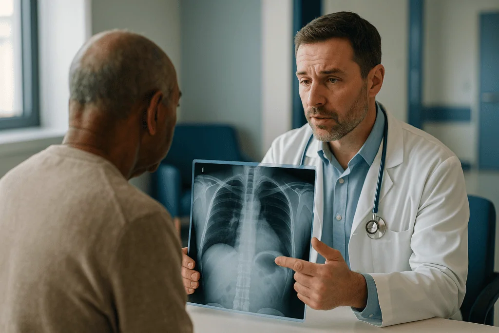
An upright X-ray needs a meaningful volume before it changes appearance; small effusions hide behind the diaphragm or along the back of the chest. If you were X-rayed lying down, as often happens in an emergency department, free fluid spreads out in a thin layer and simply makes one lung look hazier than the other. That is one reason an X-ray that is “probably clear” can still miss an early effusion, and why clinicians treat a normal film with caution when symptoms persist.
The X-ray does more than confirm fluid. It hints at cause. Effusions on both sides, with an enlarged heart shadow, point toward heart failure. A one-sided effusion with a patch of pneumonia above it suggests a parapneumonic effusion, meaning fluid reacting to infection in the adjacent lung. A lung mass, collapsed lobe or widened central chest raises the question of cancer. None of these patterns is proof; they are the first sentence of a longer conversation.
Two other first-look elements belong here. Your clinician will listen for reduced breath sounds and dullness when tapping the chest, which track with fluid, and will ask about weight change, fevers, recent travel, previous cancers and medicines. Blood tests for kidney, liver and heart function often run in parallel. Together these decide whether the next test is a repeat X-ray after treatment, an ultrasound, or a CT scan.
Chest ultrasound for pleural effusion: why it has become the first-line test
A chest ultrasound uses the same handheld probe and gel as a pregnancy scan, placed between the ribs. Fluid appears black because sound waves pass straight through it, while lung and bone bounce them back. The exam takes minutes at the bedside, involves no radiation, and can be repeated as often as needed.
Ultrasound outperforms the X-ray in three practical ways. First, it detects much smaller collections, including ones that sit behind the diaphragm. Second, it tells the clinician what the fluid looks like inside: clear and free-flowing, or filled with floating strands and walled-off pockets called loculations, which suggest infection or long-standing inflammation. A thick, nodular pleural lining visible on ultrasound raises concern for malignancy. Third, and most important for safety, it shows exactly where the fluid is deepest and where the lung, liver, spleen and diaphragm sit, so a needle can be placed without guessing.
Guidance from professional bodies including the British Thoracic Society now expects that pleural procedures be performed with ultrasound guidance, because marking a spot blind from an X-ray led to avoidable punctures of lung and abdominal organs. Cleveland Clinic’s patient information on thoracentesis describes imaging guidance as a routine part of the procedure.
Ultrasound has limits worth stating honestly. It sees only what lies directly beneath the probe, so it cannot survey the whole lung or the lymph nodes in the center of the chest. Air in the lung blocks the sound, so lung tumors or pneumonia away from the fluid stay invisible. Operator skill matters more than with CT. And ultrasound cannot say what the fluid is made of; it can only make an educated guess from its appearance. For composition, you need a sample.
Ultrasound vs CT for pleural effusion: when is a CT scan needed?
A CT scan (computed tomography) takes hundreds of X-ray slices and assembles them into a cross-sectional map of the chest. It is not a routine test for every effusion. Where heart failure or pneumonia explains the fluid and the person improves with treatment, CT adds radiation and contrast dye without changing anything.

CT earns its place when the cause is unclear, when the fluid is an exudate on analysis, when symptoms do not match the amount of fluid, or when the ultrasound shows a thickened or irregular pleura. A CT performed with intravenous contrast, ideally before all the fluid is drained so the pleural lining is stretched and visible, can reveal pleural nodules, thickening that extends around the lung, enlarged lymph nodes, a hidden lung tumor, blood clots in the pulmonary arteries, or disease in the upper abdomen that explains the fluid.
The two tests answer different questions, which is why many people have both rather than one or the other.
| Question | Chest ultrasound | CT scan |
|---|---|---|
| Is there fluid, and how much? | Excellent, detects small volumes | Excellent |
| Is the fluid free or in pockets? | Best test for this | Reasonable |
| Where is it safe to place a needle? | Essential, done in real time | Not used for live guidance in most settings |
| Is there a lung mass, clot or lymph node disease? | Poor, air blocks the view | Best test for this |
| Radiation | None | Yes, a moderate dose |
| Where it happens | Bedside, clinic | Radiology department |
One nuance people rarely hear: CT is good at suggesting malignancy but cannot confirm it. A pleura that looks suspicious still needs cells or tissue. And an unremarkable CT does not rule out cancer in the pleura, because early deposits can be flat and tiny. The scan is a guide for the next step, not a verdict.
Thoracentesis procedure: what to expect when the fluid is sampled
Thoracentesis is the medical term for drawing fluid from the pleural space with a needle. It is the pivot of the entire workup, because everything the laboratory can say about the fluid depends on this sample.
You will usually sit upright on the edge of a bed, leaning forward over a padded table with your arms resting on it. This position lets fluid pool at the back and bottom of the chest, away from the heart. The clinician scans with ultrasound to choose a spot, typically between two ribs toward the back or side, then cleans the skin and injects local anesthetic. The anesthetic stings for a few seconds; after that most people describe pressure rather than pain.
A thin needle, often followed by a small flexible catheter, passes through the chest wall into the fluid. For diagnostic purposes only a few syringes are needed, roughly the volume of a small cup. If you are also short of breath, more may be removed to relieve symptoms, though clinicians stop if you develop chest tightness or a persistent cough, and avoid draining very large volumes in one sitting because rapid re-expansion of a long-compressed lung can cause fluid to leak into the lung tissue itself, a rare complication described in the NIH StatPearls review. Cleveland Clinic’s patient guide gives a typical procedure time of about 10 to 15 minutes.
Afterward the puncture is covered with a dressing. Many centers check an ultrasound or X-ray to confirm the lung has not been nicked. The main risks are a small pneumothorax, meaning air leaking into the pleural space; bleeding; infection; and, rarely, injury to the liver or spleen, which ultrasound guidance has made uncommon. You may hear a dull ache for a day. Driving yourself home is usually discouraged if sedation was used, and your team will tell you which symptoms should prompt a call.
Who is usually asked to have a fluid sample, and who is usually asked to wait
Not every effusion is tapped, and the decision is one of the most consequential in the whole pathway. The guiding principle in most respiratory guidelines is simple: sample the fluid when the cause is not already clear, and consider waiting when it is.
People commonly offered early thoracentesis include those with a one-sided effusion of unknown cause; anyone with fever and fluid next to pneumonia, because infected pleural fluid can quickly become pus and needs drainage rather than antibiotics alone; those with a history of cancer; people whose effusion has appeared alongside weight loss, night sweats or a long cough; and anyone whose breathlessness is severe enough that removing fluid would help immediately.
People often asked to wait include those with known heart failure, kidney failure or liver cirrhosis whose effusions are on both sides, roughly equal, and fit the clinical picture. Here the plan is usually to treat the underlying condition, often with diuretics, medicines that help the kidneys remove excess water, and repeat the X-ray within days to weeks. If the fluid shrinks, no needle is needed. If it persists, grows, or stays stubbornly one-sided, sampling follows. The same applies to small effusions after chest or abdominal surgery, which commonly resolve on their own.
Some people are asked to wait for safety rather than diagnostic reasons. Very thin layers of fluid leave no safe window for a needle. Blood-thinning medicines, a very low platelet count, severe skin infection over the site, or an inability to sit still may mean the procedure is postponed or arranged in a controlled setting. None of these are reasons to stop a prescribed medicine on your own; the treating team weighs timing against risk.
Finally, some people are referred directly for a biopsy or a camera procedure rather than a simple tap, when imaging already shows a thickened, nodular pleura that a few milliliters of fluid is unlikely to explain.
What is the 3 test rule for pleural effusion? Light's criteria explained
When people search for the “3 test rule,” they almost always mean Light’s criteria, a set of three comparisons between pleural fluid and blood that have been used since the 1970s to separate transudates from exudates. The lab measures protein and an enzyme called lactate dehydrogenase (LDH), which leaks from damaged or inflamed cells, in both the fluid and a blood sample drawn the same day.
| Criterion | Threshold suggesting an exudate | What it reflects |
|---|---|---|
| Fluid protein divided by blood protein | Greater than 0.5 | Large proteins leaking through inflamed pleura |
| Fluid LDH divided by blood LDH | Greater than 0.6 | Cell damage or turnover in the pleural space |
| Fluid LDH alone | More than two-thirds of the lab’s upper normal limit for blood | Same, independent of blood level |
If the fluid meets any one of the three, it is classified as an exudate. If it meets none, it is a transudate. These thresholds are quoted in the NIH StatPearls review of pleural effusion and in MedlinePlus’s description of pleural fluid analysis.
Why does the split matter so much? A transudate says the pleura is healthy and the problem lies elsewhere: heart, kidneys, liver, or occasionally a low protein state. The search then moves to those organs and usually no further pleural testing is needed. An exudate says the pleura or adjacent lung is directly involved, and the fluid deserves the fuller panel described in the next section.
Light’s criteria are deliberately tuned to catch exudates, which means they occasionally mislabel a transudate as an exudate, particularly in people with heart failure who have been taking diuretics, because concentrating the fluid raises its protein. When the numbers sit near the cutoffs and the story screams heart failure, clinicians may look at the protein gap between blood and fluid, or at a heart-strain marker in the fluid, before committing to a label. The rule is a strong starting point, not an oracle.
Pleural fluid analysis results: what the sample actually reveals
The first clue arrives before any machine runs. Straw-colored, clear fluid is typical of transudates and many mild exudates. Cloudy or frankly pus-like fluid points to infection. Blood-stained fluid raises the question of cancer, injury or a clot in the lung, though even a small amount of blood from the needle itself can tint a sample. Milky fluid suggests lymph leaking from a damaged duct. A foul smell is a sign of bacterial infection with certain organisms.
In the laboratory, several measurements run on an exudate. A cell count with differential reports how many white cells are present and which kind dominate. Neutrophils, the rapid-response infection fighters, suggest acute infection or recent inflammation. Lymphocytes, the slower immune cells, dominate in tuberculosis, cancer and some long-standing effusions. A high proportion of eosinophils often follows air or blood in the pleural space and is less specific than people expect.
Chemistry adds texture. A low fluid glucose and a low pH, meaning the fluid has turned acidic, occur when bacteria or cancer cells consume sugar and release acid; in a parapneumonic effusion these findings push the team toward placing a drain rather than waiting. Amylase rises when the pancreas or esophagus is the source. Adenosine deaminase, an enzyme from activated immune cells, is used in some settings as a pointer toward tuberculosis.
Microbiology stains and cultures the fluid for bacteria, mycobacteria and fungi. Cultures take days, and a negative culture does not exclude infection because the fluid is often sterile even when the lung next to it is not.
Cytology examines the fluid for cancer cells. A positive result can establish a cancer diagnosis without surgery. A negative result is weaker evidence, because some tumors shed few cells; a second sample improves the yield modestly, and after that a biopsy becomes the better route. MedlinePlus’s pleural fluid analysis page summarizes these components in patient-friendly terms.
When the first tap does not explain the effusion: biopsy and thoracoscopy
A meaningful share of exudates remain unexplained after one round of fluid tests. That is not a failure of the workup; it is a known limitation of studying fluid rather than tissue. The next step depends on what the imaging showed and how unwell the person is.
If the CT or ultrasound shows pleural thickening or nodules, an image-guided pleural biopsy can be arranged. Under local anesthetic and ultrasound or CT guidance, a hollow needle takes a small core of pleural tissue from the abnormal area. This is a day procedure with risks similar to thoracentesis, and the sample goes to a pathologist who can identify cancer type, tuberculosis granulomas or other inflammatory patterns.
If the pleura looks diffusely abnormal, or a targeted biopsy is not possible, the team may offer medical thoracoscopy or video-assisted thoracoscopic surgery. In both, a camera is passed through a small cut in the chest wall into the pleural space, the fluid is drained completely, and the clinician inspects the lining directly and takes multiple biopsies from anything that looks wrong. Medical thoracoscopy is done under local anesthetic with sedation; the surgical version uses general anesthesia and allows more extensive sampling. Because the entire space is drained, the same procedure can be used to apply a treatment that encourages the lung and chest wall to stick together and prevent fluid returning, if the diagnosis warrants it. The decision to combine diagnosis and treatment in one session sits with the thoracic team.
Bronchoscopy, a camera passed down the airway, is not a routine effusion test. It is reserved for people with a lung mass, coughing up blood or a collapsed section of lung on imaging, where the airway itself may hold the answer.
Some effusions still defy explanation after biopsy. In that case watchful follow-up with repeat imaging over months is reasonable, because a few declare themselves later, and many simply resolve.
What can be mistaken for pleural effusion on imaging and in the clinic?
The chest X-ray is a shadow play, and several things cast shadows that look like fluid. Knowing them explains why your clinician may order an ultrasound to double-check something that already “looked obvious.”
A raised diaphragm is the most common impostor. If the diaphragm on one side sits high, because of nerve weakness, a swollen liver, or gas in the stomach pushing upward, the base of the lung looks white and the costophrenic angle looks blunt. Ultrasound settles it in seconds by showing the dome of the diaphragm with no black fluid above it.
Pleural thickening from an old infection, asbestos exposure or prior surgery creates a permanent white rim that mimics a small chronic effusion. On CT it appears as a dense band rather than water. Scarring after radiotherapy and a collapsed lower lobe can also pull the diaphragm upward and crowd the base of the chest.
Consolidation, the dense patch pneumonia produces, can sit low enough to resemble fluid. A pleural tumor or a large fatty pad near the diaphragm occasionally does the same. A hemidiaphragm hernia, where abdominal contents push into the chest, produces a baffling shadow until CT reveals bowel or fat.
Outside imaging, the symptoms of effusion are shared with heart failure without fluid, pulmonary embolism, pneumonia, asthma and simple deconditioning. This overlap is the reason effusion is confirmed by imaging rather than by how you feel, and why guideline-style patient information such as the NHS pleurisy page stresses getting unexplained breathlessness checked rather than guessing.
The reverse error also happens. An effusion lying flat in a person X-rayed in bed can be read as a “hazy lung” or mild pneumonia. If antibiotics bring no improvement, a repeat upright film or an ultrasound often reveals the fluid that was there all along.
What the days and weeks after pleural effusion testing usually look like
The timeline depends almost entirely on what the first tests show, so it helps to picture three common paths rather than one.
When the effusion fits heart, kidney or liver disease, the next days are about treating that organ. Breathlessness often eases as fluid comes off the whole body, and a repeat chest X-ray within a few weeks checks that the pleural fluid has followed. If it has not, or if it was always one-sided, the pathway loops back to ultrasound and a fluid sample.
When the fluid is sampled, the first results, appearance, cell count, protein, LDH, glucose and pH, usually return the same day or the next. Cultures take several days. Cytology commonly takes a few working days to a week depending on the laboratory, and tuberculosis cultures can take weeks, which is why clinicians sometimes act on the overall pattern before every result is back. The puncture site heals within days; a dull ache or a slight cough for a day or two is common, and the team will usually tell you to expect a check X-ray or ultrasound before you leave.
When infection is found, a chest drain, a soft tube left in place to empty the space continuously, may be placed for several days alongside intravenous antibiotics, with daily imaging to track progress. When cancer cells are found, the following weeks involve staging scans and a conversation with an oncology team about both the cancer and the fluid, which may need a more durable drainage solution if it keeps returning.
Across all paths, a follow-up appointment is typically arranged so results can be reviewed together rather than in fragments. Johns Hopkins’ patient information notes that treatment is directed at the cause, which is another way of saying the calendar is written by the diagnosis, not by the fluid.
What people often get wrong about pleural effusion diagnosis tests
“If the X-ray is clear, there is no fluid.” An upright X-ray needs a substantial volume before it changes, and a film taken lying down can hide an effusion entirely. Ultrasound detects far smaller collections, which is why a normal X-ray with persistent symptoms is a reason to look again, not to stop looking.
“The scan can tell if it is cancer.” CT and ultrasound can show features that raise or lower suspicion, but neither can see individual cells. Only cytology of the fluid or pathology of a biopsy can establish malignancy. A clean-looking CT does not rule it out, and a suspicious one does not confirm it.
“Draining the fluid is the treatment.” Removing fluid relieves breathlessness, but the fluid returns if the cause remains. The sample’s real value is diagnostic. Lasting control comes from treating the heart, infection, kidney, liver or tumor behind it.
“One negative cytology means no cancer.” Some tumors shed few cells into fluid. A negative result lowers the probability but does not close the question, which is why a second sample or a biopsy is often the honest next step.
“Light’s criteria are a diagnosis.” They are a sorting tool. An exudate is a category containing dozens of causes; the criteria tell the team which panel of tests to run next, not what you have.
“Thoracentesis is dangerous and should be avoided.” With ultrasound guidance the procedure is routine and complications are uncommon, as the NIH StatPearls review describes. The greater risk often lies in delaying a sample when infected fluid is quietly turning into pus.
“Fluid on both sides means it cannot be serious.” Bilateral effusions are usually transudates, but cancer, autoimmune disease and some infections can affect both sides. The whole clinical picture decides, not the number of sides.
Questions to ask your care team about pleural effusion tests
Results arrive in pieces, and clinics run on tight schedules. A short list of questions helps you leave each appointment knowing where you stand and what comes next.
- Is the fluid on one side or both, and does that change what you think is causing it?
- Do you already have a likely explanation, or is the cause unknown at this point?
- Will I need a fluid sample, and if not, how will we know whether waiting was the right call?
- Will the procedure be ultrasound-guided, and who will perform it?
- How much fluid do you plan to remove, and is that for diagnosis, symptom relief or both?
- Which results should come back today, and which will take days or weeks?
- Was the fluid a transudate or an exudate, and what does that direct you toward?
- If the first sample does not give an answer, what is the next step, and is a biopsy likely?
- Should a CT scan be done before or after the fluid is drained?
- Do any of my current medicines, especially blood thinners, affect the timing of the procedure, and who decides about pausing them?
- What symptoms after the procedure should make me call you, and which should send me to emergency care?
- Who is coordinating my results, and how will I hear about them?
Bring a written list of your medicines, previous cancers, lung conditions and any exposure to asbestos or tuberculosis, because these facts steer the laboratory panel. If you have had a prior effusion, ask that those results be pulled; a change from transudate to exudate, or a new one-sided collection in someone with a known cause, is itself a finding worth discussing.
None of these questions second-guess your team. They give the team a clearer picture of what you understand, which makes shared decisions about further testing easier for everyone at the table.
When to call your doctor: red-flag signs with a pleural effusion
Most effusions develop over days to weeks, and most testing is planned rather than urgent. A few situations change that. Seek emergency care without waiting for a scheduled appointment if you experience sudden or rapidly worsening shortness of breath, especially if you cannot speak a full sentence or must sit upright to breathe; chest pain that is severe, crushing or spreading to the arm, jaw or back; a bluish tinge to the lips or fingertips; coughing up blood; fainting or near-fainting; or a fast heartbeat with dizziness. These can signal a large effusion compressing the lung, a clot in the lung, a heart event or a collapsed lung, all of which need immediate assessment.
Call your care team the same day, rather than waiting, if you develop a fever or shaking chills while an effusion is being investigated, since this can mean the fluid has become infected; if breathlessness that had eased after drainage returns within days; if you notice new swelling in one leg, which raises the possibility of a clot; or if you lose weight or drench the sheets with night sweats while results are pending.
After a thoracentesis or biopsy, contact the team promptly for sharp chest pain that worsens with breathing, new breathlessness, a persistent dry cough that does not settle within hours, bleeding or spreading redness at the puncture site, or a fever within the following days. The NIH StatPearls review lists pneumothorax, bleeding and infection as the recognized complications, and all are more manageable when reported early.
Finally, do not stop, start or adjust any prescribed medicine on your own because of an effusion, including diuretics or blood thinners. Timing those decisions around procedures is exactly what the treating team is there to do. If something feels wrong and you are unsure which category it falls into, calling is always the safer choice; triage lines would rather hear from you than not.
Frequently asked questions
What is the best test to diagnose pleural effusion?
Chest ultrasound is the preferred first test for confirming a pleural effusion because it detects small volumes, shows whether fluid is free or loculated and guides safe sampling without radiation. A plain X-ray usually finds larger effusions first. Explaining the cause, however, generally requires pleural fluid analysis after thoracentesis, with CT reserved for unclear or suspicious cases.
What is the 3 test rule for pleural effusion?
The “3 test rule” refers to Light’s criteria, three comparisons of pleural fluid with blood. Fluid is called an exudate if its protein ratio to blood exceeds 0.5, its LDH ratio exceeds 0.6, or its LDH is above two-thirds of the lab’s upper normal limit. Meeting any one criterion makes it an exudate; meeting none makes it a transudate.
What confirms a pleural effusion?
Imaging confirms it. A chest X-ray showing fluid layering at the base of the lung is often enough, and chest ultrasound or CT confirms uncertain cases and smaller collections. Physical signs such as dullness and reduced breath sounds support the suspicion but do not confirm it. Fluid drawn by thoracentesis then confirms the type and often the cause.
What can be mistaken for pleural effusion?
A raised diaphragm, old pleural thickening or scarring, pneumonia low in the lung, a collapsed lower lobe, a pleural tumor and a diaphragmatic hernia can all mimic fluid on a plain X-ray. Ultrasound or CT usually distinguishes them. Clinically, heart failure, pulmonary embolism, pneumonia and asthma cause similar breathlessness, which is why imaging rather than symptoms confirms an effusion.
How does ultrasound vs CT compare for pleural effusion?
Ultrasound is better for detecting fluid, judging whether it is free or pocketed, and guiding needles in real time, with no radiation. CT is better for seeing the lung, central lymph nodes, pulmonary blood vessels and the whole pleural surface. They are complementary: many people with an unexplained exudate have ultrasound first and a contrast CT afterward.
What does the thoracentesis procedure feel like and how long does it take?
Most people feel a brief sting from the local anesthetic and then pressure rather than pain as the needle enters. Cleveland Clinic’s patient information gives a typical procedure time of about 10 to 15 minutes, excluding preparation and the check imaging afterward. A dull ache at the site for a day is common; worsening chest pain or breathlessness afterward should be reported promptly.
What do pleural fluid analysis results show?
They report the fluid’s appearance, protein and LDH for Light’s criteria, cell counts showing whether neutrophils or lymphocytes dominate, glucose and pH, which fall in infection and cancer, and additional tests such as amylase or adenosine deaminase when relevant. Microbiology cultures look for bacteria, mycobacteria and fungi, and cytology examines the fluid for cancer cells.
Can a pleural effusion be diagnosed without draining the fluid?
The presence of fluid can, using imaging. The cause sometimes can too: when a person has known heart failure, kidney failure or cirrhosis with matching effusions on both sides, clinicians often treat the underlying condition and repeat the X-ray rather than sampling. If the fluid persists, grows or is one-sided, a sample is usually recommended.
Why might a CT scan be done before the fluid is drained?
Fluid stretches the pleural lining, and intravenous contrast highlights it, so nodules, thickening or tumor deposits are easier to see on a CT taken while fluid is still present. After complete drainage the pleural layers collapse together and subtle abnormalities can be lost. Teams therefore often time the contrast CT before a large-volume drainage when cancer is a concern.
How long do pleural fluid test results take?
Appearance, protein, LDH, glucose, pH and cell counts usually return within hours to a day. Bacterial cultures typically take several days. Cytology often takes a few working days to about a week, depending on the laboratory. Tuberculosis cultures can take weeks, which is why clinicians sometimes act on the overall pattern before every result is final.
References
- MedlinePlus: Pleural effusion
- MedlinePlus: Pleural Fluid Analysis
- Cleveland Clinic: Thoracentesis
- NHS: Pleurisy
This article is for general information only and is not a substitute for professional medical advice. Please consult a qualified doctor about your individual situation.

More from the Blog

Who Is a Candidate for Single-Port VATS? Lung Biopsies, Collapsed Lung and Small Tumors
Single-port VATS candidates are usually people who need a lung biopsy that a needle cannot safely provide, a repair for a recurrent or persistent…

Do Antibiotics Help Acute Bronchitis? Why Most Cases Are Viral and What Treatment Looks Like
Antibiotics usually do not help acute bronchitis, because the large majority of cases are caused by viruses that antibiotics cannot touch. Pooled trials suggest…

Which Chest Conditions Suit Robotic Thoracic Surgery? Lung Nodules, Mediastinal Tumors and More
Robotic thoracic surgery is most often considered for small lung nodules and early-stage lung cancer needing a wedge resection, segmentectomy or lobectomy, for tumors…
Does Acute Bronchitis Need a Chest X-Ray? How Doctors Rule Out Pneumonia
Most people with acute bronchitis do not need a chest X-ray. Doctors usually diagnose it from the story of the illness and a physical…

Questions to Ask Your Cystic Fibrosis Team: Lung Function, Infections and Nutrition Goals
Useful questions to ask a cystic fibrosis team fall into three groups: lung function (what my FEV1 is, how it has changed, what a…

How Long Does Lung Abscess Treatment Take? Why Antibiotic Courses Run for Weeks
Lung abscess treatment usually takes several weeks, not days. Published reviews describe antibiotic courses of roughly three to six weeks, sometimes extended to eight…