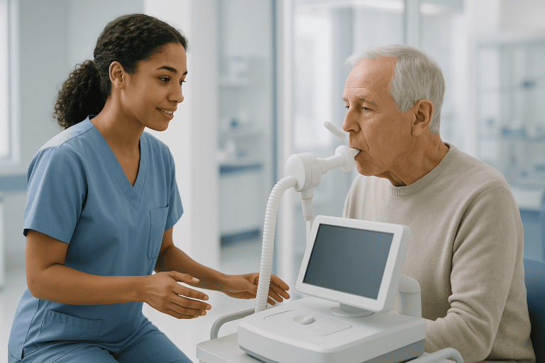
Pleural Effusion Treatment
Pleural effusion is fluid buildup around the lungs that can cause breathlessness, chest discomfort, or cough. Care focuses on finding the cause and relieving symptoms through drainage or targeted treatment.
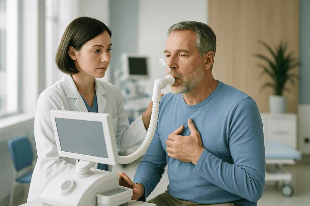
Quick answer
A pleural effusion is a build-up of excess fluid in the pleural space, the thin gap between the lung and the chest wall. Treatment depends on the cause: small effusions may simply be monitored while the underlying condition is treated, while larger or symptomatic effusions are drained by thoracentesis or a chest tube, and the fluid is analysed to identify infection, heart failure, cancer or another cause.
What Is Pleural Effusion?
A pleural effusion is a build-up of excess fluid in the pleural space, the thin gap between the lung and the inside of the chest wall. Treatment focuses on two things at once: relieving the pressure the fluid places on the lung, and finding out why the fluid collected in the first place. It is relevant to anyone whose imaging has shown fluid around a lung, whether the first sign was breathlessness, chest pressure or an incidental finding on a chest X-ray.
Normally the pleural space contains only a thin film of lubricating fluid that lets the lung glide smoothly against the chest wall as you breathe. When too much fluid accumulates, the lung cannot expand fully. That is when you may notice shortness of breath, a dry cough, chest discomfort, fatigue or reduced exercise tolerance. The larger the effusion, the more the lung is compressed and the harder ordinary activity becomes.
Being told you have a pleural effusion can be unsettling, and it is reasonable to worry that fluid around the lung means a serious disease. Sometimes it does. But pleural effusion is not one single condition. It is a finding, a sign that something else is happening, and it can develop for many reasons, ranging from infection or heart failure to cancer, inflammation, liver or kidney disease, or complications after surgery. The most important step is to understand why the fluid has collected and whether it is affecting breathing. Some effusions are small and can simply be monitored while the underlying condition is treated. Others require drainage to relieve symptoms, test the fluid or prevent complications.
Care for pleural effusion is therefore both diagnostic and therapeutic. Doctors aim to answer three questions: Is the fluid causing symptoms? What is the cause? What is the safest and most effective way to treat it? At Acibadem, evaluation is typically organised through a coordinated pathway involving pulmonology, thoracic surgery, radiology, oncology, cardiology, infectious disease and other specialists when needed. This matters because the correct treatment depends on the underlying diagnosis as much as on the amount of fluid.
What causes pleural effusion?
Pleural effusion develops when the balance between fluid production and fluid absorption in the pleural space is disturbed, either because pressure in the blood vessels rises, because protein levels in the blood fall, or because the pleural surfaces themselves become inflamed, infected or invaded by disease. Doctors broadly divide effusions into two groups. A transudate forms when systemic pressure or fluid balance problems push watery fluid into the pleural space; heart failure, kidney disease and liver disease are typical causes. An exudate forms when the pleural surfaces are directly affected by inflammation, infection, cancer or injury; the fluid is richer in protein and cells, and it usually demands a more searching diagnostic work-up.
Other recognised causes include pulmonary embolism, in which blood clots in the lungs cause pleuritic pain and effusion; autoimmune and inflammatory conditions such as rheumatoid arthritis or lupus that affect the pleura; tuberculosis, which can inflame the pleural lining; and fluid or blood that appears after chest surgery or trauma. Because the list is broad, an effusion sometimes turns out to be the first clue to a medical condition that has not yet been diagnosed. That is why a careful evaluation is essential rather than draining fluid and hoping it does not return.
What is the most common cause of pleural effusion?
Heart failure is generally the most common cause of pleural effusion overall, because raised pressure in the circulation pushes fluid into the pleural space on one or both sides. Among exudative effusions, pneumonia and cancer are the leading causes. Fluid related to heart failure often improves when the heart condition and the body’s overall fluid balance are brought under control, which is why cardiac assessment and structured fluid management can matter as much as any drainage procedure. Fluid related to pneumonia or malignancy behaves differently and usually needs sampling, and often drainage, to be managed safely.
Is pleural effusion the same as fluid in the lungs?
Not quite, what people describe as fluid in the lungs can mean two different things, and the distinction changes the treatment. A pleural effusion is fluid around the lung, sitting between the lung and the chest wall. Pulmonary oedema, by contrast, is fluid inside the lung tissue and air sacs themselves. Both can cause breathlessness, and both can occur together in heart failure, but they are assessed and treated differently. When a report mentions pleural effusion, it refers to the space around the lung, and drainage procedures target that space, not the lung tissue itself.
What is the difference between pleural effusion and pleurisy?
Pleurisy means inflammation of the pleural lining, which typically causes a sharp chest pain that worsens with deep breathing or coughing, whereas a pleural effusion is the fluid that may collect in the pleural space. The two are related: inflamed pleural surfaces often leak fluid, so pleurisy and effusion frequently occur together, particularly in infection, pulmonary embolism and autoimmune disease. Curiously, when an effusion grows, the pain of pleurisy sometimes eases, the fluid separates the inflamed surfaces, while breathlessness increases. A change in symptoms like this is one of the details doctors listen for when they take a history.
What are the three stages of pleural effusion?
The three stages usually refer to how an infected (parapneumonic) effusion evolves. In the first, exudative stage, thin, free-flowing fluid collects next to a pneumonia and can often be drained easily. In the second, fibrinopurulent stage, bacteria enter the fluid, it thickens, and strands of fibrin divide it into pockets, or loculations, that resist simple drainage; established infection of the pleural space is called empyema. In the third, organising stage, a fibrous peel forms over the lung and can prevent it from expanding. Each stage is harder to treat than the one before, which is one of the strongest arguments for assessing pleural effusion early rather than waiting to see whether it settles.
Symptoms and Who May Need Treatment
Patients may need evaluation or treatment for pleural effusion when fluid around the lung is causing symptoms, when the cause is unclear, or when imaging suggests a potentially complicated effusion. Symptoms can be mild at first and are easily mistaken for fatigue, bronchitis, asthma or simply being out of condition. In other cases, shortness of breath develops quickly and becomes difficult to ignore.
Common symptoms include:
- Shortness of breath, especially when walking, climbing stairs or lying flat
- Chest discomfort, heaviness, or a sharp pain that may worsen with deep breathing
- Persistent dry cough
- Fatigue or reduced ability to exercise
- Fever, chills or night sweats when infection is present
- Unexplained weight loss or loss of appetite in some underlying conditions
People who have cancer, pneumonia, heart failure, kidney disease, liver disease, autoimmune disease, recent chest surgery, pulmonary embolism or tuberculosis exposure carry a higher risk of developing an effusion. In these groups, new breathlessness deserves particular attention, because a growing effusion may signal a change in the underlying disease as well as a mechanical problem with breathing.
What do clinicians assess in patients with pleural effusion?
The assessment covers four broad areas: how the patient is breathing, how large and complex the fluid collection is, what is causing it, and how safe any procedure would be. In practice that means checking the respiratory rate, oxygen saturation, blood pressure and pulse; listening to the chest, where an effusion classically produces reduced breath sounds and dullness to percussion over the fluid; and reviewing the medical history for heart, kidney, liver, autoimmune or cancer conditions and for recent infection, surgery or travel. Imaging then defines the size and character of the effusion, and laboratory tests, of blood and, where sampled, of the fluid itself, narrow the cause. Before any drainage, the team also reviews bleeding risk, platelet counts and blood-thinning medication, since these affect how and when a procedure can be performed safely.
How Pleural Effusion Is Diagnosed
Pleural effusion is most often detected on a chest X-ray, ultrasound or CT scan. Ultrasound is especially useful because it confirms the presence of fluid, estimates its volume, identifies pockets or loculations, and guides safe drainage in real time. CT imaging adds detail about the lung, the pleura, lymph nodes and nearby organs, and can reveal patterns that point towards infection, embolism or malignancy. In selected patients, blood tests, echocardiography, bronchoscopy, biopsy or thoracoscopy may be needed to clarify the cause.
Fluid analysis is frequently central to diagnosis. During thoracentesis, pleural fluid can be sent for tests that evaluate protein and enzyme levels, cell counts, infection markers, culture, cytology for cancer cells, and other specialised studies. By comparing protein and enzyme levels in the fluid with those in the blood, doctors classify the effusion as a transudate or an exudate: a distinction that immediately narrows the list of likely causes. A transudate points towards systemic fluid overload and conditions such as heart, kidney or liver disease; an exudate points towards inflammation, infection, malignancy or injury to the pleural surfaces and usually prompts further investigation.
Not every effusion needs to be sampled. When the clinical picture clearly fits a known condition: a patient with established heart failure and typical bilateral effusions, for example, doctors may treat the underlying disease first and reassess with imaging. Sampling becomes important when the cause is unclear, when the effusion is on one side only, when infection or cancer is possible, or when the fluid fails to respond to treatment of the presumed cause.
Conditions and Indications Pleural Effusion Care Addresses
Pleural effusion care is used across a broad range of conditions, and the approach depends on whether the effusion is a transudate or an exudate, how large it is, and how the patient is coping. Common indications include:
- Symptomatic pleural effusion: fluid causing shortness of breath, chest discomfort, cough or low oxygen levels.
- Undiagnosed pleural effusion: fluid with no clear cause after initial examination and imaging.
- Malignant pleural effusion: fluid related to cancer involving the lung, breast, lymphoma, ovarian cancer, gastrointestinal cancers or pleural malignancies.
- Parapneumonic effusion and empyema: fluid associated with pneumonia, which may become infected or thick and require drainage.
- Heart failure-related effusion: fluid that develops because of increased pressure and fluid overload, often treated with cardiac medication and careful fluid balance, but sometimes requiring drainage.
- Tuberculous pleuritis: pleural inflammation due to tuberculosis, requiring specific antimicrobial treatment and careful follow-up.
- Pulmonary embolism: blood clots in the lungs that cause pleuritic pain and effusion.
- Autoimmune or inflammatory disease: conditions such as rheumatoid arthritis or lupus that affect the pleura.
- Postoperative or traumatic effusion: fluid or blood that develops after surgery or injury and may require observation or drainage.
- Recurrent pleural effusion: fluid that returns after drainage and may need longer-term control strategies.
Some effusions are treated as urgent: large collections causing significant breathlessness, infected pleural fluid, suspected empyema, or fluid associated with unstable oxygen levels. Others can be assessed in a planned outpatient or short-stay setting. The clinical context, not the fluid alone, determines the urgency.
How Pleural Effusion Treatment Is Performed
Treatment may involve one procedure or several, chosen according to the size of the effusion, the symptoms, the oxygen levels, the imaging findings, the medical history and the laboratory results. The guiding principle is to use the least invasive approach that will relieve symptoms, secure the diagnosis and prevent recurrence. In some cases the best treatment is not a drainage procedure at all but management of the underlying disease, diuretic therapy and cardiac care for heart failure, antibiotics for infection, or cancer-directed therapy for malignant disease, with drainage reserved for symptom relief.
Initial evaluation and preparation
The first step is a structured review of symptoms, medical history, medications, prior imaging, cancer history, infection risks, cardiac and kidney function, and any previous pleural procedures. Recent CT scans, X-rays, pathology reports, laboratory results, operation notes and oncology records all help the team reach a decision faster and with more confidence.
Doctors usually perform a physical examination and review imaging. Additional tests may include blood work, oxygen saturation measurement, electrocardiography, echocardiography, CT scanning, ultrasound or pulmonary function assessment in selected cases. If drainage is being considered, the team reviews bleeding risk, blood-thinning medication, platelet count and overall medical stability; any changes to medication around a procedure are decided by the treating doctors, not made in advance by the patient.
Before thoracentesis or tube placement, the procedure is explained in detail. You are usually positioned sitting upright or lying in a specific position, depending on the amount and location of fluid. Local anaesthetic is used to numb the area. Sedation is not always required for a simple thoracentesis, but it may be used for more complex procedures or when appropriate for comfort and safety.
Thoracentesis: removing fluid and understanding the cause
Thoracentesis is the most common procedure for pleural effusion. Using ultrasound guidance, the physician identifies a safe pocket of fluid and marks the entry point. After cleaning the skin and numbing the area with local anaesthetic, a fine needle or thin catheter is advanced into the pleural space. Fluid is removed gradually, clinicians deliberately limit how quickly and how much they drain in one sitting, because removing a very large volume too fast can stress the re-expanding lung, and samples are sent for laboratory testing when diagnostic evaluation is needed.
Many patients notice easier breathing soon after fluid removal, especially if the effusion was large. During and after the procedure, the team monitors symptoms, oxygen levels and vital signs. A follow-up chest X-ray or ultrasound may be performed to assess lung expansion and check for complications such as pneumothorax, which is air in the pleural space. Serious complications are uncommon when the procedure is performed with imaging guidance and careful monitoring, but every procedure carries some risk, and honest consent discussions cover bleeding, infection, pneumothorax and the possibility that fluid returns.
Chest tube drainage for larger, infected or complex effusions
If the fluid is infected, thick, loculated or too large to manage with a single drainage procedure, a chest tube may be placed. The tube stays in position and drains fluid over hours or days, connected to a drainage system while the medical team tracks fluid volume, appearance and lung re-expansion. Imaging is repeated as needed to confirm the effusion is resolving before the tube is removed.
For complicated infections, medication may sometimes be instilled through the tube to help break down internal pockets of fluid. If drainage remains insufficient, thoracic surgery may be considered. Minimally invasive thoracic procedures can remove infected material, free the lung from restrictive tissue and help it re-expand when a chest tube alone is not enough.
Pleurodesis and long-term control of recurrent effusion
When a pleural effusion returns repeatedly, doctors may recommend a treatment designed to reduce recurrence. Pleurodesis involves introducing an agent into the pleural space, usually after drainage, to create controlled inflammation that helps the lung lining adhere to the chest wall so there is no longer a space for fluid to occupy. It can be performed through a chest tube or during thoracoscopy, depending on the patient’s condition and the cause of the effusion.
Pleurodesis is most often considered for recurrent malignant pleural effusion when the lung is able to re-expand after drainage and the patient’s overall treatment plan supports the approach. It may reduce the need for repeated thoracentesis. Before recommending it, the team evaluates whether the lung expands well, whether the fluid is likely to recur, and whether the patient is fit for the procedure. If the lung cannot re-expand, a situation sometimes called trapped lung, pleurodesis is unlikely to work, and other options are discussed instead.
Indwelling pleural catheter
An indwelling pleural catheter is a soft tube placed into the pleural space that stays in position and allows periodic drainage outside the hospital. It may suit patients with recurrent effusions, especially when repeated hospital visits would be difficult or when the lung does not fully re-expand. The catheter can usually be managed with trained nursing support and clear instructions covering drainage, dressing care and the signs of infection to watch for.
This option can control symptoms while the broader medical condition, cancer, advanced heart disease or another chronic illness, continues to be managed. The decision is individualised and weighs the patient’s goals, home environment, travel plans, expected recurrence and overall treatment strategy. For some people, regular drainage at home restores a degree of independence that repeated hospital procedures cannot.
Technology used in modern pleural effusion care
Modern pleural effusion care relies on accurate imaging and careful procedural guidance. Ultrasound helps physicians locate fluid and avoid nearby organs, removing the guesswork of unguided procedures. CT imaging evaluates the lung, pleura, lymph nodes and underlying disease patterns. Laboratory testing of pleural fluid supports diagnosis, while cytology and pathology can identify malignant or inflammatory conditions. In selected cases, thoracoscopy allows direct visualisation of the pleural surfaces and targeted biopsy when fluid analysis alone has not answered the question.
The value of the technology lies not in its availability but in how it is applied. A small effusion may need monitoring rather than drainage. A complex effusion may need interventional radiology, pulmonology, thoracic surgery and infectious disease working together. A malignant effusion needs coordination with oncology so that symptom relief and cancer treatment are planned as one strategy rather than two competing ones.
How long does treatment take?
A diagnostic or therapeutic thoracentesis is usually a same-day or short-stay procedure; the needle or catheter is in place only briefly, with additional time for preparation, monitoring and follow-up imaging. Chest tube drainage typically requires a longer observation period or hospital admission, particularly when infection or complex fluid is being treated, because the tube stays until drainage slows and the lung has re-expanded. Pleurodesis and thoracoscopic procedures generally involve a hospital stay whose length depends on recovery, drainage volume, pain control and lung expansion. The honest answer is that duration is set by the cause and the response to treatment, not by the procedure alone.
Why Acting Early Matters
Pleural effusion is not always an emergency, but it should not be ignored. Fluid around the lung can worsen gradually, limiting the lung’s ability to expand and making daily activity harder week by week. Patients often begin avoiding movement, sleeping propped upright, or feeling anxious because they cannot take a full breath, changes that are easy to attribute to age or fitness until imaging reveals the real cause. Early assessment can relieve symptoms and prevent unnecessary deterioration.
Delay can also make diagnosis harder and allow complications to develop. Infected pleural fluid can thicken, divide into pockets and become far more difficult to drain, progressing through the stages described earlier towards empyema, prolonged hospitalisation or surgery. If the effusion is related to cancer, earlier diagnosis helps the oncology team plan appropriate staging and treatment. If it is related to heart failure, kidney disease or liver disease, timely medical management and attention to overall fluid management can reduce recurrence and protect general health.
Clinicians treat certain situations with greater urgency: increasing breathlessness, fever alongside an effusion, sharp or worsening chest pain, low oxygen levels, or a new effusion in someone with a known cancer diagnosis. In these settings, early imaging and specialist review clarify whether drainage, antibiotics, anticoagulation, cancer treatment or another intervention is the right next step.
Benefits of Pleural Effusion Treatment
The benefits of treatment depend on the cause, but the main goals are consistent: improve breathing, establish a diagnosis, and prevent complications.
| Benefit | What It Means for You |
|---|---|
| Relief of breathlessness | Removing fluid may allow the lung to expand more fully, making breathing and daily activity easier. |
| Clearer diagnosis | Fluid analysis can help identify infection, inflammation, cancer-related causes, or systemic conditions such as heart failure. |
| Targeted treatment planning | Once the cause is understood, doctors can choose the most appropriate medication, procedure, cancer therapy or monitoring plan. |
| Reduced risk of complications | Timely drainage and treatment may help prevent infected fluid from becoming more organised and difficult to manage. |
| Better symptom control in recurrent effusion | Options such as pleurodesis or an indwelling catheter may reduce repeated emergency visits or repeated needle drainage. |
Recovery Timeline After Pleural Effusion Treatment
Recovery varies by procedure and diagnosis, but many patients follow a broadly similar pattern after drainage or related treatment.
| Time Period | What Patients Can Expect |
|---|---|
| Day 1 | After thoracentesis, breathing may improve quickly. The team monitors vital signs, oxygen levels and the puncture site. Mild soreness can occur. |
| First Week | Patients usually resume light activities as advised. If a chest tube was placed, drainage and imaging guide the timing of tube removal and discharge. |
| First Month | Follow-up focuses on test results, recurrence risk, wound healing, and treatment of the underlying cause, such as infection, heart failure or cancer. |
| Longer Term | Some patients need ongoing surveillance, repeated imaging, cancer-directed treatment, medication adjustment, pleurodesis follow-up or catheter care. |
After thoracentesis, most people return to light activity quickly, though strenuous exertion is best avoided for a short period and the puncture site needs simple care as instructed. After chest tube drainage or pleurodesis, soreness, fatigue and breathing capacity take longer to settle. If the effusion is due to pneumonia, heart failure, cancer or another systemic condition, recovery also depends on how well that condition is brought under control: the drainage relieves the symptom, but the underlying disease sets the pace.
Factors That Influence Outcomes
A good result in pleural effusion care is not measured only by how much fluid is removed. It depends on whether the lung re-expands, whether symptoms improve, whether the diagnosis is accurate, and whether the underlying condition is treated effectively. Several factors shape this.
The underlying cause is the single most important determinant. Effusions caused by heart failure often improve when fluid balance and cardiac function are controlled. Parapneumonic effusions may resolve with antibiotics and drainage if treated before they become highly organised. Malignant effusions can usually be managed for symptom relief, but they require coordinated cancer care and realistic planning about what drainage can and cannot achieve.
The size and complexity of the effusion also matter. Free-flowing fluid is generally straightforward to drain; loculated or thick fluid is not. If the lung has been compressed for a long time, or if the pleura is affected by tumour or fibrosis, the lung may not fully expand after drainage, the trapped lung situation, which changes the options available.
The patient’s overall health influences both procedural risk and recovery. Age, lung function, heart health, kidney function, nutrition, immune status and blood-thinning medication all play a role. A medically fragile patient may still benefit from symptom-relieving drainage, but the plan has to be adapted rather than applied by formula.
Accuracy of diagnosis is essential. Draining fluid without understanding why it collected invites recurrence and can delay targeted care. This is why fluid analysis, imaging review and specialist input are combined, and why complex cases, particularly those involving cancer, infection or possible thoracic surgery, may be reviewed by a multidisciplinary board before a treatment path is fixed.
Follow-up is the final major factor. Pleural effusion can recur, and early follow-up detects returning fluid before symptoms become severe. Where long-term monitoring is needed, follow-up can be coordinated with the patient’s own physician at home.
Can you recover from pleural effusion?
Yes, many pleural effusions resolve once the cause is treated, and the outlook depends far more on that cause than on the fluid itself. An effusion linked to pneumonia commonly clears with antibiotics and drainage. One linked to heart failure often settles as cardiac treatment and fluid balance take effect, though it can return if the heart condition worsens. Effusions related to cancer are different: drainage reliably relieves symptoms, but the fluid tends to recur unless the cancer itself responds to treatment, which is why longer-term strategies such as pleurodesis or an indwelling catheter exist. Recovery, in other words, is realistic for most patients, but what recovery looks like ranges from complete resolution to well-controlled, well-managed symptoms.
How long can you live with pleural effusion?
There is no single answer, because a pleural effusion is a finding rather than a disease, and life expectancy is determined by whatever is causing it. Someone whose effusion stems from a treatable pneumonia or well-managed heart failure can live for many years with appropriate care; the effusion itself, once treated, may never return. A malignant pleural effusion is a more serious sign, because it usually indicates that a cancer has spread to the pleura, and the outlook then depends on the cancer type, its stage, the treatments available and the patient’s overall condition. Even in that setting, controlling the fluid can meaningfully improve breathing, comfort and quality of life. Any honest discussion of prognosis has to be individual: it belongs in a conversation with the treating team, who know the diagnosis, the test results and the treatment options for that specific person.
How Care Is Coordinated for Complex Effusions
Pleural effusion sits at the intersection of several specialties, and the quality of care depends heavily on how well those specialties work from the same diagnostic picture. A patient with suspected malignant pleural effusion may need pulmonology, thoracic surgery, radiology, pathology and medical oncology aligned on one plan. A patient with infection may need pulmonology, infectious disease, interventional radiology and sometimes thoracic surgery. A patient with cardiac or kidney-related fluid may need medical management and structured fluid balance rather than repeated drainage. At Acibadem, these decisions are reviewed through multidisciplinary collaboration and, when appropriate, specialist boards that align care with evidence-based international protocols.
The same principle applies to technology: imaging-guided procedures identify safer access points for drainage; CT and ultrasound characterise the amount and nature of the fluid; laboratory and pathology services define the cause; and minimally invasive thoracic procedures are available when biopsy, drainage or surgical management is needed. The clinical aim is to use the right tool at the right time, neither over-treating a small, stable effusion nor under-treating an infected or malignant one.
Treatment planning is personalised throughout. Some patients need urgent drainage and rapid testing. Others need a considered opinion on recurrent fluid, unclear cytology, suspected pleural malignancy, or whether pleurodesis or an indwelling catheter fits their situation. The team weighs symptom burden, diagnosis, treatment goals, cancer stage where relevant, lung re-expansion and the patient’s own preferences, because two people with identical scans can reasonably choose different paths.
Living Well After Diagnosis
A pleural effusion diagnosis can be frightening, but it is a condition that can usually be evaluated and managed with a clear, step-by-step plan. The priorities are consistent: relieve breathing difficulty when present, identify the cause, and choose the least invasive effective treatment. For some patients, that means a single drainage procedure and medical therapy, after which the fluid never returns. For others, it means longer-term fluid control, infection management, cancer care or thoracic surgery, carried out in stages and adjusted as results come in.
What helps most, in practice, is understanding the logic of the plan: why the fluid is being sampled or drained, what the tests are looking for, what the realistic options are if the fluid returns, and how follow-up will detect recurrence early. Patients who understand these steps tend to find the process less alarming: the fluid stops being a mystery and becomes a problem with a defined pathway. Whatever the cause turns out to be, pleural effusion is a finding that modern respiratory medicine understands well, with a range of options that runs from simple observation to definitive long-term control.
Preparation
- Evaluation usually includes physical examination, chest imaging, blood tests, and sometimes ultrasound or CT to assess the fluid. Tell your doctor about blood thinners, allergies, infections, and existing heart, lung, liver, or kidney disease. You may be asked to fast for a short period if sedation or an additional procedure is planned.
Aftercare
- After drainage, vital signs and breathing are monitored, and a chest X-ray may be performed to check lung expansion. Mild soreness at the puncture site is common and usually improves quickly. Seek urgent care for increasing shortness of breath, fever, severe chest pain, or bleeding from the site.
Frequently Asked Questions
What affects the cost of pleural effusion treatment?
The final cost depends on the cause of the effusion, the diagnostic tests needed, whether drainage is required, the treatment method, hospital stay, anaesthesia, pathology review and any care needed for the underlying condition.
How can I get a personalised quote from Acibadem?
You can request a free consultation and share your medical reports, imaging, test results and current symptoms. The international patient team can coordinate specialist review and provide a personalised treatment plan and quote.
Is drainage always required for pleural effusion?
No. Some effusions can be monitored while the underlying condition is treated, while others need diagnostic or therapeutic drainage. A specialist decides based on symptoms, imaging findings, fluid characteristics and overall health.
What is usually included in an international patient package?
Depending on the case, a package may include specialist consultation, diagnostic coordination, the planned procedure, hospital services, interpreter support, transfer assistance and follow-up coordination. The exact inclusions should be confirmed before travel.
Can pleural effusion come back after treatment?
Yes, recurrence can happen if the underlying cause continues. In recurrent cases, specialists may discuss options such as repeat drainage, pleurodesis, an indwelling pleural catheter or treatment directed at the cause.
Medically reviewed by the Acıbadem International Medical Board September 1, 2026
See our medical review board →
Update history
- PublishedJune 8, 2026
- Medical review approvedSeptember 1, 2026
- Last content updateSeptember 8, 2026
References2
- Pleural Disorders medlineplus.gov
- Pleural effusion my.clevelandclinic.org
Trusted care for international patients
From the Acibadem Blog

How Pleural Fluid Is Drained: What Happens During a Thoracentesis, Step by Step
A thoracentesis drains fluid from the space between the lung and chest wall using a thin needle and small catheter, usually while you sit…