

Which Tests Shape a Brain Tumor Treatment Plan? From the First MRI to the Pathology Report

Key Takeaways
- An MRI narrows the possible diagnoses and maps the tumor for surgery, but in most cases only tissue examined by a pathologist can name the tumor and assign its grade.
- Contrast enhancement on MRI marks areas where the blood-brain barrier is leaking, which helps surgeons target the most active region but does not by itself prove a tumor is high grade.
- A stereotactic biopsy reaches deep or sensitive tumors through a pencil-width opening using navigation linked to the MRI, and several cores are taken because tumors vary from one millimeter to the next.
- The frozen section gives a preliminary answer during surgery; the final report, including molecular results, can refine or occasionally revise that first impression.
- IDH mutation, 1p/19q codeletion and MGMT promoter methylation are now part of the standard diagnosis for adult gliomas and can change both the tumor's name and the treatment offered.
- Spine MRI and lumbar puncture are routine staging for tumors that spread through cerebrospinal fluid, such as medulloblastoma and primary central nervous system lymphoma, rather than signs of a worse outlook.
A brain tumor treatment plan is built from several tests that work in sequence: a CT scan if symptoms arrive suddenly, an MRI with contrast to map the tumor's size and position, advanced MRI sequences to study blood flow and nearby brain function, and a biopsy or surgical sample that pathologists examine under the microscope and with molecular tests. The pathology report, combining tumor type, grade and genetic markers, usually carries the most weight.
The envelope from radiology sits on the kitchen table for a full day before anyone opens it. Inside is a single page describing a “mass” in the right frontal lobe, with a measurement in centimeters and a sentence about enhancement that nobody in the house can parse. The appointment with the neurosurgeon is still four days away. In the meantime, there is the internet, and the internet is loud.
This is the moment most families meet the phrase brain tumor MRI and biopsy for the first time, and it is also the moment when the order of events feels most opaque. Why another scan when there is already a picture? Why operate just to take a sample? Why does the report take so long when the surgeon has already seen the tissue?
The answer is that a treatment plan is not built from one test. It is assembled, step by step, from imaging that shows where and how big, then from tissue that shows exactly what. Understanding that sequence takes some of the noise out of the waiting.
How do a brain tumor MRI and biopsy work together to build a treatment plan?
Think of the process as two questions asked in order. Imaging answers the first: where is the abnormality, how large is it, and what is it doing to the brain around it? Tissue answers the second: what is it made of, how fast is it likely to grow, and which genetic features does it carry? A treatment team needs both before it can recommend surgery, radiation, medicines, observation, or some combination.
Magnetic resonance imaging, or MRI, uses a strong magnet and radio waves rather than X-rays to build detailed cross-sections of the brain. It is the workhorse of brain tumor diagnosis because it separates soft tissues better than any other routine scan, and it can be repeated to track change over time (Mayo Clinic). A biopsy is the removal of a small piece of the tumor so that a pathologist, a doctor who diagnoses disease by examining tissue, can study it directly.
Here is the part that surprises people. Even an excellent MRI cannot reliably name a tumor. Several very different tumors can look alike on a scan, and the same tumor can look different from one person to the next. The scan narrows the possibilities and guides the surgeon’s hand; the microscope and the molecular laboratory make the call. That is why guidelines treat tissue diagnosis as the standard for most suspected brain tumors, with imaging-only diagnosis reserved for specific situations where the picture is characteristic or surgery is unsafe (NCI PDQ).
Everything downstream flows from these two pillars. Radiation planning borrows the MRI map. Medicine choices depend on molecular results. Follow-up scans are compared against the first one. Understanding this sequence is the single most useful thing a newly diagnosed person can do before the first consultation.
Why is the first scan often a CT, and what does the MRI add?
Many brain tumors are found in an emergency department after a seizure, a sudden severe headache, or a spell of confusion. In that setting the first picture is usually a computed tomography scan, or CT, which uses X-rays taken from many angles to produce a quick cross-sectional image. A CT of the head takes minutes, is available around the clock in most hospitals, and is very good at spotting bleeding, swelling, or a shift of brain structures that might need urgent attention (Mayo Clinic).
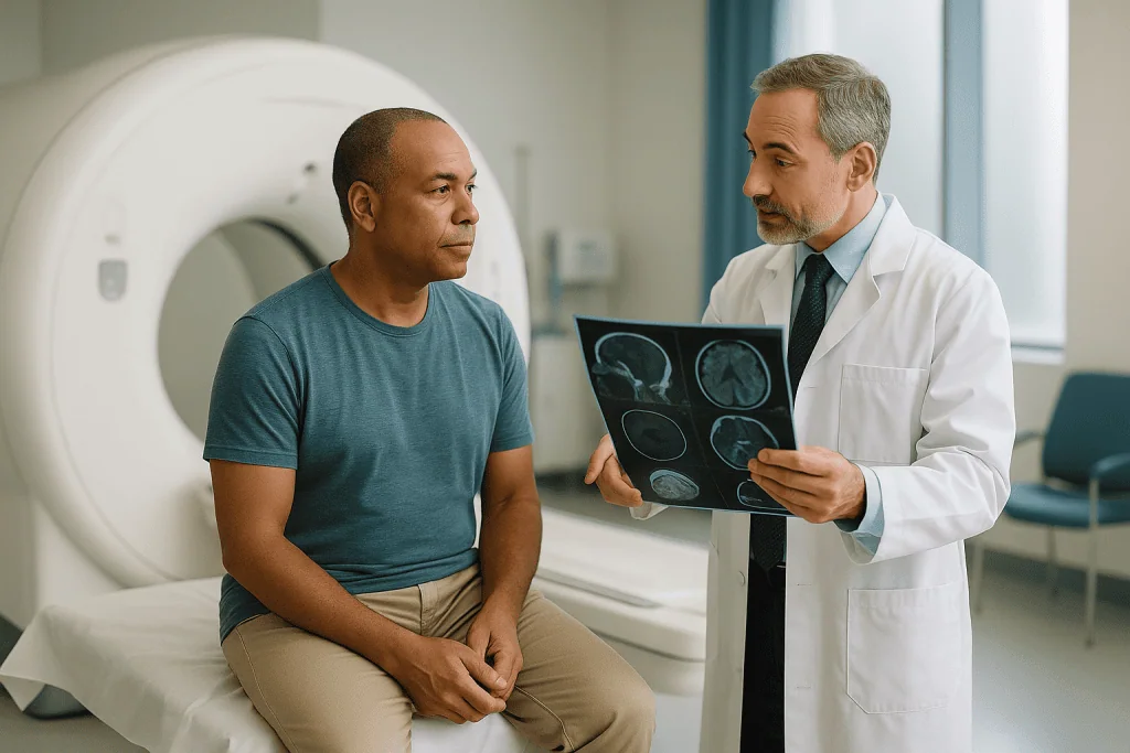
CT has limits, though. It shows bone brilliantly and soft tissue only moderately well. Small tumors, tumors deep in the brainstem, and tumors that blend into surrounding brain can hide on CT. So almost everyone with a suspicious CT, and most people whose symptoms developed slowly enough to skip the emergency room, will be sent for an MRI.
The MRI adds three things. First, resolution: the boundary between tumor and normal brain is far clearer. Second, multiple “sequences,” which are different ways of weighting the signal so that fluid, blood products, swelling and solid tumor each stand out in turn. Third, contrast behavior, which we cover in the next section. An MRI examination takes longer than a CT, anywhere from about 15 minutes to more than an hour depending on how many sequences are needed (Mayo Clinic), and the scanner is loud, so earplugs or headphones are standard.
If you have a pacemaker, cochlear implant, metal fragments, or certain older surgical clips, tell the radiology staff before the scan. Many modern devices are MRI-conditional, meaning they are safe under specific settings, but the team must know in advance. People who find enclosed spaces difficult can ask about a wider-bore scanner or about a mild sedative; that decision rests with the prescribing clinician.
What does a brain MRI with contrast show, and what do advanced sequences add?
Halfway through most tumor MRIs, a technologist injects a contrast agent into a vein. For MRI this is a gadolinium-based compound, not the iodine dye used for CT. Gadolinium shortens the magnetic signal of water nearby, which makes any area it reaches appear bright on certain sequences.
Why does this matter? Healthy brain is protected by the blood-brain barrier, a tightly sealed lining of blood vessels that keeps most substances in the bloodstream out of brain tissue. Many tumors grow leaky, abnormal vessels that let contrast through. So a region that “enhances” after injection is a region where the barrier is broken. Enhancement often, though not always, points to a faster-growing tumor, and it helps the surgeon see the most active part to target for biopsy (NCI PDQ). Some slow-growing tumors do not enhance at all, which is one reason imaging alone cannot settle the diagnosis.
Beyond the standard pictures, radiologists may add sequences that measure function rather than shape:
- Perfusion imaging estimates how much blood is flowing through the tumor, a rough proxy for how aggressively it is growing.
- Diffusion imaging measures how freely water molecules move, which can hint at how densely packed the cells are.
- Magnetic resonance spectroscopy reads the chemical makeup of a small region, distinguishing, for example, markers of rapid cell turnover from those of normal brain.
- Functional MRI maps which areas light up during speech or hand movement, so a surgeon knows where not to cut.
- Diffusion tensor imaging traces the major white-matter pathways that carry signals between regions.
None of these replaces a biopsy. Together they refine the surgical approach, and functional and tractography maps can be loaded into the operating room navigation system so that the plan on screen matches the brain on the table (Mayo Clinic).
Who has a biopsy first, who goes straight to surgery, and who is asked to wait and rescan?
Once the MRI is complete, the team sorts people into three broad paths. The sorting depends on location, the likely tumor type, the person’s overall health, and what the surgery would cost in terms of brain function.
Straight to resection. If the tumor sits in a region that can be reached without damaging speech, movement or vision, and if imaging suggests that removing most of it would help, surgeons often plan to take out as much as is safe in one operation. The removed tissue becomes the diagnostic sample. Here the “biopsy” and the treatment happen on the same day (Mayo Clinic).
Biopsy first. When a tumor lies deep, near the brainstem, in the thalamus, or spread across both hemispheres, a full removal may not be possible or wise. A needle biopsy provides the diagnosis with far less disruption. The same applies when imaging raises the possibility of lymphoma or an infection, conditions treated with medicines rather than surgery, where a large operation would offer little and could delay the right treatment (NCI PDQ).
Watch and rescan. Some findings are so characteristic on MRI, such as a small, calm-looking meningioma arising from the brain’s covering membranes, that teams may recommend observation with repeat imaging rather than immediate tissue sampling, particularly if there are no symptoms (Mayo Clinic). People with serious heart or lung disease, or those taking blood-thinning medicines that cannot be safely paused, may also be asked to wait while those issues are addressed.
Being placed in the waiting group can feel like being ignored. It usually means the opposite: the team judges that the risk of acting now outweighs the risk of looking again in a defined interval. Ask what change on the next scan would prompt a different decision, and when that scan is scheduled.
What happens during a stereotactic brain biopsy?
Stereotactic simply means guided by three-dimensional coordinates. In a stereotactic brain biopsy, the surgeon uses the MRI as a map and a navigation system as the GPS, directing a thin needle to a precise point inside the skull through an opening roughly the diameter of a pencil (Mayo Clinic).
The day typically unfolds like this. A fresh MRI or CT is taken, sometimes with small adhesive markers on the scalp, so that the images are registered to the person’s actual head position. In the older frame-based technique, a lightweight metal frame is attached to the skull under local anesthetic before scanning; in frameless systems, the navigation camera tracks the head without a frame. The procedure may be done under general anesthesia or with sedation and local numbing, a choice the anesthesia team makes based on the individual (NHS).
In the operating room, a small area of scalp is shaved and cleaned. The surgeon makes a short incision, drills a burr hole in the bone, and advances the needle along the planned trajectory, watching its position on screen. Several tiny cores of tissue are withdrawn, usually from more than one depth, because tumors are not uniform and the most informative region may be only millimeters from a less helpful one. A pathologist often examines a preliminary sample within minutes to confirm that the needle has reached abnormal tissue before the surgeon closes.
Risks are real but uncommon: bleeding at the biopsy site, infection, swelling, a seizure, or a new neurological symptom depending on the path the needle takes. There is also the possibility of a non-diagnostic sample, where the tissue obtained does not answer the question and the procedure or an open operation must be considered (NHS). Your surgeon will put these risks into context for your particular tumor location.
Inside the lab: how is a brain tumor pathology report built?
The sample leaves the operating room and begins a second journey most people never see. It splits almost immediately into two tracks.
The frozen section track is fast. A sliver of tissue is frozen solid, cut into slices thinner than a hair, stained and placed under a microscope while the person is still on the table. The pathologist phones the surgeon with a preliminary impression: tumor present, likely category, enough tissue or not. This is a working answer, not the final one. Frozen tissue distorts cells, and the pathologist is deliberately cautious about committing to a precise diagnosis at this stage.
The permanent section track is slow and far more detailed. The remaining tissue is fixed in preservative, embedded in paraffin wax, sliced, and stained with the classic hematoxylin and eosin dyes that color nuclei blue and cytoplasm pink. Under the microscope the pathologist studies cell shape, how closely the cells resemble their normal counterparts, how many are dividing, whether new blood vessels are forming, and whether any areas have died from outgrowing their blood supply. These features feed the histological grade.
Then come special stains called immunohistochemistry, in which antibodies tagged with color bind to specific proteins. One stain might light up if cells carry a particular mutated protein; another counts dividing cells. Finally, portions of tissue go to the molecular laboratory for genetic sequencing and tests of chromosome structure (NCI PDQ).
Each step adds time, and the molecular tests take longest. Turnaround varies by laboratory and by which tests are needed, so the most reliable estimate comes from your own team rather than a general figure. Some centers release a preliminary report and then an addendum with molecular results, which is why a diagnosis can shift slightly in wording between the first conversation and the final version.
How do you read a brain tumor pathology report? Type, grade and markers explained
The final report usually has three layers that together form what the World Health Organization classification calls an integrated diagnosis: the tumor’s cell of origin, its grade, and its molecular signature (NCI PDQ). Reading them in that order makes the document far less intimidating.
Type names the family. Gliomas arise from the brain’s supporting glial cells; meningiomas from the membranes covering the brain; medulloblastomas from the cerebellum, mostly in children; metastases are cancers that began elsewhere and spread. Grade, written as a number from 1 to 4, describes how abnormal the cells look and how quickly the tumor is expected to grow, with 1 the slowest and 4 the fastest (Mayo Clinic). Markers are the genetic and protein findings that refine both.
| Report section | What it describes | Why it shapes the plan |
|---|---|---|
| Histological type | Which brain cell the tumor most resembles | Different families respond to different treatments |
| WHO grade (1–4) | Cell abnormality, division rate, vessel growth, dead tissue | Guides how urgently and how intensively to treat |
| Immunohistochemistry | Proteins detected by antibody stains | Confirms type; some stains reflect key mutations |
| Molecular markers | Specific mutations, gene deletions, methylation | Can reclassify the tumor and predict response to certain medicines |
| Margins or extent | Whether tumor cells reach the edge of resected tissue | Interpreted alongside the post-surgery MRI |
Two cautions. A report may say “consistent with” or “favor” when tissue is limited; that is honest uncertainty, not evasion. And grade alone no longer tells the whole story. The current classification allows some molecular findings to assign a higher grade even when the cells look relatively calm, because those findings predict faster behavior (NCI PDQ). If a sentence in your report does not make sense, ask for it to be read aloud and explained; neuro-oncology teams expect this request.
Why does molecular testing for glioma matter so much?
A generation ago, two gliomas that looked identical under the microscope were given the same name and often the same treatment, yet one person did well for years while another declined within months. Molecular testing explained much of that gap, and it has since moved from research curiosity to a required part of diagnosis for adult gliomas (NCI PDQ).
Three markers come up repeatedly in conversations about adult diffuse gliomas:
- IDH mutation. IDH stands for isocitrate dehydrogenase, an enzyme involved in cell metabolism. A mutation in the IDH1 or IDH2 gene marks a biologically distinct group of gliomas that, as a group, grow more slowly than those without the mutation. Glioblastoma is now defined as IDH-wildtype, meaning the gene is normal; a tumor with the mutation receives a different name even if it looks similar.
- 1p/19q codeletion. This is the loss of a segment of chromosome 1 and a segment of chromosome 19 together. Combined with an IDH mutation, it defines oligodendroglioma, a type that tends to respond to particular chemotherapy regimens.
- MGMT promoter methylation. MGMT is a DNA-repair gene. When chemical tags called methyl groups silence it, tumor cells repair chemotherapy damage less effectively, which helps predict response to alkylating chemotherapy, the class of medicines that damages tumor DNA.
Other tests, such as histone H3 mutations in some childhood and midline tumors, TERT promoter changes, or chromosome 7 gain with chromosome 10 loss, may also appear depending on the tumor and the laboratory.
What this means in practice: your oncologist may hold off finalizing the medicine component of the plan until molecular results return, and a clinical trial you are offered may be open only to people with a specific marker. The results do not change what was removed; they change how the team expects the remaining disease to behave, and what is most likely to help.
Which tests look beyond the head? Spine MRI, lumbar puncture, body scans and blood work
Not every test in a brain tumor workup looks at the brain. Which extras are ordered depends on the suspected type, and understanding the reasoning stops them feeling random.
Spine MRI. Some tumors spread through the cerebrospinal fluid, the clear liquid that bathes the brain and spinal cord. Medulloblastoma, ependymoma, germ cell tumors and primary central nervous system lymphoma are examples. For these, imaging the entire spine is part of staging, the process of defining how far a tumor has spread, because deposits along the cord change the radiation plan (NCI PDQ).
Lumbar puncture. Also called a spinal tap, this draws a small amount of cerebrospinal fluid from the lower back through a thin needle so that a laboratory can look for tumor cells and measure proteins. It is used for the same fluid-spreading tumors and is deliberately delayed if a large mass is causing pressure, because changing fluid dynamics in that situation can be dangerous. Your team decides the timing.
Body imaging. When the MRI pattern suggests a metastasis, a cancer that began elsewhere, a CT of the chest, abdomen and pelvis or a PET scan is ordered to find the primary site. Sometimes the body scan identifies a more accessible place to biopsy than the brain itself.
Blood tests. Routine panels check clotting, kidney function (relevant to contrast agents) and general fitness for anesthesia. Specific blood markers matter for specific tumors: alpha-fetoprotein and beta-hCG are raised in some germ cell tumors, and a full set of hormone levels is standard when a mass sits in or near the pituitary gland, which regulates many of the body’s hormones (Mayo Clinic).
Hearing and vision assessments round out the picture for tumors near the auditory nerve or optic pathways, giving a baseline against which the team can measure the effect of treatment.
How does a tumor board turn test results into one treatment plan?
Most hospitals that treat brain tumors hold a regular multidisciplinary meeting, often called a tumor board. Around the table, physically or on screen, sit the neurosurgeon, a neuroradiologist who specializes in brain imaging, the neuropathologist, radiation and medical oncologists, and frequently a specialist nurse, a neurologist and a rehabilitation physician. Each person brings one piece of your workup (NHS).
The radiologist projects the MRI and walks through size, location, enhancement, swelling and relationship to critical structures, pointing out anything the advanced sequences added. The pathologist presents the integrated diagnosis, including which molecular results are back and which are pending. The surgeon describes what was removed and what the post-operative scan shows. Then the group debates: observe, operate again, radiation alone, radiation with chemotherapy, a clinical trial, or supportive care first while the person recovers strength.
The point of this ritual is to prevent one specialty’s instinct from dominating. A tumor that looks surgically tempting may, on molecular grounds, be better served by a smaller operation followed by medicines. A tumor that looks grim on a scan may carry markers that justify a more hopeful plan. The board also checks the plan against national guidelines and current trial availability.
What you receive afterward is a recommendation, not an order. Your own priorities, work, family, how much function you are willing to risk for a larger resection, belong in the conversation, and a good team will ask about them. If a recommendation does not sit right, you can ask for the reasoning behind each alternative that was considered. Seeking a second opinion from another multidisciplinary team is routine in neuro-oncology, and reports and images can be shared electronically; the treating team remains the group that carries out and adjusts the plan.
What do the days and weeks after a brain tumor biopsy usually look like?
After a needle biopsy, most people wake in a recovery area with a small dressing on the scalp and a headache that is managed with routine pain relief chosen by the anesthesia team. Nurses perform neurological checks at regular intervals through the first night, asking you to squeeze their hands, name objects and follow a light with your eyes, because the main early concern is bleeding at the biopsy site, which usually declares itself within hours (NHS). A CT scan may be done before discharge to confirm no bleeding has occurred.
Many people are home within a day or two of a needle biopsy; recovery after a larger resection takes longer and often includes a few days on a neurosurgical ward, where physiotherapists and speech therapists assess function early. Ask your own team for their typical timings rather than relying on general figures.
Corticosteroids, a class of anti-inflammatory medicines, are commonly prescribed around brain surgery to reduce swelling; they can cause restlessness, appetite changes and raised blood sugar, and they are tapered gradually by the prescribing clinician rather than stopped abruptly. Antiseizure medicines may be continued if a seizure was the presenting symptom. Never adjust either on your own.
The hardest part of this phase for most people is not physical. It is the wait for the final pathology report and molecular results, during which the plan feels suspended. Use that window to arrange practical support, ask about driving restrictions (which exist in most jurisdictions after a seizure or brain surgery), and request a named contact for questions.
Follow-up includes a wound check, a results appointment where the integrated diagnosis is explained, and a baseline MRI after surgery that becomes the reference point for every future scan. Hair regrows over the shaved area; the small scar from a burr hole usually fades into the scalp line.
What people often get wrong about brain tumor MRI and biopsy
The myths in this area are not foolish. Most grow from a reasonable idea stretched one step too far.
“The MRI already showed what it is, so the biopsy is unnecessary.” Imaging gives a probability, not a diagnosis. Infections, inflammatory lesions, lymphoma and several glioma types can mimic one another on a scan, and two tumors with identical pictures can carry different molecular markers that demand different treatment (NCI PDQ). Tissue is the exception only when imaging is truly characteristic or when sampling would be unsafe.
“Cutting into a tumor makes it spread.” This fear is widespread and understandable. Brain tumors do not behave like a spilled liquid; needle tract seeding is exceptionally rare for the common tumor types, and the information gained directs treatment that would otherwise be guesswork (Mayo Clinic).
“If the frozen section said one thing, the final report cannot change it.” The frozen section is a rapid first look at distorted tissue. The final report, especially after molecular testing, can refine or occasionally revise the name. That is the process working, not a mistake.
“A low grade means nothing needs to be done.” Lower-grade tumors can still cause seizures and may change over time; the classification now allows molecular findings to raise the expected behavior of a calm-looking tumor (NCI PDQ). Observation, when chosen, is active, with scheduled scans.
“Contrast dye is toxic and should be refused.” Gadolinium agents have a long safety record; your team screens kidney function and uses the lowest effective amount. Tiny deposits have been detected in the brain after repeated doses without evidence of harm, and regulators continue to monitor the question (Mayo Clinic). Discuss concerns rather than declining a test that changes surgical planning.
“More tests mean the team is worried.” Usually they mean the team is thorough. A spine MRI or lumbar puncture is standard staging for certain tumor types regardless of how things look.
Questions to ask your care team
Consultations after a brain tumor diagnosis are dense, and most people remember a fraction of what was said. Bring someone to take notes, and bring questions in writing. The following are the ones neuro-oncology nurses say they wish more people asked.
- What does my MRI show, and can you point to it on the images with me?
- Which parts of the brain is the tumor near, and what functions do those parts control?
- Are you recommending a biopsy, a resection, or observation, and what tipped the decision?
- If surgery: how much do you expect to remove, and what are the specific risks for this location?
- Which of the advanced MRI sequences, if any, were used to plan the approach?
- When will the preliminary pathology be available, and when will the molecular results follow?
- Which molecular tests are being run, and how might each result change the plan?
- Will my case be discussed at a multidisciplinary tumor board, and will I receive a summary?
- Are there clinical trials for which my tumor type and markers might qualify?
- Do I need a spine MRI, lumbar puncture or body scan, and why or why not?
- Which medicines will I be taking around surgery, what are they for, and who adjusts them?
- What restrictions apply to driving, work and exercise, and for how long?
- Who is my named contact for questions between appointments, and how do I reach them out of hours?
- Can I have copies of my imaging and pathology reports, and will they be shared if I seek a second opinion?
You are entitled to ask the same question twice, and to ask for an answer in plainer words. If a term in your report has not been explained, ask for it to be defined in one sentence. Teams that treat brain tumors regularly field these questions every day, and a thoughtful question usually improves the plan rather than slowing it.
Keep a single folder, paper or digital, with every report, scan disc and appointment letter. In neuro-oncology, the comparison between today’s scan and the first one is often the most important piece of information in the room.
When to call your doctor
Between tests, after a biopsy, and during the long stretch of waiting for results, there are symptoms that should prompt a same-day call to your neurosurgical team, and a smaller set that call for emergency services without waiting.
Call emergency services immediately if someone has a seizure lasting more than a few minutes or repeated seizures without recovering in between, a sudden severe headache described as the worst ever, sudden weakness or numbness on one side of the body, sudden difficulty speaking or understanding speech, a sudden loss of vision, or a rapid decline into drowsiness or unresponsiveness. These can signal bleeding or dangerous pressure inside the skull and are treated as emergencies regardless of a known tumor (Mayo Clinic; NHS).
Call your care team the same day for a headache that is new or steadily worsening, especially one that wakes you from sleep or is worse on lying down or coughing; vomiting without an obvious cause; new or worsening confusion or personality change; a first seizure of any kind; progressive clumsiness or balance problems; or new double vision.
After a biopsy or surgery, also report fever, increasing redness, swelling or discharge at the wound, clear fluid leaking from the incision or the nose, a stiff neck with light sensitivity, or any new neurological symptom that was not present when you left hospital (NHS). If you are taking corticosteroids and develop marked thirst, frequent urination or very high readings on a home glucose monitor, let the prescribing clinician know; raised blood sugar is a recognized effect that the team can manage.
None of these signs tells you what is happening on its own, and most calls turn out to be reassurance. The point is that the people who know your scan and your surgery are the ones best placed to judge. Keep the out-of-hours number where everyone in the household can find it.
Frequently asked questions
Can a brain tumor be diagnosed from an MRI alone?
Sometimes, but not usually. Certain tumors with a highly characteristic appearance, such as some small meningiomas, may be diagnosed and monitored on imaging alone. For most suspected brain tumors, guidelines call for tissue examined by a pathologist, because different tumors can look identical on a scan and molecular results that affect treatment can only come from a sample.
What does a brain MRI with contrast show that a plain MRI does not?
It shows where the blood-brain barrier is leaking. Gadolinium contrast normally stays inside blood vessels; where tumor vessels are abnormal, it seeps into tissue and appears bright. That enhancement often marks the most active part of a tumor, helps separate tumor from surrounding swelling, and gives the surgeon a target for biopsy. Some slow-growing tumors do not enhance at all.
Is a stereotactic brain biopsy done awake or asleep?
Either, depending on the person and the center. Some stereotactic biopsies are performed under general anesthesia; others use sedation with local numbing of the scalp, since the brain itself has no pain receptors. The anesthesia team makes that choice based on your health, the tumor location and the planned technique, and will explain what you can expect to feel.
How long does a brain tumor pathology report take?
It varies by laboratory and by which tests are required, so your own team’s estimate is the reliable one. A frozen section gives a preliminary impression during surgery within minutes. The final report with routine stains follows later, and molecular testing adds further time, which is why some centers release a preliminary report and then an addendum once genetic results return.
Why is molecular testing for glioma needed if the pathologist already saw the cells?
Because tumors that look alike under the microscope can behave very differently. Markers such as IDH mutation, 1p/19q codeletion and MGMT promoter methylation define distinct tumor types, help predict how the tumor is likely to grow, and in some cases predict response to specific medicine classes. Current classification requires these results for an integrated diagnosis of adult diffuse gliomas.
Does a brain biopsy cause the tumor to spread?
Spread along a needle track is exceptionally rare for the common types of brain tumor, and it is weighed against the value of a precise diagnosis. Without tissue, treatment would be based on probability alone. Surgeons plan the trajectory to avoid critical structures and sample the most informative region, and the information gained directs the rest of the plan.
What is the difference between a frozen section and the final pathology result?
A frozen section is a rapid look at tissue frozen and sliced during surgery, used to confirm that abnormal tissue was obtained and to give a working category. Freezing distorts cells, so the pathologist stays cautious. The final result uses properly fixed tissue, special antibody stains and molecular tests, and it can refine or occasionally change the preliminary name.
Why would I need a spine MRI or lumbar puncture for a brain tumor?
Some tumors spread through cerebrospinal fluid rather than through blood, including medulloblastoma, ependymoma, germ cell tumors and primary central nervous system lymphoma. For these, imaging the spine and examining the fluid are standard staging steps that shape radiation planning. A lumbar puncture is delayed if a mass is causing pressure, because removing fluid in that situation can be unsafe.
What does the grade on a brain tumor pathology report mean?
Grade, from 1 to 4, describes how abnormal the cells appear and how quickly the tumor is expected to grow, with 1 slowest and 4 fastest. It is based on features such as cell division rate, new blood vessel growth and dead tissue. Under current classification, certain molecular findings can raise the grade even when cells look relatively calm.
Who decides my treatment plan after the MRI and biopsy results are in?
A multidisciplinary team, often called a tumor board, reviews the imaging, pathology and molecular results together and produces a recommendation. That recommendation is then discussed with you, and your priorities are part of the decision. You can ask for the reasoning behind each alternative, and seeking a second opinion is routine; the treating team carries out and adjusts the final plan.
References
This article is for general information only and is not a substitute for professional medical advice. Please consult a qualified doctor about your individual situation.

More from the Blog

Living Well With Epilepsy: Sleep, Driving Rules, Work and Keeping a Seizure Diary
Most people with epilepsy live full lives, but three areas need planning. Driving is usually paused after any seizure until a legally set seizure-free…

Living With Essential Tremor: Eating, Writing and Caffeine Habits Your Neurologist May Discuss
Living with essential tremor usually means managing a long-term, non-life-threatening shaking of the hands, head or voice rather than treating a disease that shortens…

When Does a Traumatic Brain Injury Need Surgery? How Pressure and Imaging Guide the Decision
A traumatic brain injury usually needs surgery when bleeding or swelling raises pressure inside the rigid skull enough to threaten brain tissue. Surgeons weigh…

Myasthenia Gravis Follow-Up: Which Breathing or Swallowing Changes Need Urgent Care
In myasthenia gravis, seek urgent care the same day if you notice new or worsening shortness of breath, trouble lying flat, a weak cough,…

Can Sleep, Hydration and Regular Meals Change a Migraine Treatment Plan’s Results?
Sleep, hydration and regular meals do not replace migraine treatment, but they can change how well a plan appears to work. Irregular sleep, dehydration…
Orthotics, Walkers and Standing Frames in Cerebral Palsy: How Equipment Supports Movement
Orthotics and equipment for cerebral palsy do not change the brain injury itself; they change what the body can do with the movement it…