

Why a Dental Assessment Comes Before Head Cancer Radiotherapy and What It Involves
Key Takeaways
- Extracting a doubtful tooth before radiotherapy is routine dentistry; extracting the same tooth from irradiated jawbone afterward carries a lifelong risk of osteoradionecrosis, bone that fails to heal.
- NIH guidance advises a dental examination before head and neck radiation, with extractions completed early enough for sockets to close, typically a matter of weeks set by the oncologist.
- Radiation to the major salivary glands can reduce saliva within the first weeks of treatment, and the resulting dryness drives fast, gumline decay known as radiation caries.
- Custom fluoride trays are made from impressions at the assessment visit because daily fluoride is one of the few measures with consistent support for protecting enamel in a dry mouth.
- Mouth soreness usually begins around the second or third week of treatment and settles over the weeks after it ends, while saliva recovers slowly or, for some, only partly.
- The irradiated jaw never regains its full healing capacity, so every future dentist needs a written record of which area was treated before any extraction or implant is considered.
A dental assessment before head and neck radiotherapy is a specialist check of the teeth, gums and jaw carried out before treatment starts. Radiation reduces saliva and the blood supply to the jawbone, so decay accelerates and extractions afterward can trigger poor bone healing known as osteoradionecrosis. Removing at-risk teeth first, allowing healing time, and setting up fluoride and hygiene plans lowers those risks. The radiotherapy team decides the timing.
The letter lists five appointments in eleven days: a planning scan, a mask fitting, a meeting with the dietitian, a speech therapist, and, oddly, a dentist. Most people reading that letter have just been told they have a cancer in the mouth, throat or salivary glands. The dentist feels like the one appointment they could skip.
It is the one they should not. A dental assessment before head cancer radiotherapy is not a courtesy check-up bolted onto a serious treatment. It is part of the treatment plan, and the order matters. Work done on teeth in the weeks before the first radiation session can be routine; the same work done a year afterward can turn into a slow, painful problem with the jawbone that some people carry for the rest of their lives.
This article explains why the dental visit comes first, what the team is looking for, how the timing is worked out, and what the mouth usually goes through in the weeks that follow.
Why does a dental assessment come before head cancer radiotherapy?
Radiotherapy for cancers of the mouth, throat, voice box, nose and salivary glands cannot avoid healthy tissue entirely. The beams are shaped with remarkable precision, but the teeth, jawbone and salivary glands sit within millimeters of most of these tumors, so they receive part of the dose whether anyone wishes it or not. The National Institutes of Health describes the result plainly: radiation to the head and neck can permanently reduce saliva, thin the lining of the mouth, weaken tooth enamel and reduce the jawbone’s ability to heal.
That last effect is the heart of the matter. Bone that has been irradiated has fewer working blood vessels and fewer bone-building cells. Pull a tooth from that bone and the socket may simply fail to close. The medical term is osteoradionecrosis, which means bone in an irradiated area that has died and will not repair itself. Once it develops, it is difficult to manage and can require surgery.
Timing therefore does the heavy lifting. A tooth that will clearly cause trouble later is far safer to remove while the bone is still healthy and can heal in the normal way. Guidance from the National Cancer Institute (NCI) recommends that people due to have radiation to the head and neck see a dentist before treatment begins, ideally with enough lead time for any dental work to heal first.
There is a second, quieter reason. Radiotherapy reshapes the mouth’s chemistry for years. Saliva that used to rinse sugars away, buffer acids and deliver minerals back into enamel becomes scarce and thick. Teeth that were coping fine begin to decay quickly, often at the gumline and along the edges of fillings. The pre-treatment visit is the moment to set up the defenses, fluoride, hygiene coaching and a follow-up schedule, before the damage starts rather than after.
What actually happens at a dental assessment before head cancer radiotherapy?
Expect a longer appointment than a routine check-up, usually with a dentist who works closely with the cancer team. Some centers call this a pre-radiotherapy dental clearance; the word clearance simply means the dentist confirms the mouth is ready, or lists what must happen first.
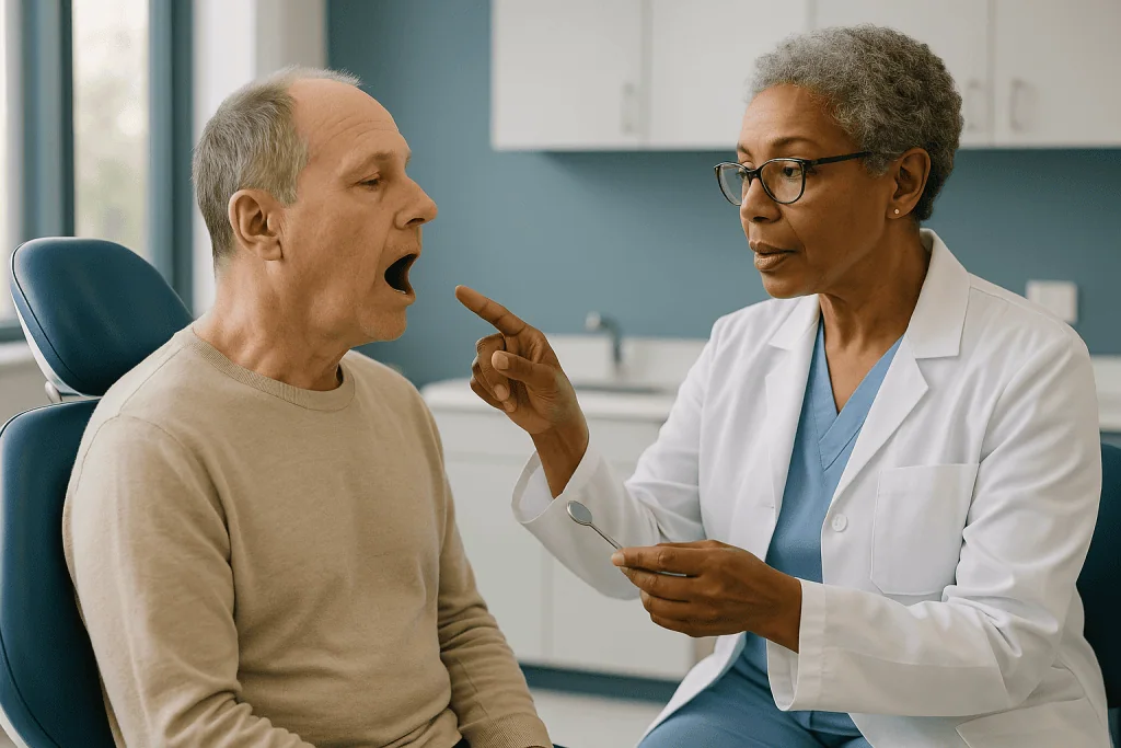
The visit typically begins with a conversation. The dentist will want to know where the tumor is, which side of the jaw and which salivary glands will receive the most radiation, whether chemotherapy or surgery is also planned, and how you have managed your teeth so far. Those answers shape everything that follows, because a tooth near the beam path is judged more harshly than one well outside it.
Then comes the examination itself. A full-mouth set of X-rays, often including a panoramic film that shows both jaws in one image, allows the dentist to look for decay hidden between teeth, infection at the root tips, impacted wisdom teeth, cysts and bone loss from gum disease. Each tooth is checked for mobility and pocket depth, the small measurement of how far the gum has pulled away from the root. Existing crowns, bridges and dentures are inspected for fit and for sharp edges that could rub an increasingly fragile mouth lining.
The dentist may also take impressions, molds of the teeth, to make custom soft trays for fluoride gel and, in some plans, a positioning device the radiotherapy team uses to hold the tongue or jaw steady during treatment. Cleaning of tartar and polishing is often done the same day.
At the end you should leave with a written plan: which teeth stay, which go, what work will be done and by when, and how oral care will change during treatment. That plan goes back to the radiation oncologist, who fits the dental dates into the overall schedule.
How does radiation change the mouth, saliva and jaw?
It helps to picture the mouth as a system that depends on flow. Roughly a liter of saliva moves through it every day in healthy adults, rinsing food debris away, neutralizing acids produced by bacteria, and carrying calcium and phosphate back into enamel that acids have softened. Radiation to the parotid and submandibular glands, the large glands in front of the ears and under the jaw, damages the cells that make saliva. According to the NCI, changes in saliva can start within the first week or two of treatment, and some reduction is often permanent when the major glands lie in the treatment field.
Without that flow, the mouth becomes acidic and sticky. Bacteria that cause decay thrive, while the protective species that prefer a neutral environment fall back. The lining of the cheeks and tongue, which normally renews itself every few days, is hit by radiation while it is dividing, so it thins and ulcerates. The medical term is mucositis, meaning inflammation and soreness of the mouth’s lining.
Bone is slower to show change but changes more permanently. Radiation damages the fine blood vessels that supply the jaw, particularly the lower jaw, which has a less generous blood supply than the upper jaw to begin with. Over months the bone becomes what specialists call hypovascular and hypocellular: fewer vessels, fewer living cells. It can carry normal loads and looks unremarkable on an X-ray, yet it has lost much of its ability to respond to injury.
Muscles matter too. The chewing muscles and the joint just in front of the ear can stiffen and scar, a condition called trismus, which means a reduced ability to open the mouth. Trismus makes brushing and dental treatment harder, which is another reason the team wants the mouth in good order before it develops.
What is osteoradionecrosis of the jaw, and why does it drive the timing?
Osteoradionecrosis, often shortened to ORN, is an area of jawbone that has been exposed to radiation and then failed to heal after an injury, most commonly a tooth extraction, though ill-fitting dentures, deep gum infection and occasionally no obvious trigger at all can set it off. The bone becomes exposed through the gum, may feel rough to the tongue, and can ache, discharge or, in advanced cases, fracture.

What makes ORN different from an ordinary infection is that it does not resolve with time. Healthy bone remodels itself constantly; irradiated bone has lost much of that capacity. Management ranges from careful cleaning and antibiotics for small, stable areas to surgical removal of dead bone and reconstruction for larger ones. The NCI notes that the risk persists for the rest of a person’s life, which is why the phrase “irradiated jaw” should follow you to every future dentist.
Two facts about ORN shape the pre-treatment dental plan. First, extractions are the commonest trigger, so removing doubtful teeth before radiation converts a high-risk future procedure into a low-risk present one. Second, healing takes time and healing is what protects the bone. A socket that has closed over and started to fill with new bone before the first fraction of radiation is delivered is in a far stronger position than one that is still open.
People sometimes ask whether the risk is worth worrying about if the tooth feels fine now. The honest answer is that a tooth with deep decay, a failing root filling or advanced gum disease is unlikely to stay fine in a mouth that has lost most of its saliva. The dental team is not predicting the present; it is predicting the mouth as it will be two or three years after treatment, and choosing the safer moment to act.
Which teeth are usually removed before radiotherapy?
No two plans look alike, but dentists working with radiation oncologists tend to apply the same logic. A tooth is more likely to be recommended for removal when it has poor long-term prospects and sits in bone that will receive a high radiation dose. A tooth in the same condition well outside the treatment field may be treated and kept.
Teeth that most often come out include those with decay too deep to restore reliably, those with infection at the root tip that has not responded to root canal treatment, and teeth loosened by advanced gum disease, where the bone that should hold them has already been lost. Partially erupted wisdom teeth are a frequent candidate because the flap of gum over them traps food and bacteria and is hard to keep clean once the mouth is dry and sore. Root fragments and teeth with large cracks are also usually removed.
The threshold is stricter than in everyday dentistry, and this catches people off guard. A tooth that a dentist might watch for a few years in an otherwise healthy patient is judged against a very different future here. The NIH’s dental research institute frames the aim as eliminating sources of infection and irritation before treatment, because problems that arise afterward are harder and riskier to fix.
What usually stays: sound teeth, teeth with small fillings that can be repaired, and teeth with mild, stable gum disease that the person can keep clean. Where possible the team preserves teeth that anchor a denture or help with chewing, since nutrition during treatment is a real concern. The decision is a discussion, not a decree; you are entitled to ask why each tooth is on the list and what would happen if it were kept.
Who is asked to wait, and who is cleared to go straight ahead?
Not everyone needs dental work before radiotherapy begins. Someone with a well-maintained mouth, no active decay and healthy gums may be cleared at the first visit and simply given fluoride trays, hygiene instructions and a follow-up schedule. For them the assessment adds an appointment but no delay.
A short pause is more likely when several extractions are needed, when gum disease is extensive, or when a tooth requires a root filling that must be completed and checked. The radiotherapy team weighs that pause against the behavior of the cancer. Fast-growing tumors and those already causing pain, bleeding or airway concerns may not allow much waiting, and in those cases the team may accept a shorter healing window, limit extractions to the teeth in the highest-dose area, or plan to manage lower-risk teeth conservatively after treatment.
Some situations change the calculation in the other direction. People who have already had surgery to remove the tumor sometimes have extractions performed during that same operation, under the same anesthetic, so the healing clock for both starts together. People whose radiotherapy field will spare the jaw and major salivary glands, for example some cancers of the voice box or thyroid, may need a lighter assessment because the jawbone is not in the beam path. And people who have no natural teeth are not exempt: dentures are checked for pressure points, since a sore spot on irradiated gum can become an entry point for bone exposure.
The pattern across guidance from the NIH and the NHS is consistent. Almost everyone due to receive radiation to the head and neck should be assessed; a smaller group needs treatment first; the timing of that treatment is a joint decision between dentist and oncologist, made against the urgency of the cancer itself.
How long before radiotherapy should extractions be done?
The short answer is: long enough for the gum to close over the socket, and ideally for early bone repair to begin. The NCI’s patient guidance advises seeing a dentist before radiation begins and completing dental work with time to heal, and many teams aim to finish extractions two to three weeks before the first session, with some preferring closer to a month when the cancer allows. Those are typical ranges drawn from published guidance, not a promise; your own window is set by your radiation oncologist.
Why not simply wait as long as possible? Because the tumor is also on a clock. Delaying curative-intent radiotherapy by several weeks is not a neutral decision, and teams are reluctant to extend the wait for dental reasons alone. The usual compromise is to book the dental appointment as early as possible in the diagnostic pathway, often in the same week as the planning scan, so that any extractions happen while the rest of the preparation is underway. The dental work then runs in parallel rather than in series.
A few practical things help the window work in your favor. Extractions are often done with a technique that smooths sharp bone edges and closes the gum snugly, which speeds healing. Smoking slows wound healing, and both the NHS and the NCI encourage stopping before treatment for reasons that go well beyond the mouth. Careful, gentle cleaning around the healing site with the mouth rinses the team suggests keeps bacteria down without disturbing the clot.
If the dentist finds problems late, days before radiotherapy is due to start, the plan changes rather than the start date. The team may remove only the most urgent teeth, defer the rest with a tight monitoring plan, or, rarely, adjust the treatment field. That is a conversation to have openly with both clinicians in the room, or at least in the same email chain.
The pre-treatment dental timeline at a glance
The sequence below reflects a typical pathway described in NIH and NHS patient guidance. Every center arranges it a little differently, and the urgency of the cancer can compress the early stages considerably.
| Stage | Typical timing | What usually happens | Why it matters |
|---|---|---|---|
| Referral to dental team | As soon as radiotherapy is planned | Appointment booked alongside planning scan and mask fitting | Gives the longest possible healing window |
| Assessment visit | Several weeks before first session | History, examination, X-rays, cleaning, impressions for trays | Identifies teeth at risk and sets up preventive care |
| Dental treatment | Immediately after assessment | Extractions, fillings, smoothing of sharp edges, denture adjustment | Removes future infection sources while bone can still heal |
| Healing window | Roughly two to four weeks, set by the oncologist | Sockets close; fluoride and hygiene routine begins | Closed gum before radiation lowers osteoradionecrosis risk |
| Radiotherapy course | Commonly six to seven weeks of daily sessions | Fluoride trays continue; soreness managed with the team | Protects enamel as saliva falls |
| Early recovery | Weeks after the last session | Mouth lining heals; dental review scheduled | Catches early decay and denture problems |
| Long term | Lifelong | Frequent dental checks; extractions only with specialist input | Jawbone remains at risk indefinitely |
Two points in this table deserve emphasis. The healing window is the part most often squeezed, and it is the part the whole assessment exists to protect. The long-term row is the one most often forgotten: the NCI is explicit that the risk to irradiated bone does not fade with the years, so the phrase “I had radiotherapy to my jaw” belongs at the top of every future dental record.
What are fluoride trays for radiotherapy, and do I really need them?
Fluoride trays are thin, soft, custom-made mouthguards that hold fluoride gel against the teeth for a few minutes each day. Fluoride works by being taken up into the outer layer of enamel, where it makes the mineral more resistant to acid and helps softened areas re-harden. In a mouth with normal saliva, toothpaste and diet usually supply enough. In a mouth where saliva has fallen sharply, the NIH’s dental research institute and the NCI both describe daily fluoride, whether as a prescription-strength toothpaste, a gel in trays or a rinse, as a central part of preventing the rapid decay that follows head and neck radiation.
That decay has a distinctive pattern. It attacks the necks of the teeth at the gumline, wraps around the tooth, and can weaken it enough to snap off within months. Dentists call it radiation caries. It develops in teeth that were previously sound, and it moves faster than ordinary decay because none of the mouth’s normal repair chemistry is working.
The trays are made from impressions taken at the assessment visit and are usually ready before treatment starts. The routine the dental team gives you, how long to wear them and which product to use, is a prescription decision that sits with them, not something to change on your own. What matters from the patient’s side is consistency: fluoride only protects the enamel it touches, and it needs to touch it every day, including the days when the mouth is sore and the last thing you want is a tray in it. Many people find it easier to use the trays at a fixed time, often at night after brushing, so the fluoride sits undisturbed.
Are they truly necessary for everyone? People whose radiotherapy spares the major salivary glands may need less intensive protection. For those whose saliva will fall, the evidence summarized by the NIH favors treating fluoride as non-negotiable, in the same category as the mask you wear on the treatment couch.
What do the mouth and jaw usually feel like during and just after treatment?
The first week of radiotherapy is often uneventful for the mouth. Then, according to the NCI, soreness of the mouth lining typically begins around the second or third week, builds through the course, and peaks in the final weeks. Taste often flattens or turns metallic at about the same time. Saliva thickens and then dwindles, and many people describe waking at night with a mouth that feels lined with paper.
Mucositis is the side effect people find hardest. Ulcers can make brushing painful, and this is exactly when the teeth need cleaning most, because bacteria are flourishing in the dry environment. The NHS advises a soft-bristled brush, gentle technique and the rinses your team recommends, and cautions against alcohol-based mouthwashes and against products that sting. Dental teams often teach a modified routine before treatment starts so that it is already a habit when the mouth becomes sore.
Jaw stiffness can begin during treatment or months later. Physiotherapy-style stretching exercises are often introduced early, before the muscles tighten, on the reasoning that it is easier to keep an opening than to regain one. A speech and language therapist or the dental team usually shows you how.
In the weeks after the last session the mouth lining recovers, generally within a few weeks according to Mayo Clinic guidance on treatment-related mouth sores, and taste often begins to return over the following months. Saliva is the slow one. Some people see gradual improvement over a year or more; where the major glands received a high dose, dryness may be permanent, which is why long-term dental protection is planned from the outset rather than reconsidered later.
Nothing in this timeline is a guarantee in either direction. It describes the common pattern reported in mainstream guidance, and your own team will tell you what to expect for your particular treatment field.
What does dental care after head and neck radiotherapy involve?
After radiotherapy, dentistry changes in two directions at once. Preventive care becomes more intensive: the NCI recommends more frequent dental visits than the usual routine, daily fluoride for as long as dryness persists, careful attention to diet, and prompt repair of small areas of decay before they spread. Invasive care becomes more cautious: extractions, implants and deep gum surgery in irradiated bone are approached only after a specialist has weighed the risk of osteoradionecrosis and, where possible, avoided the procedure altogether by saving the tooth.
This is the point at which the pre-treatment assessment pays off. A mouth cleared of doubtful teeth before radiation has fewer emergencies afterward. When a tooth does fail, the options are root canal treatment to remove the infection while leaving the root in the bone, smoothing a broken tooth to gum level rather than pulling it, or a carefully planned extraction with additional precautions. Hyperbaric oxygen therapy, breathing pure oxygen in a pressurized chamber to encourage new blood vessel growth in bone, is sometimes used around extractions in irradiated jaws; the NCI describes the evidence as mixed, and it is not offered universally.
Dry mouth is managed rather than fixed. Mayo Clinic guidance lists frequent sips of water, sugar-free gum or lozenges to stimulate any remaining gland function, humidified air at night and saliva-substitute gels or sprays as the mainstays. Medicines that stimulate salivary flow exist and act on receptors in the glands; whether one is appropriate is a decision for the treating team, weighed against other conditions and medicines.
Dentures need rechecking, because gum tissue shrinks and any pressure sore on irradiated bone is a risk. Diet advice from the dietitian and the dentist sometimes pulls in different directions, with high-energy foods often being sugary, and it is worth asking both to agree on a plan. Above all, every future dentist needs to know which part of the jaw was irradiated, so a written note from the radiotherapy team is worth keeping.
What people often get wrong about the dental check
The first misunderstanding is that the dental visit is optional or cosmetic. It is neither. Guidance from the NIH, the NCI and the NHS treats it as part of preparing for radiotherapy, in the same category as the planning scan.
The second is that healthy-feeling teeth are safe teeth. Radiation does not care how a tooth feels today; it changes the conditions the tooth will live in for years. A tooth with a deep filling and no symptoms can decay under that filling within a year of losing its saliva bath. The dentist is assessing prognosis, not current pain.
The third is that extractions are being done to save time or money. They are done because an extraction from irradiated bone can lead to osteoradionecrosis, and an extraction from healthy bone rarely does. The recommendation to remove a tooth before treatment is a risk trade, and you can ask to see the reasoning for each one.
The fourth is that once treatment ends, ordinary dentistry resumes. The jaw does not recover its full healing capacity, and the NCI is explicit that the risk is lifelong. Any dentist who does not know your history needs to be told before touching an irradiated area.
The fifth is that fluoride is only for children. In a dry mouth, daily high-strength fluoride is one of the few measures with consistent support in mainstream guidance for preventing radiation caries.
The sixth is that a very dry mouth means something has gone wrong. Dryness is an expected consequence of treating tumors near the salivary glands, not a sign of error. The realistic goal is protecting the teeth and keeping the mouth comfortable while the glands recover whatever function they can.
The last is the belief that asking for a delay to finish dental work is a failure of nerve. Raising the question is reasonable; deciding the answer is the oncologist’s job, made with the dentist and with the behavior of the cancer in view.
Questions to ask your care team
Good questions do two things: they help you understand the plan, and they make sure the dentist and the radiation oncologist are working from the same information. The list below is a starting point; bring it, and add your own.
- Which parts of my jaw and which salivary glands will receive the most radiation, and which teeth sit in that area?
- For each tooth you recommend removing, what is the specific concern, and what would happen if we kept it?
- How much healing time does the radiation oncologist want between extractions and the first session, and what happens if healing runs late?
- Will any dental work be done during my cancer surgery, if I am having one?
- What fluoride routine should I follow, when should it start, and how long should it continue?
- How should I brush and rinse once my mouth becomes sore, and which products should I avoid?
- Do I need jaw-stretching exercises, and who will teach me?
- How often should I see a dentist after treatment, and should it be a specialist or my usual dentist?
- If I ever need a tooth removed in the future, who should do it and what precautions will be needed?
- Can I have a written summary of my radiotherapy field to give to any future dentist?
- Are my dentures likely to fit differently after treatment, and who will adjust them?
- Who do I contact if I have jaw pain, a loose tooth or exposed bone months or years from now?
Write the answers down or ask for them in writing. People often report that the days around diagnosis blur, and the dental plan is one of the parts most easily lost. A single page listing the teeth to be removed, the dates, the fluoride routine and the follow-up interval is worth more than any amount of reassurance in the moment.
When to call your doctor
Most mouth problems during radiotherapy are expected and are handled at the regular review visits built into the treatment schedule. A few signs should prompt a same-day call to the radiotherapy team or your dentist rather than waiting for the next appointment.
Call promptly if a tooth extraction site is bleeding heavily or has reopened, if pain around a healing socket is worsening after the first few days rather than easing, or if you develop a fever or swelling of the face or neck. During treatment, seek help if mouth soreness is stopping you from drinking enough, if you notice white patches that may indicate a fungal infection, or if pain is not controlled by the measures your team has given you. After treatment, and at any point in the years that follow, contact the team if you can see or feel exposed bone through the gum, if a tooth becomes loose or breaks, if there is a persistent bad taste or discharge from one area, if your jaw becomes difficult to open, or if you have numbness of the lip or chin that was not there before. These can be early signs of osteoradionecrosis or infection, and both are easier to manage when caught early.
Seek emergency care immediately for difficulty breathing or swallowing saliva, bleeding that will not stop with firm pressure, or rapidly spreading swelling of the face, neck or floor of the mouth.
Every decision about dental treatment before, during and after radiotherapy sits with the team treating you. This article describes what mainstream guidance says usually happens; your own plan is written for your tumor, your mouth and your circumstances, and the people to change it are the clinicians who know all three.
Frequently asked questions
Why do I need to see a dentist before radiotherapy if my teeth feel fine?
Because radiation changes the conditions your teeth will live in, not how they feel today. Saliva falls, acid rises and decay accelerates, so a tooth with a deep filling or early gum disease can fail within a year or two. Removing or repairing such teeth before treatment, while the jawbone can still heal normally, avoids risky extractions from irradiated bone later. The dentist is judging each tooth’s future, not its present comfort.
Which teeth are removed before radiotherapy, and can I refuse?
Teeth most often recommended for removal are those with deep decay, infected roots, advanced gum disease, cracks or partially erupted wisdom teeth, especially when they sit in the part of the jaw receiving the highest dose. You can ask the reasoning for each and discuss keeping a tooth; the dentist and oncologist will explain the trade-off. The final plan is a shared decision, but the risk of later bone problems is the reason the threshold is strict.
How long before radiotherapy should dental extractions be done?
Long enough for the gum to close over the socket, which is typically a matter of weeks. Many teams aim for two to three weeks between extraction and the first session and prefer closer to a month when the cancer allows, in line with NIH patient guidance. The radiation oncologist sets the actual window against the urgency of the tumor, and dental appointments are booked as early as possible so they run alongside planning rather than delaying it.
What is osteoradionecrosis of the jaw?
Osteoradionecrosis is jawbone that has been exposed to radiation and then fails to heal after an injury, most often a tooth extraction. Irradiated bone has fewer blood vessels and fewer living cells, so a socket can stay open, become painful, discharge and in advanced cases fracture. Management ranges from cleaning and antibiotics to surgery. The risk lasts for life, which is the central reason doubtful teeth are removed before treatment rather than after.
What are fluoride trays for radiotherapy?
They are soft, custom-made mouthguards that hold fluoride gel against the teeth for a few minutes each day. Fluoride is absorbed into enamel and makes it more resistant to the acid that builds up when saliva is scarce. The trays are made from impressions taken at the pre-treatment dental visit and are usually ready before radiotherapy starts. The routine for using them is set by the dental team and continues for as long as dryness persists.
Will my mouth be dry forever after head and neck radiotherapy?
It depends on how much of the major salivary glands lay within the treatment field. Some people see gradual improvement over a year or more; where the glands received a high dose, dryness can be permanent. Modern planning tries to spare at least one gland when the tumor’s position allows. Because recovery is uncertain, long-term protection with fluoride, frequent dental checks and saliva substitutes is planned from the start rather than added later.
What does dental care after head and neck radiotherapy involve?
More frequent check-ups than usual, daily fluoride while the mouth stays dry, careful diet, prompt repair of small decay and great caution about any extraction or implant in irradiated bone. Dentists often try to save a failing tooth with root canal treatment or by smoothing it to gum level rather than pulling it. Every future dentist should be told which part of the jaw was irradiated before any treatment there.
Can I have dental implants after radiotherapy?
Sometimes, but only after specialist assessment. Placing an implant means surgery into bone, and irradiated bone heals poorly, so the risk of osteoradionecrosis and implant failure is higher than in untreated jaws. Some centers place implants during reconstruction surgery or in areas outside the high-dose field. The decision rests with a team that knows your radiation plan, and it is worth raising the question before treatment if implants matter to you.
Does the dental check delay the start of my cancer treatment?
Usually not, because the dental visit is booked alongside the planning scan and mask fitting so that any extractions heal while the rest of preparation is underway. If several teeth need removing late in the pathway, the team may treat only the most urgent ones or shorten the healing window rather than push back radiotherapy. Any pause is a joint decision between the dentist and the radiation oncologist, weighed against the behavior of the cancer.
I have no natural teeth. Do I still need the assessment?
Yes. The dentist checks your dentures for pressure points and sharp edges, because a sore spot on irradiated gum can become an opening for infection and bone exposure. Gum tissue also shrinks during and after treatment, so dentures often need adjusting or relining. The assessment also looks for buried root fragments or cysts on X-ray that would be safer to deal with before radiation rather than after.
References
- Cancer Treatments and Oral Health, National Institute of Dental and Craniofacial Research (NIH)
- Mouth cancer – Treatment, NHS
- Radiotherapy – Side effects, NHS
This article is for general information only and is not a substitute for professional medical advice. Please consult a qualified doctor about your individual situation.

More from the Blog

What Happens After TURBT? How Pathology Results and Follow-Up Cystoscopy Guide the Next Steps
After TURBT, the removed tissue goes to a pathologist, whose report on stage, grade and the presence of bladder muscle in the sample decides…

Bone Marrow Transplant in Older Adults: Reduced-Intensity Conditioning and What Age Changes
Older adults can and do receive bone marrow transplants; fitness, organ function and the specific blood cancer matter more than a birthday. Reduced-intensity conditioning…

Who May Be a Candidate for HIPEC? Peritoneal Spread, Imaging and the Surgical Evaluation
People considered for HIPEC usually have cancer confined to the lining of the abdomen, a primary tumor type where the approach is supported by…

Your First Chemotherapy Infusion Day: Check-In, Blood Work, Pre-Medication and the Infusion
On a first chemotherapy infusion day, most people check in, have weight and vital signs recorded, give a blood sample, and wait for results…

Working, Eating and Resting During Lymphoma Treatment: A Practical Guide for Patients
Many people keep some work, meals and routines going during lymphoma treatment, but the pattern usually follows the treatment cycle: a few harder days…
Preparing for Neck Cancer Treatment: Dental Review, Nutrition Planning and Fitness Tests
Preparing for neck cancer treatment usually means three things done in the weeks before therapy starts: a dental review to treat or remove teeth…