

Why Is Cross-Linking Discussed Early for Teenagers With Keratoconus? Timing and Follow-Up

Key Takeaways
- Keratoconus most often begins in the late teens to early twenties and tends to progress for a decade or more before slowing, which is why adolescent corneas are watched most closely.
- Cross-linking uses riboflavin (vitamin B2) activated by ultraviolet A light to add bonds between corneal collagen fibers; its aim is to stop steepening, not to sharpen vision.
- The standard protocol requires a minimum corneal thickness, generally around 400 micrometers, so very thin corneas may not be eligible.
- Progression is judged from repeated corneal maps, not from symptoms, and definitions of a meaningful change vary between practices, so ask which thresholds your team uses.
- The Cochrane review found low-quality trial evidence suggesting cross-linking may slow progression, with longer observational follow-up broadly consistent; it is not effective in every eye.
- About 1 in 10 people with keratoconus have a parent with the condition, and vigorous eye rubbing and allergic eye disease are the main modifiable associations.
Cross-linking is discussed early for teenagers with keratoconus because the cornea tends to steepen fastest during adolescence, and the procedure is designed to stiffen the cornea and slow that change rather than reverse it. Eye specialists therefore track a young patient's corneal shape closely and may recommend treatment once measurable progression appears. The timing, and the final decision, always rest with the treating team.
The eye chart looked fine in ninth grade. By eleventh, a new pair of glasses lasted about four months before the letters started to smear again, and the optometrist paused a little too long over the corneal map before saying the word out loud. Keratoconus in teenagers often arrives like this: not as a dramatic loss of sight, but as a prescription that will not sit still.
Then comes the second surprise. Almost in the same breath as the diagnosis, the specialist mentions a procedure called corneal cross-linking, and a family that walked in expecting a stronger lens walks out with a referral and a lot of questions. Why so soon? Is this an emergency? Would waiting be reasonable?
None of those are silly questions. The answers turn on one fact about adolescent corneas, and on how carefully the eye is measured in the months that follow.
What is keratoconus in teenagers, and why does age change the conversation?
The cornea is the clear, dome-shaped window at the front of the eye, and it does most of the eye’s focusing. Keratoconus is a condition in which that dome gradually thins and bulges forward into a cone-like shape, so light is bent unevenly and vision becomes blurred and distorted. According to Mayo Clinic, it typically begins in the late teens to early twenties and tends to progress slowly for ten years or longer before it stabilizes.
Older textbooks put the frequency at roughly 1 in 2,000 people, a figure MedlinePlus and Cleveland Clinic still quote. Newer school-based screening studies in adolescents have found it more often than that, particularly among teenagers with high or changing astigmatism, which is why some eye-care professionals now look for it deliberately rather than waiting for it to announce itself.
Age matters because keratoconus is not the same disease at 15 as it is at 45. A cornea diagnosed in adulthood may already be near the end of its active phase. A cornea diagnosed at 14 is, statistically, at the start of the steepest stretch of change, with more years ahead in which the shape can shift. Younger patients also tend to rub their eyes more, have more allergic eye disease, and are less likely to notice gradual blur in one eye when the other still compensates.
That combination is the whole reason the conversation starts early. Nobody is rushing a teenager toward the operating chair on day one. Instead, the specialist is signalling that this eye needs watching on a shorter leash, and that a treatment exists whose entire purpose is to hold the line while the cornea is still changing.
How does corneal cross-linking actually work?
Think of the cornea as a tightly woven fabric of collagen, the same structural protein found in tendons and skin. In keratoconus that weave is weaker than it should be, and normal eye pressure slowly pushes it outward. Cross-linking, in plain language, adds extra chemical bonds between the collagen fibers so the fabric holds its shape better.

The procedure uses two ingredients. The first is riboflavin, which is vitamin B2, applied to the cornea as drops until the tissue is saturated. The second is ultraviolet A light, a wavelength that on its own does very little, but that activates the riboflavin and triggers the bond-forming reaction inside the cornea. Mayo Clinic and Johns Hopkins describe the goal in the same terms: to stiffen and stabilize the cornea and slow or stop further bulging.
Two details help families set expectations. Cross-linking is not designed to flatten the cone back to normal or to sharpen vision. Some patients see a modest improvement in corneal shape afterward, some see no change in their glasses prescription, and a few still need contact lenses fitted or refitted later. The treatment is a stabilizer, not a refractive correction.
The second detail concerns the corneal surface. The outer skin of the cornea, called the epithelium, is a barrier to the riboflavin. In the standard approach studied in the clinical trials, that surface layer is removed so the drops can soak in; this is often called epithelium-off cross-linking. Techniques that leave the surface intact exist and are less uncomfortable, but the evidence for them is less mature, and your specialist will explain which approach they use and why.
Cross-linking also has a thickness requirement. The Cochrane review of the procedure notes that standard protocols require the cornea to be thick enough, generally around 400 micrometers, to protect the deeper layers of the eye from the light.
Who is usually offered cross-linking, and who is usually asked to wait?
The classic candidate is a young person whose corneal measurements have demonstrably changed between two visits: a steeper cone, a thinner point, or a prescription that has drifted in a way that matches the maps. That documented progression is what turns a watchful diagnosis into a treatment discussion, because cross-linking can only protect what is still there. It cannot undo change that has already happened.
Some specialists take a more proactive stance with the youngest patients, on the reasoning that a 13- or 14-year-old with keratoconus is so likely to progress that waiting for proof risks losing corneal tissue. Others prefer to document change first, even in adolescents, to be sure the procedure is needed. Both positions are defensible, and the difference often comes down to how advanced the cone already is, how thin the cornea is, and how reliably the family can return for measurements.
Who is generally asked to wait, or is not offered the procedure at all? Mayo Clinic and Cleveland Clinic guidance, together with the Cochrane review, point to a consistent list:
- Corneas already too thin for the standard protocol, where the light could reach deeper structures.
- Significant central scarring, since stiffening a scarred cornea does not restore the clarity that has been lost.
- Active eye infection or severe surface disease, which must settle first.
- Stable measurements over a meaningful period, particularly in older patients, where the balance of benefit and risk shifts.
- Pregnancy, during which specialists commonly defer the procedure.
For a minor, the pathway also includes a proper consent conversation with the teenager as well as the parents, and many surgeons want the young person themselves to understand what the day involves. Whichever way the decision falls, it belongs to the treating team, made with the family and revisited as the measurements come in.
How do doctors track keratoconus progression in teens, and how often is follow-up needed?
Progression is a measurement, not a feeling. A teenager may say vision is fine while the maps say otherwise, or may complain of blur that turns out to be an allergy flare or a dirty lens. So the follow-up visit rests on instruments.
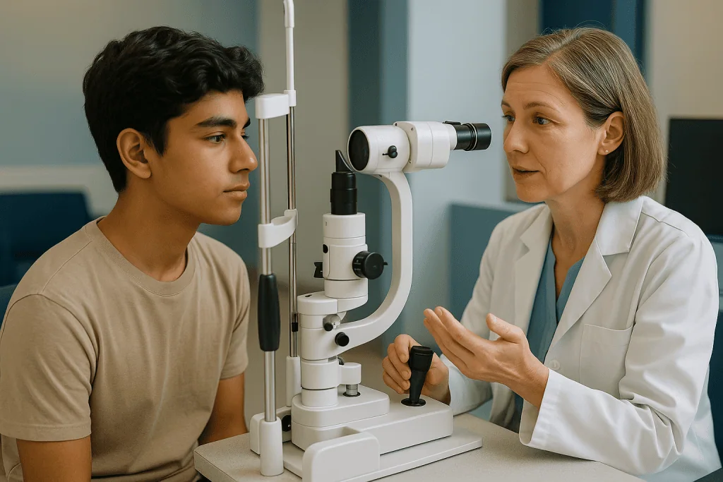
The workhorse is corneal topography or tomography: a painless scan that photographs the front of the eye and produces a color-coded map of its curvature and thickness, rather like an elevation map of a hill. From that map the clinician reads the steepest point, often labeled Kmax, the location and value of the thinnest point, and how the shape compares with the previous scan. Alongside that come the familiar chart, a refraction, and a slit-lamp look at the cornea for early scarring or fine stress lines.
Progression is usually defined as a meaningful worsening in one or more of those numbers between visits, beyond the small wobble every instrument shows from day to day. Definitions vary between practices, and a good specialist will tell you which thresholds they use so that the words “stable” and “progressing” have a clear meaning in your child’s file.
How often should a teenager be scanned? There is no single guideline interval, and the honest answer is that the schedule follows the rate of change. A newly diagnosed adolescent is typically seen more frequently, often every few months, so that two or three sets of measurements exist before any decision is made. Once the cornea appears steady, or after cross-linking has been done and the early healing phase has passed, visits usually stretch toward yearly, with the option to tighten again if anything shifts.
Both eyes are measured every time. Keratoconus is usually bilateral but rarely symmetrical, and the quieter eye deserves the same attention as the one that prompted the diagnosis.
What actually happens on the day of the procedure?
Cross-linking is an outpatient procedure, done in a clinic room or minor-procedure suite rather than a hospital operating theatre, and the patient goes home the same day. Cleveland Clinic patient information puts the total visit at roughly an hour, most of it spent lying still while drops soak in.
Here is the sequence a teenager can expect, in order:
- Numbing drops are placed in the eye, so the cornea itself does not feel the procedure. A small holder keeps the eyelids from blinking.
- In the standard approach, the surgeon gently removes the thin surface layer of the cornea. This is the step that later causes the scratchy feeling during healing.
- Riboflavin drops are applied repeatedly until the cornea is saturated, which takes a substantial part of the appointment and is the point where good headphones and a favorite playlist earn their keep.
- The corneal thickness is checked again before the light is switched on.
- The ultraviolet A light is directed at the cornea for a set period while the surgeon monitors and continues drops.
- A soft bandage contact lens is usually placed over the eye to protect the healing surface, and a clear shield or dark glasses are provided for the trip home.
Most surgeons treat one eye at a time, partly for safety and partly so the young person has a functioning eye during the uncomfortable first days. Where both eyes need treatment, the second is commonly scheduled once the first has settled.
Preparation for a teenager is mostly practical: a parent or adult to drive, a note for school covering a few days off, sunglasses, and a clear understanding that the eye will feel gritty and sore for a short while. The surgeon will give written instructions on the prescribed drops to be used afterward; those instructions, not this article, govern how they are taken.
What does corneal cross linking recovery look like in the following days and weeks?
The first two to four days are the hard part, and it helps to say so plainly. With the surface layer removed, the eye behaves like a scraped knee: it waters, it stings, it hates bright light, and it wants to be shut. Over-the-counter comfort measures and any prescribed drops are used exactly as the surgeon directs; a family should never adjust or add eye medicines on their own, because some common products are unsafe on an open corneal surface.
The epithelium regrows quickly. Cleveland Clinic describes the surface healing within a few days, after which the bandage lens is removed at a check-up and the sharp discomfort eases. Vision, however, lags behind comfort. It is normal for the treated eye to be blurrier than before the procedure for several weeks, and Mayo Clinic notes that it can take weeks to a few months for vision to settle. A faint haze in the cornea is a recognized part of the healing response and usually fades over the following months.
A typical timeline, drawn from the sources above and expressed as ranges rather than promises:
- Days 1–4: pain, light sensitivity and watering; rest, dark glasses, no screens if they hurt.
- Days 4–7: bandage lens out, surface healed, return to school possible for many.
- Weeks 2–8: blurry but improving vision; glasses may be temporarily wrong.
- Months 1–3: vision stabilizing; contact lens fitting or refitting, if needed, is usually considered only once the surgeon confirms the shape has settled.
Through all of it, the two non-negotiables are no eye rubbing and no water in the eye from pools, lakes or hot tubs until the surgeon clears it. Swimming, contact sports and dusty environments are typically paused for the early weeks. School can resume when the teenager is comfortable, with a heads-up to teachers that reading may be slow for a while.
What does the evidence actually show about cross-linking for progressive keratoconus?
Here the honest answer is more nuanced than most patient leaflets suggest. The Cochrane systematic review of corneal collagen cross-linking for keratoconus, published on PubMed, examined the randomized trials available at the time and concluded that the evidence was limited in quantity and of low quality, largely because the trials were small, short and had methodological weaknesses. Within those limits, the review found that cross-linking may slow progression compared with no treatment, but it called for larger, longer studies before firm conclusions could be drawn.
Since then, a large body of observational follow-up, some of it now extending well beyond a decade, has been broadly consistent with the trial signal: in most treated eyes the steepest measurements stop rising, and in a proportion they flatten slightly. Regulators in several countries have approved the procedure for progressive disease on that basis, and Mayo Clinic, Johns Hopkins and Cleveland Clinic all now describe it as the established option for slowing progression. Observational data are not the same as trial data, though, and a careful clinician will say so.
Three points deserve emphasis. First, cross-linking does not reliably improve vision, and families who expect it to will be disappointed; it is judged a success if the cornea stops changing. Second, it does not work in every eye. A minority continue to progress and may be offered a second treatment or other options. Third, it carries real, if uncommon, risks: infection of the healing surface, delayed healing, persistent haze, scarring that reduces vision, and, rarely, damage to deeper layers if the cornea was thinner than measured.
Set against that is the alternative of doing nothing while a young cornea steepens, which in advanced cases can end with contact lenses that no longer fit and a discussion about corneal transplant. That trade-off is the core of the decision, and it is one the treating team should walk through with the family in specific, individual terms.
Keratoconus treatment options for teenagers compared
Families often imagine the choice as cross-linking versus everything else. In practice the options do different jobs, and most teenagers use more than one. Correction helps the young person see today; stabilization protects the cornea for tomorrow; transplant is reserved for the small minority whose cornea has become too scarred or too irregular for lenses.
| Option | What it does | Who it usually suits | What it does not do |
|---|---|---|---|
| Glasses or soft contact lenses | Correct mild blur and astigmatism | Early, mild disease | Cannot correct an irregular cone well; do not slow progression |
| Rigid gas-permeable or scleral lenses | Create a smooth new optical surface over the cone | Moderate to advanced irregularity | Do not stop progression; need skilled fitting and hygiene |
| Corneal cross-linking | Stiffens the cornea to slow or halt steepening | Documented or highly likely progression, adequate thickness | Does not reliably sharpen vision or remove the need for lenses |
| Corneal ring segments | Small implants that flatten part of the cornea | Selected cases where lenses are not tolerated | Do not treat the underlying weakness |
| Corneal transplant | Replaces scarred or very steep tissue with donor cornea | Advanced disease, lens intolerance or central scarring | Long recovery; lenses often still needed; graft risks |
Mayo Clinic describes progression through these stages: glasses and soft lenses early, specialty lenses as the cone advances, cross-linking when progression is shown, and transplant only when scarring or extreme thinning leave no other route. The order is not fixed. A teenager may have cross-linking while still in glasses, or be fitted for scleral lenses years after a stable cross-linked cornea.
What the table cannot show is the value of time. Cross-linking sits apart from the others because it is the only row aimed at the disease process itself rather than at its optical consequences, and that is precisely why it comes up early in adolescence.
Can you live a normal life with keratoconus as a teenager?
For the large majority, yes, and it is worth saying that before anything else. Keratoconus is a condition that shapes daily logistics, not one that closes doors. Johns Hopkins and Cleveland Clinic both describe most people managing well with corrective lenses throughout life; total loss of sight from keratoconus is rare, and even advanced disease is usually addressed with lenses or surgery rather than accepted.
School is the first practical arena. Distorted vision makes small print and whiteboards tiring, and glare from screens or headlights can be worse at night. Sitting near the front, using larger fonts, and having spare lenses and solution at school are simple fixes. Formal accommodations for exams are reasonable to ask about if reading speed is affected, and a letter from the eye specialist usually settles it.
Sport is fine, with two caveats. Contact lenses of any kind, and especially rigid or scleral lenses, should be protected by proper sports eyewear in ball and contact sports. After cross-linking, the surgeon will set a specific pause on swimming and contact sports during healing. Beyond that, there is no evidence that ordinary physical activity worsens keratoconus.
Screens do not cause or accelerate keratoconus, despite what a teenager may be told by well-meaning relatives. Long screen sessions can dry the eyes and provoke rubbing, which is the real hazard, so regular breaks and lubricating drops recommended by the eye team are sensible.
Driving and careers deserve a frank word. Licensing depends on corrected visual acuity, and most people with keratoconus meet the standard with their lenses. A few careers with strict uncorrected vision requirements, such as some aviation or military roles, apply their own rules; a teenager with those ambitions should ask the specialist early rather than late.
What to avoid with keratoconus, and why eye rubbing tops the list
If a family takes away one behavioral change from the diagnosis, it should be this: stop rubbing the eyes. Mayo Clinic lists vigorous eye rubbing among the factors associated with keratoconus and its progression, and the mechanism is intuitive. A cornea that is already weaker than average is being pressed and sheared, sometimes dozens of times a day, exactly where it is thinnest. Rubbing is also a habit that is easy to underestimate; ask a teenager how often they do it and the honest answer is usually “I didn’t notice.”
Because most rubbing is driven by itch, the second item is untreated allergy. Allergic eye disease is common in adolescents with keratoconus, and controlling it, whether through avoidance of triggers, cool compresses or medicines chosen by the eye or allergy specialist, is a treatment for the cornea as much as for the itch. This is a point to raise explicitly at follow-up, because the two problems are often managed by different clinicians.
Other habits and choices worth avoiding, drawn from Mayo Clinic and Cleveland Clinic guidance:
- Sleeping face-down or with a hand or pillow pressing on the eye.
- Poor contact lens hygiene, including tap water and over-wearing, which raises infection risk in an eye that may later need surgery.
- Laser vision correction such as LASIK, which removes corneal tissue and is considered unsafe in keratoconus; any refractive surgery consultation must know about the diagnosis.
- Skipping follow-up because vision “seems fine,” since progression is silent until it is not.
- Unproven supplements or eye exercises marketed as strengthening the cornea; there is no evidence they alter the disease.
None of this is about blame. Eye rubbing did not cause the teenager’s keratoconus, which has genetic and structural roots; Mayo Clinic notes that about 1 in 10 people with the condition have a parent who also has it. Avoidance is simply one of the few levers a young person can pull themselves.
Can keratoconus cause anxiety, and does it count as a disability?
Yes, it can, and the worry is not irrational. A teenager who hears that their eye is changing shape, that a procedure may be needed, and that the future is measured in scans, is being asked to carry uncertainty at an age when most peers are not. Distorted night vision and the fatigue of constant refocusing add a daily grind. Anxiety, low mood and a reluctance to talk about the eye are common enough that specialists in adolescent eye care expect them.
Two things help. The first is information delivered without drama: keratoconus is manageable, progression slows with age, and cross-linking exists precisely to reduce the chance of the outcomes people fear. The second is naming the feeling to someone. A school counsellor, family doctor or the eye team itself can point toward support, and a young person who is struggling should be told that this is part of care, not a distraction from it.
The disability question comes up because families are trying to plan. In the United States, eligibility for Social Security disability benefits, including SSI, is not decided by diagnosis but by measured visual function, principally best-corrected visual acuity and visual field, against published criteria. Because most people with keratoconus see well with lenses, the condition on its own does not usually meet those thresholds; a small number with advanced disease, scarring or lens intolerance may. Educational accommodations under school disability provisions are a separate matter and are often granted on the basis of functional difficulty with reading or boards, regardless of benefit eligibility.
This is not legal advice, and criteria differ between countries and change over time. The eye specialist can document visual function accurately; a benefits adviser or school special-education coordinator can explain how that documentation is used.
What people often get wrong about cross-linking and keratoconus in teenagers
Misunderstandings cluster around this diagnosis, partly because “laser” and “cornea” appear in the same sentence and partly because the internet blends keratoconus with the far more common conversation about vision correction. A few corrections, in order of how often they come up:
- “Cross-linking will fix my child’s vision.” It is designed to stop the cornea steepening. Any improvement in sharpness is a bonus, not the aim, and lenses are usually still needed.
- “It’s basically LASIK.” It is close to the opposite. LASIK removes tissue to reshape the cornea and is contraindicated in keratoconus. Cross-linking removes nothing structural; it strengthens what is there.
- “Once it’s done, we’re finished.” Follow-up continues, initially to check healing and then to confirm stability over years. A minority progress despite treatment.
- “Glasses or contacts make it worse.” Correction does not change the cornea. Poorly fitted rigid lenses can cause surface problems, which is why fitting matters, but wearing appropriate correction is protective of a teenager’s schooling and safety.
- “Screens caused it.” There is no evidence for this. Genetics, connective-tissue factors, allergy and rubbing are the associations supported by Mayo Clinic and MedlinePlus.
- “Keratoconus means eventual blindness or a transplant.” Most people never need a transplant, and legal blindness from keratoconus is uncommon. Transplant is reserved for advanced, scarred or lens-intolerant eyes.
- “If one eye is fine, it’s only one eye.” The condition is usually in both eyes, unevenly. The better eye still needs scanning.
- “Waiting until 18 is safer.” Not necessarily. Adolescence is often when progression is fastest, so delay can mean treating a thinner, steeper cornea later. Equally, treating a stable eye offers no benefit. The measurements, not the birthday, should drive timing.
The thread through all of these is that keratoconus is a structural, progressive, usually manageable condition, and cross-linking is a stabilizer. Hold onto those two ideas and most of the noise falls away.
Questions to ask your care team before deciding on cross-linking
A good consultation leaves a family with fewer questions than they arrived with, and the best way to get there is to bring a written list. The teenager should hold the list, not just the parent; it is their eye, and surgeons generally welcome a young patient who asks directly. Consider these:
- What exactly has changed in my measurements since the last visit, and by how much? Which numbers are you using to define progression?
- How thick is my cornea at its thinnest point, and does that affect which technique you would use?
- Are you recommending treatment now, or a further period of monitoring first? What would change your mind either way?
- Will you treat one eye or both, and how far apart?
- Which technique do you use, surface-off or surface-on, and what does the evidence say about each?
- What will the first week feel like, and who do I call, day or night, if something worries me?
- What are the specific risks in my case, and how often do you see them in patients my age?
- How long before I can return to school, sport, swimming and contact lenses?
- What does follow-up look like over the next two years, and how will we know the treatment has done its job?
- What happens if the cornea keeps progressing despite treatment?
- How should I manage my allergies and eye rubbing alongside this?
- Are there any activities, careers or future eye surgeries I should know are affected by this diagnosis?
It is also fair to ask for a copy of the corneal maps and to request that future scans be compared with them, so the family can see the same data the specialist sees. Bring a notebook or record the answers with permission. Decisions of this kind are better made across two conversations than one, and no reputable team will object to a family taking a few days to think.
When to call your doctor: red-flag signs after cross-linking and during follow-up
Most of the recovery from cross-linking is uncomfortable but predictable, and most of the years of follow-up are uneventful. The point of knowing the warning signs is to be able to tell the difference between expected soreness and a problem that needs same-day attention. Contact the surgeon or the emergency number they provided, without waiting for the next scheduled visit, if any of the following occur after the procedure:
- Pain that is getting worse after the first few days instead of easing, or pain that returns after it had settled.
- Increasing redness, a thick or colored discharge, or a white or gray spot visible on the cornea; these can indicate infection of the healing surface, which is uncommon but urgent.
- A sudden drop in vision in the treated eye, or a new dense cloudiness, beyond the gradual blur the surgeon warned about.
- The bandage contact lens falling out or becoming dislodged before its planned removal.
- Severe light sensitivity that persists beyond the early days or worsens.
- Any injury to the eye, including a poke or a rub, during the healing weeks.
Outside the recovery period, the signals are quieter and easier to ignore. Arrange an earlier review than planned if a teenager notices vision worsening between visits, if glasses or lenses that were comfortable become unhelpful, if one eye is clearly deteriorating faster than the other, if there is new sensitivity to light or halos around lights at night, or if a rigid or scleral lens becomes painful or repeatedly pops out. Very sudden, severe blurring in a keratoconic eye, sometimes with pain and marked watering, can indicate an acute swelling event called hydrops and warrants prompt assessment.
None of these signs is a diagnosis in itself, and most turn out to have straightforward explanations. The treating team would far rather see a teenager for a false alarm than miss an early problem, and they will tell you exactly which route, clinic line, urgent-care eye service or emergency department, to use for which situation.
Frequently asked questions
Can you live a normal life with keratoconus?
Yes, most people with keratoconus live full, ordinary lives using glasses or specialty contact lenses, and complete loss of sight from the condition is rare. School, sport, driving and most careers remain open, with some practical adjustments such as protective eyewear during contact sports and good lens hygiene. A small minority with advanced scarring or lens intolerance may need surgery, and a few professions with strict uncorrected vision standards apply their own rules.
Is keratoconus considered a disability by the SSI?
Not on the basis of diagnosis alone. In the United States, disability benefits including SSI are assessed on measured visual function with best correction, mainly visual acuity and visual field, against published criteria. Because most people with keratoconus see well with lenses, the condition itself rarely meets those thresholds, though advanced cases may. School accommodations are a separate, more accessible route. This is general information rather than legal advice; a benefits adviser can review individual circumstances.
What should you avoid with keratoconus?
Avoid rubbing the eyes above all, since Mayo Clinic links vigorous rubbing to keratoconus and its progression. Manage allergies that drive the itch, do not sleep with pressure on the eye, keep contact lens hygiene strict, and never undergo LASIK or similar tissue-removing laser surgery, which is contraindicated. Skipping follow-up because vision seems stable is another common mistake, because progression is often silent until the prescription changes.
Can keratoconus cause anxiety in teenagers?
It can. A progressive diagnosis, uncertainty about treatment, and the daily fatigue of distorted vision are real stressors for a young person. Anxiety and low mood are common enough that eye teams expect them and can help find support. Clear information usually eases the worry: keratoconus is manageable, progression slows with age, and cross-linking exists to reduce the chance of the outcomes people fear most. Struggling emotionally is a reason to talk to the care team, not a distraction from care.
Does corneal cross-linking improve vision?
Not reliably, and it is not designed to. The goal is to stiffen the cornea so it stops steepening. Some patients see a small flattening of the cone and a modest improvement in glasses prescription afterward, while others notice no change, and vision is usually blurrier than before for several weeks during healing. Most people still need glasses or contact lenses after treatment. The procedure is judged successful when the corneal measurements stop changing.
How long does corneal cross linking recovery take?
The most uncomfortable phase is the first two to four days, when the eye is sore, watery and light-sensitive. The corneal surface typically heals within a few days, after which a bandage lens is removed. Vision remains blurry and fluctuates for several weeks, and Mayo Clinic notes it may take weeks to a few months to settle. A faint corneal haze often appears and fades over months. Swimming and contact sports are paused until the surgeon clears them.
Why is keratoconus progression in teens faster than in adults?
The cornea is still maturing in adolescence, and the collagen weave that keratoconus weakens has not yet reached its adult stiffness. Teenagers also tend to have more allergic eye disease and rub their eyes more, both associated with progression. Mayo Clinic describes the condition beginning in the late teens to early twenties and progressing for a decade or longer before stabilizing, so a younger diagnosis simply means more years of potential change ahead.
Is cross-linking painful for a teenager?
The procedure itself is not painful because numbing drops are used, though lying still for about an hour is tedious. Discomfort comes afterward, when the removed surface layer heals: the eye feels gritty and sore, waters, and dislikes bright light for a few days. Surgeons provide prescribed drops and instructions for comfort, which should be followed exactly. Pain that worsens after the first few days rather than easing is a reason to call the team promptly.
Can cross-linking be repeated if keratoconus keeps progressing?
In some cases, yes. A minority of treated eyes continue to steepen, and a second treatment may be considered if the cornea remains thick enough and free of significant scarring. Alternatively the team may move toward specialty lenses or, in advanced disease, discuss a corneal transplant. The decision depends on repeated measurements over time, which is why follow-up continues for years after the procedure rather than ending once healing is complete.
At what age can a teenager have cross-linking for keratoconus?
There is no fixed minimum age in guideline terms; suitability rests on documented or highly likely progression, adequate corneal thickness and a clear central cornea rather than on a birthday. Younger adolescents are often considered because their disease tends to progress fastest. Minors need a proper consent process involving both the young person and their parents or guardians. The treating specialist weighs these factors individually and may recommend monitoring first.
This article is for general information only and is not a substitute for professional medical advice. Please consult a qualified doctor about your individual situation.

More from the Blog

Preparing for Femto LASIK: Contact Lens Breaks, Corneal Mapping and Makeup Rules
Femto LASIK preparation usually means pausing contact lenses for a period set by your surgeon, commonly about two weeks for soft lenses and longer…

Diabetic Retinopathy Stage by Stage: How Retinal Damage Advances and When Treatment Begins
Diabetic retinopathy is graded in four stages: mild, moderate and severe nonproliferative retinopathy, in which small retinal blood vessels leak and close, followed by…

Who Can Wear Contact Lenses? Dry Eye, Astigmatism and Allergies at the Fitting Exam
Most people who need vision correction can wear contact lenses, including many with dry eye, astigmatism and seasonal allergies, provided a fitting exam confirms…

Preparing for LASEK: Contact Lens Breaks, Corneal Thickness Checks and Planning Days Off
LASEK preparation usually involves three things: pausing contact lenses for a period your surgeon sets (often a few weeks, longer for rigid lenses) so…

How Eye Alignment Is Measured: The Tests Behind a Strabismus Disorder Diagnosis
Eye alignment is measured with a short set of clinic tests. The cover test, in which one eye is covered while the examiner watches…

What PRK Results Usually Look Like Once Healing Finishes and How Long They Hold
PRK reshapes the cornea after its surface layer is removed, so vision is blurry for the first several days and sharpens gradually rather than…