

Why Routine Eye Exams Are Part of Juvenile Idiopathic Arthritis Treatment

Key Takeaways
- In young children with JIA, anterior uveitis is typically painless and the eye stays white, so the absence of complaints tells you nothing about whether the eye is inflamed.
- The ACR guideline sets screening intervals from every 3 months for the highest-risk children to roughly every 12 months for the lowest, based on subtype, ANA status, age at onset and years since diagnosis.
- The mildest-looking arthritis, a single swollen knee in a young ANA-positive child, often carries the highest risk of silent eye inflammation.
- A slit lamp exam looks for white blood cells and leaked protein in the clear fluid at the front of the eye; a school vision chart cannot detect either.
- Uveitis can flare while the joints are in remission and is especially likely when immune-modifying medicines are being reduced, which is why both specialists tighten the schedule at that point.
- Untreated eye inflammation can lead to glaucoma, cataract, corneal calcium deposits and macular swelling, and in preschoolers to amblyopia, where the brain stops using a blurred eye.
Routine eye exams are built into juvenile idiopathic arthritis treatment because the same immune process that inflames a child's joints can inflame the front of the eye (uveitis), usually without pain, redness or any complaint from the child. A slit lamp exam by an eye specialist can detect it early, before it damages vision. Guidelines set exam intervals of roughly every 3 to 12 months depending on the child's risk profile.
The rheumatology appointment is winding down. The knee that started all this has settled, the morning limp has faded, and the parent in the chair is already thinking about the car park. Then the doctor says something that lands oddly: “And you have the eye appointment booked for next month, yes?” The child’s eyes look perfectly fine. Nobody has complained about them once.
That small moment of confusion is where most families first meet the idea of a juvenile idiopathic arthritis eye exam, and it deserves a proper explanation rather than a reminder slip. In this condition, the eye can be inflamed for months while looking completely normal from the outside and feeling completely normal to the child.
What follows is a plain account of why the eye specialist is part of the arthritis team, what actually happens in that darkened room, how often children are seen, and which signs should prompt a call rather than a wait.
Can juvenile idiopathic arthritis affect the eyes?
Yes, and the link is close enough that eye care is written into every major treatment pathway for the condition. Juvenile idiopathic arthritis, usually shortened to JIA, is joint inflammation that begins before a child’s 16th birthday, lasts at least six weeks, and has no other identifiable cause; “idiopathic” simply means the cause is unknown. The immune system that mistakenly attacks the lining of a knee or ankle can also attack the uvea, the pigmented middle layer of the eye made up of the iris, the ciliary body behind it, and the choroid at the back.
In JIA the inflammation almost always sits at the front: the iris and ciliary body. Doctors call this anterior uveitis, or sometimes iridocyclitis. Both eyes are often involved, though not always at the same time. The guideline on screening and treatment from the American College of Rheumatology summarises published estimates placing JIA-associated uveitis in roughly 10 to 20 percent of children with the condition, which makes it the most common serious complication outside the joints.
Two features make it unusual. First, the eye disease and the joint disease do not move in step. Uveitis can appear years after the arthritis, or occasionally before it, and it can flare while the joints are calm. Second, in young children it is typically painless and the eye stays white. Adults with anterior uveitis usually arrive with a red, aching, light-sensitive eye. A four-year-old with the same inflammation may show nothing at all.
That combination, a condition that is common, disconnected from joint symptoms and silent, is the whole reason a scheduled exam exists.
Why a juvenile idiopathic arthritis eye exam is treatment, not an optional extra
It is tempting to file the eye appointment under “monitoring” and the rheumatology prescription under “treatment.” In this condition the distinction collapses. The exam is the intervention, because there is no other reliable way to know whether the eye is inflamed.
Consider what the alternative looks like. Without screening, chronic anterior uveitis in a young child is usually discovered only when it has already produced something visible: a pupil that has become irregular because the iris has stuck to the lens, a cloudy lens, a squint that appeared out of nowhere, or a child who has quietly started holding books closer. Each of those signs represents months of unchecked inflammation. The National Health Service describes glaucoma, cataracts and swelling at the centre of the retina among the complications of untreated uveitis, and in a developing visual system those changes can leave permanent gaps.
The exam also changes what the rheumatologist does. Finding inflammatory cells in the eye may move a child onto a disease-modifying medicine sooner than the joints alone would justify, and a clean exam can support a decision to hold steady. Findings travel in both directions: the eye doctor needs to know when arthritis medicines are being reduced, because the eye often flares when the immune system is given more room.
Seen this way, the rheumatologist and the ophthalmologist, an eye doctor trained in medical and surgical eye disease, are two halves of one treatment plan. Skipping the eye half does not leave a child half-treated. It leaves one of the two targets of the disease entirely unwatched.
What actually happens during a slit lamp exam for children
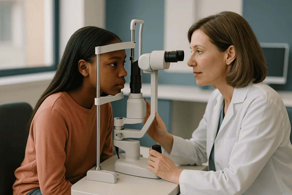
The room is dim, and the central instrument looks a little like a pair of binoculars mounted on a table. That is the slit lamp: a microscope that shines a narrow, very bright beam of light through the front of the eye so the examiner can see each layer in cross-section, much as a thin slice of light through a glass of water reveals specks floating in it.
A school-age child rests their chin and forehead against the frame and looks at the doctor’s ear or a sticker on the wall. A toddler usually sits on a parent’s lap, and many clinics also keep a handheld version for children who will not sit still. The examiner is looking for two things. “Cells” are white blood cells drifting in the aqueous humour, the clear fluid filling the front chamber of the eye; they show up as tiny bright dots in the beam. “Flare” is leaked protein that makes the beam look hazy, like car headlights in fog. Both are graded on a standard scale, and that grade is what the two specialists compare from visit to visit.
The doctor will also check the iris for adhesions, the lens for early clouding, and the pressure inside the eye. Dilating drops are sometimes used to widen the pupil and view the lens and the back of the eye. According to MedlinePlus, these drops take around 15 to 20 minutes to work and leave near vision blurred and eyes light-sensitive for several hours, which is why sunglasses and a skipped afternoon of homework are sensible plans.
None of it hurts. The brightness is the main complaint, followed by boredom. Practising the chin-rest position at home as a game helps more than any amount of explanation.
Who needs JIA uveitis screening most often, and how often?
Not every child with JIA carries the same risk, and screening intervals reflect that. The American College of Rheumatology guideline on JIA-associated uveitis builds its schedule around four questions: which subtype of arthritis the child has, whether blood tests show antinuclear antibodies (ANA, proteins the immune system makes against the body’s own cell nuclei), how old the child was at onset, and how long since the arthritis began.

| Risk profile (per ACR guideline) | Typical screening interval |
|---|---|
| Oligoarticular, polyarticular RF-negative, psoriatic or undifferentiated JIA; ANA positive; onset at age 6 or younger; within 4 years of diagnosis | Every 3 months |
| Same subtypes but ANA negative, or onset after age 6, or more than 4 years since diagnosis | Every 6 to 12 months, depending on how many risk factors remain |
| Systemic JIA, or polyarticular RF-positive JIA | Roughly every 12 months |
| Enthesitis-related arthritis | Roughly every 12 months, with prompt review if the eye becomes red or painful |
The pattern is counterintuitive to many parents. The child with a single swollen knee and no other obvious illness, the mildest-looking arthritis, is frequently the one who needs the eye doctor every three months. That is because oligoarticular JIA (four or fewer joints in the first six months) in a young ANA-positive child is exactly the profile in which silent uveitis is most common.
Children already diagnosed with uveitis follow a different, tighter schedule set by the eye specialist until the inflammation is quiet. And whenever an immune-modifying medicine is being reduced or stopped, both specialists usually agree to see the child more often for a period, because that is when the eye is most likely to flare.
Who is usually screened less often, and does anyone stop?
The schedule in the table is not a life sentence of quarterly visits. Risk falls with time and with age, and intervals stretch accordingly. A child who was diagnosed at four and screened every three months may, after passing the four-year mark without any eye inflammation, move to every six months, and later to yearly. The decision belongs to the eye specialist and rheumatologist together, based on the child’s accumulated record of clean exams.
Some groups start on longer intervals. Children with systemic JIA, the form that begins with high spiking fevers and a salmon-coloured rash, carry a comparatively low risk of silent uveitis, and the guideline places them at around a yearly check. Those with rheumatoid factor-positive polyarticular disease, a pattern more like adult rheumatoid arthritis, sit in a similar bracket.
Enthesitis-related arthritis, which involves inflammation where tendons attach to bone and often affects older boys, is a special case. Its uveitis tends to be the adult type: sudden, red, painful and light-sensitive. Because the child will usually notice and complain, routine screening can be yearly, but families are taught to treat a red sore eye as an urgent appointment rather than something to watch.
Does screening ever end? The guidance is cautious. Uveitis can appear years into the disease and can recur after long quiet spells, so most pathways keep some form of periodic eye review in place through adolescence, even after the arthritis has gone into remission and medicines have stopped. The interval lengthens; the habit stays. Adults who had JIA as children are generally advised to tell any new eye care provider about their history, because flares in adulthood, while uncommon, do happen.
JIA associated uveitis symptoms: why "silent" is the whole point
Parents sensibly ask what to watch for, and the honest answer has two parts: here is what inflammation can eventually produce, and here is why waiting for it is a poor strategy.
Older children and teenagers sometimes report blurred vision, floaters (small drifting shapes in the field of view), or discomfort in bright light. Occasionally they describe a dull ache. Younger children rarely say anything, partly because the inflammation is painless and partly because a child who has always seen the world through a slight haze has no comparison to make.
The signs a parent might eventually notice are, almost by definition, late ones. A pupil that is no longer perfectly round can mean synechiae, adhesions between the iris and the lens that form when inflamed tissue sticks together. A whitish or cloudy look to the pupil can mean a cataract, a clouding of the lens, or band keratopathy, a greyish band of calcium deposits across the cornea. A new squint, a tilt of the head, sitting unusually close to the screen, or losing interest in detailed play can all reflect a drop in vision in one eye.
This is not a checklist to run through at home each evening. Its purpose is the opposite: to show that by the time any of it is visible across a kitchen table, the inflammation has typically been active for a long while. The Cleveland Clinic describes uveitis as capable of causing permanent vision loss when untreated, and the developing eye is especially vulnerable. The scheduled slit lamp exam exists precisely so that the first sign of trouble is a few cells in a beam of light, not a cloudy pupil.
What are the common symptoms of juvenile idiopathic arthritis in children?
Because the eye story begins with a joint diagnosis, it helps to understand how JIA usually announces itself, and why it is so often missed for a while.
The Mayo Clinic lists the core features as joint pain, swelling and stiffness, with stiffness typically worst in the morning or after a nap and easing as the child moves. Affected joints may feel warm. Yet children with JIA frequently do not complain of pain at all. What parents notice instead is behaviour: a toddler who suddenly wants to be carried, a child who limps for the first half hour of the day, a reluctance to climb stairs, or a knee that looks puffy compared with the other one. Teachers sometimes spot it before parents, because they see the child at the stiffest time of the morning.
Doctors group JIA into subtypes according to the first six months of illness. Oligoarticular disease involves four or fewer joints, often a knee or ankle, and is the most common form in young children. Polyarticular disease involves five or more joints and is split by whether rheumatoid factor, an antibody also seen in adult rheumatoid arthritis, is present. Systemic JIA starts with daily fevers and a fleeting rash alongside joint symptoms. Psoriatic arthritis pairs joint inflammation with the skin condition psoriasis or with nail changes; enthesitis-related arthritis targets tendon attachments and the lower spine. Anything that fits none of these is labelled undifferentiated.
The subtype matters for the eyes. It is one of the four variables that set the screening interval, and it is the reason two children with the same diagnosis on paper can have very different appointment calendars.
At what age is juvenile idiopathic arthritis diagnosed, and why timing shapes eye care
By definition JIA begins before the age of 16, the cut-off used by the Mayo Clinic and the National Institute of Arthritis and Musculoskeletal and Skin Diseases alike. Within that range, the oligoarticular form clusters in toddlers and preschoolers, while enthesitis-related and rheumatoid factor-positive polyarticular disease tend to appear in older children and adolescents.
There is no single test. Diagnosis rests on a doctor examining the joints and finding inflammation that has persisted for at least six weeks with other causes ruled out. Blood tests contribute pieces: markers of inflammation, rheumatoid factor, the ANA result, and sometimes a genetic marker called HLA-B27 associated with enthesitis-related disease. Imaging such as ultrasound or MRI can confirm fluid and inflamed lining in a joint. Infections, injuries and, rarely, blood disorders have to be excluded first, which is one reason the road to a diagnosis can be slow.
Age at diagnosis then feeds straight into eye care. The youngest children are the ones at highest risk of silent uveitis and the least able to report a change in their vision. They are also the ones in whom unchecked inflammation does the most lasting harm, because the brain’s visual pathways are still being wired; an eye that delivers a blurred image during those years can be permanently “turned down” by the brain, a condition called amblyopia.
For that reason, the first eye exam should follow the arthritis diagnosis promptly rather than waiting for a convenient gap. Most rheumatology services make the referral at the same visit as the diagnosis, and families are encouraged to treat that first appointment as the baseline against which every later exam is measured.
If uveitis is found: what the following weeks usually look like
Hearing that there are cells in your child’s eye when the eye looks perfectly normal is disorienting. The weeks that follow tend to have a recognisable shape, even though the details are set by the treating team.
The first step is grading. The eye specialist records how many cells and how much flare are present, whether adhesions have started to form, and whether the eye pressure and lens are normal. This grade becomes the yardstick. Treatment usually begins the same day, most often with anti-inflammatory eye drops and sometimes drops that keep the pupil wide so the iris cannot stick to the lens.
Follow-up is then frequent, typically a matter of weeks rather than months, so the specialist can see whether the cell count is falling. The two specialists talk. If the rheumatologist has been considering a disease-modifying medicine for the joints, the eye finding often settles the question. The American College of Rheumatology guideline advises that when eye drops alone cannot bring inflammation under control within a few months, or when the eye needs them at high frequency to stay quiet, a systemic medicine that treats the immune system as a whole should be added.
For the child, daily life changes less than parents fear. School continues. Sport continues unless the eye doctor says otherwise. Drops become part of the morning routine, and a sticker chart does more than any lecture.
Many children reach a quiet eye within weeks to a few months; others need medicine adjustments along the way. The honest framing is that uveitis in JIA is a condition to be controlled over time, with visits that become less frequent as the eye stays calm, rather than a one-off event with a clean finish line.
How JIA uveitis is treated: drops, disease-modifying medicines and biologics
Treatment works in layers, and understanding what each layer does makes the specialists’ decisions less mysterious. Nothing here is a recommendation; the choice and sequence belong to the prescribing clinicians.
The first layer is local. Corticosteroid eye drops deliver an anti-inflammatory medicine directly to the front chamber and are the usual starting point. They act quickly, but prolonged use carries its own eye risks: raised pressure inside the eye and, over time, cataract. That is why the goal is always the lowest exposure that keeps the eye quiet, and why specialists are reluctant to leave a child on frequent drops for long. Dilating drops are often added early; by keeping the pupil wide and moving they reduce the chance of the iris sticking to the lens.
The second layer treats the immune system. Methotrexate, a conventional disease-modifying antirheumatic drug (DMARD), dampens the activity of the immune cells driving inflammation in both joints and eyes. It is taken regularly and is slow to show its effect, typically over a period of weeks to months, so eye drops usually continue while it takes hold. The ACR guideline positions it as the first systemic step for most children.
The third layer is biologic. Tumour necrosis factor (TNF) is a signalling protein that acts like a megaphone for inflammation; monoclonal antibodies such as adalimumab and infliximab bind to it and block the signal. The guideline favours these over the related medicine etanercept for uveitis, based on comparative evidence. Other biologics targeting different signals are reserved for children whose eyes stay inflamed despite the first choices.
Each step involves monitoring, including blood tests and attention to infection risk, which is exactly why the eye doctor and rheumatologist share one plan rather than two.
Can JIA uveitis cause permanent vision loss?
It can, and this is the fact that justifies the whole screening programme, so it deserves to be stated plainly rather than softened. It also deserves context: the risk is concentrated in inflammation that goes undetected or uncontrolled, which is precisely what scheduled exams are designed to prevent.
The damage comes through several mechanisms. Synechiae, the adhesions between iris and lens, can block the normal flow of fluid inside the eye and push the pressure up, leading to glaucoma, a condition in which raised pressure damages the optic nerve. Chronic inflammation itself, and the corticosteroid drops used to treat it, both encourage cataract, a clouding of the lens. Band keratopathy lays down calcium across the cornea and can scatter light. Cystoid macular oedema, swelling at the centre of the retina, blurs the sharp central vision used for reading and faces. The NHS lists glaucoma, cataracts and macular swelling among the complications of uveitis that is not treated promptly.
Children face one extra hazard adults do not. If one eye delivers a blurred image during the early years, the brain learns to rely on the other eye and may never fully re-engage the weaker one, even after the eye itself is treated. This is amblyopia, and it is why early detection in preschoolers carries such weight.
What does the evidence say about outcomes? Observational studies summarised in the ACR guideline associate earlier detection and earlier systemic treatment with fewer complications, and the broad direction of that evidence is consistent across studies. It is not a guarantee for any individual child, and no responsible clinician will offer one. It is, however, a strong argument for keeping every appointment, including the ones that feel unnecessary because the eyes look fine.
What people often get wrong about eye exams in juvenile arthritis
Some misunderstandings come up in almost every clinic. Correcting them matters because each one leads to a missed appointment.
“No eye symptoms means no eye problem.” In young children with JIA, inflammation is typically painless and invisible from outside. The absence of complaint is the normal state of early uveitis, not reassurance against it.
“The joints are quiet, so the eyes must be too.” Eye and joint inflammation run on loosely connected timetables. Uveitis can begin or flare while the arthritis is in remission, including after joint medicines have stopped.
“The arthritis medicine protects the eyes.” Some systemic medicines do reduce eye inflammation, which is why the specialists coordinate. But protection is not guaranteed, children on treatment still need screening, and the period when a medicine is being reduced is when the eye is most likely to flare.
“The school vision screening or a glasses check covers it.” A vision test measures how well a child reads a chart. Early uveitis does not change the chart result. Only a slit lamp exam by a clinician experienced in paediatric uveitis can see cells in the front chamber.
“Mild arthritis means low eye risk.” Often the reverse. A young ANA-positive child with a single swollen knee is in the highest-risk group.
“Eye drops are harmless, so a few more weeks won’t matter.” Corticosteroid drops carry real risks of raised pressure and cataract with long use, which is why clinicians try to limit them.
“Once the eye is quiet, we’re done.” Uveitis recurs. Intervals lengthen as the record of clean exams grows, but periodic review usually continues through adolescence.
Questions to ask your care team about your child's eye care
A list in your hand is the best defence against the appointment that ends before you have asked anything. These questions are phrased so that the answers come from the people who know your child’s chart, not from a magazine.
- Which JIA subtype does my child have, and what is the ANA result? Those two facts set the screening interval, so it is worth knowing them yourself.
- How often should the eye exams be right now, and what would make that interval shorter or longer over the next year?
- Who is doing the exam, and are they experienced in examining young children for uveitis? Who will I call if I cannot get the next appointment within the recommended window?
- How will the eye specialist and the rheumatologist share results with each other, and with us? Is there one summary we can keep?
- Will dilating drops be used at every visit, and how should we plan the rest of that day for school or nursery?
- If inflammation is found, what is the first treatment likely to be, how soon would you expect to recheck, and what would prompt a change of plan?
- If my child is on a medicine that treats the immune system, does it change anything about vaccinations or how we handle a fever?
- When a medicine is being reduced or stopped, will the eye exams become more frequent for a time?
- Which eye signs should make us call the same day rather than wait for the next scheduled visit?
- As my child gets older, when will they take over understanding their own screening schedule, and how does this transfer to adult services?
Write the answers down during the visit. Memory under mild stress is unreliable, and the screening schedule is one thing that should never depend on it.
When to call your doctor: red-flag signs in a child with JIA
Most of this article argues for calm, scheduled care. This section is about the exceptions, when the right move is a phone call that day rather than the next appointment. The eye specialist or rheumatology team will tell you how they prefer to be contacted; keep that number where you can find it.
Call the same day, or seek urgent care, if your child develops a red, painful eye, especially with sensitivity to light; a sudden drop in vision, blurring, or a new complaint of floaters; a pupil that has changed shape or no longer looks round; a white, grey or cloudy appearance in the pupil; a new squint or eye turn, or a sudden habit of closing or covering one eye; or severe eye pain with headache, nausea or vomiting, which can signal a sharp rise in pressure inside the eye.
Call the rheumatology team promptly if a joint becomes hot, swollen and very painful, particularly alongside fever, since that pattern needs infection ruled out; if your child is on a medicine that dampens the immune system and develops a fever, persistent cough, unusual rash or signs of chickenpox or shingles after exposure; or if a medicine has been missed or stopped for any reason, so the team can advise rather than guess.
Call to rebook, and say why, if a scheduled eye exam has been missed or delayed beyond its interval. A missed quarterly exam in a high-risk child is a clinical event, not an administrative one.
None of these signs is a diagnosis in itself. Each is a reason for someone with a slit lamp to look, soon. Every treatment decision that follows rests with the team that knows your child.
Frequently asked questions
Can juvenile arthritis affect the eyes even when the joints feel fine?
Yes. Eye inflammation in JIA, called anterior uveitis, runs on a separate timetable from the joints and can appear or flare while the arthritis is in remission or after joint medicines have stopped. Published estimates summarised in the ACR guideline put it in roughly 10 to 20 percent of children with JIA, which is why eye exams continue on a schedule regardless of how the joints are doing.
What is JIA uveitis screening and how often is it done?
JIA uveitis screening is a scheduled slit lamp examination by an eye specialist to look for inflammation that the child cannot feel or see. The ACR guideline recommends every 3 months for the highest-risk children (oligoarticular or RF-negative polyarticular, ANA positive, onset at 6 or younger, within 4 years of diagnosis) and every 6 to 12 months for others. Your child’s team sets the exact interval.
What are JIA associated uveitis symptoms parents might notice?
Often none, which is the central problem. Older children may mention blurred vision, floaters or discomfort in bright light. Signs a parent might spot, such as an irregular or cloudy pupil, a new squint, or holding things unusually close, usually mean the inflammation has been active for some time. These are reasons to call the eye doctor, not a checklist to replace scheduled exams.
At what age is juvenile idiopathic arthritis typically diagnosed?
JIA is by definition diagnosed before the age of 16, after joint inflammation has lasted at least six weeks with other causes excluded. The oligoarticular form, which carries the highest uveitis risk, most often begins in toddlers and preschoolers, while enthesitis-related and RF-positive polyarticular disease tend to appear in older children and teenagers. The youngest patients need the closest eye follow-up.
Is a slit lamp exam for children painful or frightening?
It is painless. The child rests their chin on a frame, or sits on a parent’s lap for a handheld version, while a bright narrow beam of light is shone through the front of the eye. The brightness is the main complaint. Dilating drops, when used, sting briefly and blur near vision for several hours according to MedlinePlus, so plan a quieter afternoon and bring sunglasses.
Does juvenile arthritis go away, or is it lifelong?
The course varies. Many children reach remission, a period with no active inflammation on or off medicine, and some stay there into adulthood, while others have disease that persists or returns. There is no single treatment that guarantees a permanent end to it. Because uveitis can recur even during joint remission, most pathways keep periodic eye review in place through adolescence, with longer gaps as exams stay clear.
Does my child need dilating drops at every eye appointment?
Not necessarily. The key part of the screening exam, looking for cells and flare in the front chamber, is done with the slit lamp before any drops. Dilation is used when the specialist wants a clear view of the lens and the back of the eye, or to check for adhesions. How often that happens depends on the child’s history and is the eye specialist’s call.
Can a regular optometrist or school vision screening check for JIA uveitis?
A vision chart cannot detect early uveitis, because the inflammation does not change how well a child reads letters until damage has occurred. Screening needs a slit lamp exam by a clinician experienced in examining young children for inflammation, usually an ophthalmologist working alongside the rheumatology team. Ask your team who should perform the exams and how results will be shared.
If the arthritis is in remission and medicines have stopped, do we still need eye exams?
Yes. Eye inflammation in JIA can begin or flare after the joints have settled, and the period when medicines are being reduced or stopped is one of the higher-risk windows. The ACR guideline advises continued screening, with intervals that lengthen over time as risk falls and exams stay clear. Your eye specialist and rheumatologist will agree the schedule together.
Does finding uveitis mean the arthritis treatment is not working?
Not necessarily. Joint and eye inflammation respond somewhat independently, and uveitis can appear even when the joints are well controlled. A new eye finding does often prompt the rheumatologist to review the overall plan, sometimes adding or changing a systemic medicine so that both targets are covered. That decision rests with the treating team based on the grade of inflammation and your child’s history.
References
- NHS: Uveitis
- Cleveland Clinic: Uveitis
- NIH NIAMS: Juvenile Arthritis
- MedlinePlus: Standard ophthalmic exam
This article is for general information only and is not a substitute for professional medical advice. Please consult a qualified doctor about your individual situation.

More from the Blog

When Rheumatic Fever Care Needs Urgent Review: Chest Pain, Fainting or New Joint Swelling
In someone with a history of rheumatic fever, new chest pain, fainting or near-fainting, or fresh joint swelling should prompt same-day medical review rather…
On Biologics for Psoriatic Arthritis: Blood Tests, Vaccination Review and Infection Checks
Psoriatic arthritis biologics monitoring usually means a set of checks before the first dose and at intervals afterward: blood tests such as a full…

Polymyalgia Rheumatica and Giant Cell Arteritis: Why New Headache or Jaw Pain Is Urgent
Polymyalgia rheumatica (PMR) and giant cell arteritis (GCA) are linked inflammatory conditions that almost always begin after age 50. NIAMS estimates roughly 15% of…

Systemic Lupus Erythematosus in Young Adults: Building a Treatment Routine That Lasts
Lupus treatment for young adults usually combines an antimalarial medicine taken long term, short courses of other medicines during flares, regular blood and urine…
Lupus and Pregnancy Planning: Why Your Rheumatologist Wants to Know Early
Lupus and pregnancy planning means meeting your rheumatologist before you try to conceive, ideally after about six months of quiet disease, so your medicines…

Preparing for a Sarcoidosis Consultation: Chest Scans, Biopsy Reports and Medication History
For a sarcoidosis specialist appointment, bring the actual chest imaging (X-ray and CT files or a disc, not just the written summary), the full…