

Why Surgeons Remove a Margin of Healthy Skin in Wide Local Excision for Melanoma
Key Takeaways
- Melanoma cells can sit in skin that looks normal, which is why the excision margin is measured in centimeters around the biopsy scar rather than hugging the visible edge.
- The margin width is set by Breslow thickness, the tumor's depth in millimeters, with guideline ranges from about 0.5 cm for melanoma in situ up to 2 cm for tumors thicker than 2 mm.
- The excision goes through the full thickness of skin and the fat beneath, usually down to but not through the fascia, so the wound is deeper than a biopsy but does not involve muscle.
- Randomized trials since the 1980s showed that today's narrower margins do not increase melanoma deaths compared with the 4 to 5 cm margins once routine, which is why scars are far smaller than they used to be.
- The MSLT-I trial found sentinel lymph node biopsy improves staging and regional control but did not show a clear survival benefit across all patients, making it a genuine decision to discuss rather than an automatic step.
- Nodular melanoma can deepen by a millimeter in weeks while lentigo maligna may spread on the surface for years, so there is no fixed timeline from stage 1 to stage 4.
Surgeons remove a rim of healthy-looking skin around a melanoma, usually between 0.5 and 2 centimeters wide depending on how thick the tumor is, because melanoma cells can spread microscopically beyond the visible edge. Wide local excision is typically a day procedure under local anesthetic. Expect soreness for a few days, stitches removed within a few weeks, and a pathology report confirming whether the margins are clear.
The mole is already gone. That is the part that confuses people. A dermatologist shaved or cut it out last week, the pathology report came back with the word melanoma, and now a surgeon is drawing a circle on your arm that is noticeably bigger than the scar. Healthy skin, by the look of it. Skin that has never bothered anyone.
If you are searching for wide local excision for melanoma and what to expect, that circle is probably the thing you most want explained. Why take more when the visible problem has been removed? How much more? How deep does it go, how much will it hurt, and how long until you can lift a grandchild or go back to the gym?
The honest answers are more reassuring than the size of the marker line suggests, and they rest on decades of trial data rather than surgical habit. Here is what actually happens, and why.
Why remove healthy-looking skin at all?
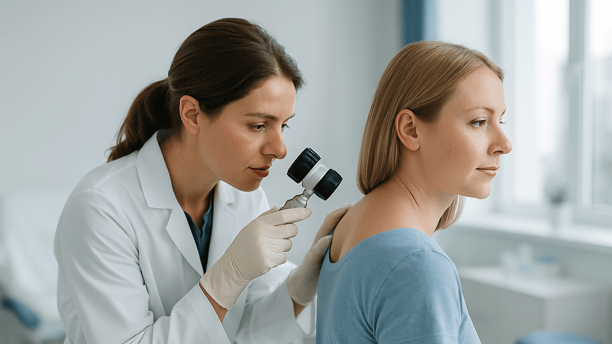
Melanoma does not stop where it looks like it stops. The pigmented spot you or your doctor noticed is the part of the tumor dense enough to show color. Around it, single melanoma cells and tiny clusters can sit in skin that appears completely normal to the naked eye and even under a dermatoscope, the handheld magnifier dermatologists use.
Pathologists have known this for a long time. When a melanoma is removed with only a millimeter or two of surrounding skin, the microscope sometimes shows tumor cells reaching or touching the cut edge. Left behind, those cells can regrow as a local recurrence, and a recurrence in the scar is harder to treat than the original lesion. Mayo Clinic and the NHS both describe the wide local excision as a deliberate second step: the biopsy makes the diagnosis, the excision removes the safety zone.
The zone is called a margin. In plain terms, a margin is the measured distance between the edge of the tumor and the edge of what the surgeon removes. It is planned in two directions, outward across the skin and downward through the fat beneath, so the tumor comes out inside a block of tissue rather than as a thin disc.
Why not take a huge margin and be done with it? For most of the twentieth century, surgeons did exactly that, removing 4 or 5 centimeters around every melanoma and leaving large, disfiguring wounds. Randomized trials from the 1980s onward compared wide and narrow margins and found that, within the ranges now used, narrower margins did not increase the risk of dying from melanoma. That evidence is why today’s margins are measured in millimeters of tumor thickness rather than in the surgeon’s nerves.
Melanoma margins by thickness: the table surgeons work from
The single number that decides your margin is Breslow thickness: the depth of the melanoma in millimeters, measured by a pathologist from the top of the skin’s granular layer to the deepest tumor cell. It comes from the original biopsy, which is why a good initial biopsy matters so much; a shave that clips the bottom of the tumor can leave the thickness uncertain.
Guidelines in the United States and the United Kingdom use very similar tiers. The NHS describes the margin as depending on how thick and how deep the melanoma is, and Mayo Clinic notes that surgeons remove more surrounding tissue for thicker or more aggressive tumors. The ranges below are typical guideline values; your surgeon may adjust them for the site, for example near an eyelid or on a finger.
| Breslow thickness | Typical clinical margin around the tumor | What that means on the skin |
|---|---|---|
| Melanoma in situ (confined to the top layer) | About 0.5 cm, sometimes up to 1 cm | Roughly the width of a pencil eraser on each side |
| Up to 1 mm | 1 cm | About a fingertip’s width on each side |
| Over 1 mm and up to 2 mm | 1 to 2 cm | Surgeon’s judgment within this range |
| Over 2 mm | 2 cm | Roughly the width of two fingertips on each side |
Notice what the table does not contain. It does not widen the margin for larger diameter, darker color or longer history. Depth, not surface size, predicts where stray cells are likely to be. A flat 2 centimeter melanoma in situ may need a smaller margin than a 4 millimeter nodule that is thicker than it is wide.
Keep in mind that the margin is measured from the edge of the tumor, or from the biopsy scar if the tumor has already been removed, so the final ellipse of skin is always wider than the margin figure alone suggests.
How deep is a wide local excision for melanoma?
People picture the excision as skin-deep. It is not. The surgeon removes the full thickness of the skin plus the layer of fat underneath, typically down to the fascia, which is the thin, tough sheet of connective tissue wrapping the muscles. Cleveland Clinic and Mayo Clinic both describe the procedure as removing the tumor along with a margin of normal skin and the tissue beneath it.
The fascia itself is usually left in place. It acts as a natural barrier, and studies have not shown a benefit to removing it routinely. Muscle is not involved unless the tumor has grown into it, which is uncommon for a primary skin melanoma.
So how deep is that in practice? It varies enormously by body site. On the back, where fat is generous, the wound may be 1 to 2 centimeters deep. On the shin or the scalp, where skin sits almost directly on bone or fascia, the depth is a few millimeters and the surgeon may need a skin graft because there is little tissue to pull together.
The shape matters too. Most excisions are cut as an ellipse, roughly three times as long as it is wide. That looks alarming on the consent diagram, but the length is what allows the two sides to meet in a flat line rather than bunching into puckered ‘dog ears’ at each end. A 1 centimeter margin around a 1 centimeter scar therefore produces a wound about 3 centimeters wide and 9 centimeters long. The scar you are left with is that long line, and it fades considerably over 12 to 18 months as collagen remodels.
Ask your surgeon to draw the planned ellipse on your skin before you sign. Seeing it once, calmly, removes a lot of the surprise on the day.
Who is usually offered wide local excision, and who is asked to wait
Almost everyone with a confirmed melanoma of the skin is offered a wide local excision, regardless of stage. The NHS lists it as the main treatment for early melanoma, and even when a melanoma has already spread, removing the primary tumor with a margin is usually still recommended to control disease at the skin.
The exceptions are about timing and sequence rather than whether to operate.
- Staging comes first for thicker tumors. If the biopsy shows a melanoma more than about 1 millimeter thick, or a thinner one with ulceration, your team may arrange a sentinel lymph node biopsy for the same operation. That requires a nuclear medicine scan beforehand, so the excision date may shift by a week or two.
- Uncertain thickness. When a shave biopsy has cut through the base of the tumor, the pathologist may report thickness as ‘at least’ a figure. Some teams re-examine the biopsy or discuss the case at a multidisciplinary meeting before setting the margin.
- Blood thinners and other medicines. Anticoagulants, which are medicines that reduce clotting, raise bleeding risk during surgery. Most skin excisions go ahead without stopping them, but that decision belongs to the prescribing clinician and the surgeon together. Never stop a prescribed medicine on your own before surgery.
- Difficult sites. Melanomas on the face, ear, fingertip or genitals often involve a plastic or specialist surgeon and a reconstruction plan, which can add planning time.
- Pregnancy. Excision under local anesthetic is generally considered safe in pregnancy; it is the imaging and node procedures that need discussion.
Waiting a few weeks for these steps is normal. Melanoma is treated urgently, but a short planned delay to get the operation right is different from a delay in diagnosis.
Wide local excision for melanoma: what to expect on the day
For most people this is an outpatient procedure lasting 30 to 60 minutes, done while you are awake. You will be asked to eat normally unless a general anesthetic or sentinel node biopsy is planned, in which case you will get fasting instructions.
The surgeon begins by measuring. Using a ruler and a skin marker, they draw the margin around the biopsy scar, then extend it into the ellipse. Photographs are often taken for the record. The area is cleaned with antiseptic, which feels cold, and then local anesthetic is injected around the drawn shape. The injection stings and burns for perhaps 10 to 20 seconds; that is genuinely the most uncomfortable moment of the day for most patients, and it passes as the numbness spreads.
Once the skin is numb you will feel pressure, tugging and sometimes an odd sensation of movement, but not sharp pain. If you do, say so; more anesthetic can be added. The surgeon cuts along the marked line, lifts the block of skin and fat away from the fascia, and places it in a labeled container oriented with a stitch or ink so the pathologist knows which edge is which. Small blood vessels are sealed with a cautery device, which makes a faint buzzing and a smell like a blown fuse. Then the wound is closed, usually in two layers: dissolving stitches beneath the skin to take the tension, and finer stitches or glue on the surface.
A dressing goes on, you are given written aftercare instructions, and you can generally go home within an hour. Bring someone to drive if the excision is on your dominant arm or anywhere that makes seatbelts or gear changes awkward. Wear loose clothing that will not rub the dressing.
Is wide local excision painful?
Less than most people fear, and more than a biopsy. During the procedure the local anesthetic blocks pain completely in the great majority of cases. Afterward, the numbing typically lasts 2 to 4 hours, and as it wears off you feel an aching, bruised sensation rather than a sharp one.
Where the wound is makes the biggest difference. Excisions on the back, shoulder or thigh are often surprisingly comfortable because the skin is thick and does not move much. Wounds on the lower leg, foot, hand or scalp tend to throb more, partly because these areas are under tension and partly because gravity pools fluid in a leg that is standing or a hand that is hanging down. Raising a leg on cushions for the first two or three days makes a measurable difference to swelling and to pain.
Simple over-the-counter pain relievers are usually enough; your team will advise which ones suit you, since some, such as anti-inflammatory medicines, can affect bleeding or interact with other prescriptions. Follow their instructions rather than a general rule, and never combine products without checking.
Two sensations often alarm people and are usually normal. The first is numbness or tingling around the scar, caused by tiny skin nerves being cut. It can persist for months and sometimes permanently in a small patch. The second is a pulling tightness when you stretch, which eases as the deep stitches dissolve over 6 to 8 weeks. Pain that gets steadily worse after the second day, rather than better, is not normal and is covered in the section on when to call your doctor.
Why a sentinel lymph node biopsy is sometimes done at the same time
Melanoma spreads first through lymphatic channels to the nearest lymph nodes, the bean-sized filters in the armpit, groin and neck. The sentinel node is the first node that drains the skin where the melanoma sat. A sentinel lymph node biopsy finds that node with a tracer and removes it for examination, so it can be checked for cells that are too few to feel or see on scans.
Guidelines generally consider offering it when the melanoma is around 1 millimeter thick or more, or thinner with high-risk features such as ulceration. The NHS describes it as an option for people whose melanoma is at a stage where node spread is possible, and Mayo Clinic frames it as a staging tool.
What does it actually change? The largest randomized trial, MSLT-I, followed more than 2,000 patients and found that sentinel node biopsy gave more accurate staging and reduced later recurrence in the lymph node basin, but did not show a clear improvement in overall melanoma-specific survival across the whole trial population. That is the honest evidence: the procedure tells you and your team more about your risk, and that information now guides who is offered further treatments such as immunotherapy, which are medicines that help the immune system recognize melanoma cells.
Practically, it means a radioactive tracer injection around the scar a few hours before surgery, a scan to locate the node, a second small incision in the armpit, groin or neck, and usually a general anesthetic. Recovery is longer than for excision alone, and a seroma, a pocket of fluid under the node scar, is a common and usually harmless complication. Whether to have it is a genuine decision with trade-offs, and one to talk through carefully with your team.
How is the wound closed? Stitches, flaps and grafts
Most excisions on the trunk and limbs close in a straight line, a technique surgeons call primary closure. The two edges of the ellipse are undermined slightly, lifted free of the fascia so they slide, and brought together with layered stitches. This gives the neatest scar and the quickest healing.
Sometimes the skin will not stretch that far. The shin, scalp, face and foot have little spare tissue, and a 2 centimeter margin there can leave a gap that cannot be pulled shut without strangling the blood supply. Two alternatives exist.
A local flap moves a tongue of neighboring skin, still attached at one end, to cover the defect. Because it keeps its own blood supply it matches color and thickness well, at the price of a longer, sometimes geometric scar. A skin graft takes a thin sheet of skin from elsewhere, often the thigh or behind the ear, and lays it over the wound like a patch. Grafts heal more slowly, tend to look paler and slightly sunken, and create a second wound at the donor site that can be more uncomfortable than the graft itself. Mayo Clinic notes that grafts are used when a large area must be removed.
Surface stitches are usually removed by a nurse; the timing depends on the site and how much tension the wound is under, and your team will give you a date. Facial wounds generally have stitches out sooner than wounds on the back or legs. Dissolving stitches under the skin simply disappear, though a knot occasionally works its way to the surface weeks later as a small spitting sore, which is a nuisance rather than a problem.
Whichever method is used, the surgeon’s first job is a clear margin. Cosmetic outcome is planned around that, never instead of it.
What to expect after wide local excision for melanoma: the first two weeks
Recovery has a fairly predictable rhythm, though the pace depends on where the wound is and how it was closed. Cleveland Clinic and the NHS both describe healing over a period of weeks, with restrictions easing gradually.
Days 1 to 3. Keep the dressing dry and in place unless told otherwise. Expect aching, mild swelling and some bruising, which can spread surprisingly far under the skin and turn yellow-green before it fades. A small amount of blood staining on the dressing is normal; a dressing that soaks through is not. Rest the area, keep a leg wound elevated, and avoid lifting anything heavier than a kettle with the affected arm.
Days 4 to 7. Most people are back at desk work within a few days if the wound is not on a hand or foot. Showering is usually allowed after 24 to 48 hours, with the wound patted dry; baths and swimming wait until the skin has fully sealed. Itching begins as nerves recover.
Week 2. Surface stitches are typically removed around now for many sites. The scar looks red and raised at this stage, which is normal; it is at its worst appearance roughly six weeks after surgery before it starts to soften and fade.
Weeks 3 to 6. Strenuous exercise, stretching and sport are usually reintroduced gradually, later for wounds on the back and legs than on the arm. Wound strength is still only a fraction of normal skin at this point, so a hard stretch can widen the scar even if it does not open it.
The pathology report typically arrives within one to two weeks of surgery, and its content decides what happens next.
What the pathology report will tell you, and what happens if margins are not clear
The excised block goes to a pathologist who slices it, stains it and examines every edge. The report answers two questions. Was there any residual melanoma in the specimen? And how far is the nearest tumor cell from each cut edge?
‘Clear margins’ means no melanoma cells were found at any edge. In most cases after a properly planned excision, that is exactly what the report says, and the surgical part of your treatment is complete. Follow-up then shifts to skin checks and, for higher-stage disease, discussion of additional treatment.
Occasionally the report is less tidy. Sometimes the original biopsy turns out to have removed the entire tumor, so the wide excision specimen contains only scar. That is good news, not a sign of unnecessary surgery: the margin was still the right thing to do because the microscope, not the eye, decides where melanoma ends.
Less often, tumor cells reach a margin. This happens more with melanoma in situ of the lentigo maligna type, a slow-growing form on sun-damaged skin of the face that spreads outward along the surface in a way that is hard to see. The usual response is a further excision of the involved edge, sometimes after mapping biopsies to define the extent. A second operation is disappointing, but it is a known and planned-for possibility, not a failure.
Your report may also mention mitotic rate, ulceration, regression or lymphovascular invasion. These are microscopic features that refine the stage. Ask your team to walk through the report line by line; you are entitled to a copy, and understanding it makes the follow-up plan feel far less arbitrary.
How long does it take melanoma to go from stage 1 to stage 4?
This is one of the most-searched questions about melanoma, and the honest answer is that there is no clock. Stage 1 means a thin melanoma confined to the skin; stage 4 means it has spread to distant organs. Whether, and how fast, a tumor moves between those points varies from lesion to lesion in ways that no one can predict for an individual.
What the evidence does support is this. Thickness is the strongest single predictor of risk, and thickness increases with time, so an untreated melanoma is more dangerous the longer it is left. Some subtypes behave differently. Superficial spreading melanoma, the most common type, often grows outward across the skin for months or years before growing downward. Nodular melanoma, by contrast, grows vertically from the start and can gain a millimeter of depth in a matter of weeks; Cleveland Clinic and Mayo Clinic both describe it as fast-growing. Lentigo maligna can spread on the surface of the face for a decade before invading.
Once a melanoma has been completely excised with clear margins, it does not ‘continue’ to progress in the skin. The risk that remains is that cells left the primary site before surgery. For a thin stage 1 melanoma that risk is low, which is why guidelines do not recommend routine scans; for thicker tumors the sentinel node biopsy and follow-up schedule are designed to catch spread early.
If you are reading this while waiting for your excision date, the useful takeaway is not to calculate a timeline but to keep the appointment and, in the meantime, note any new lump near the scar or in the nearby lymph node areas so you can report it.
What people often get wrong about wide local excision
‘Cutting into it will make it spread.’ Surgery does not seed melanoma through the body. The concern comes from a misunderstanding of how cancer cells travel; excision removes the source and is the foundation of melanoma treatment in every major guideline.
‘The biopsy already took it all out, so this is just cosmetic.’ The reverse is true. The biopsy is diagnostic; the excision is the treatment. Even when the pathologist finds no residual tumor, the margin removes the zone where invisible cells are most likely to lie.
‘A bigger scar means the cancer was worse.’ Scar length is driven by margin width, which is driven by thickness, and by the ellipse needed to close flat. Two people with identical melanomas can have quite different scars depending on site and skin laxity.
‘If I had waited for it to change more, I would have known it was serious.’ Many melanomas that are treated early looked unremarkable. Thinness at diagnosis is the goal, not a sign the surgery was excessive.
‘I will need chemotherapy.’ For melanoma confined to the skin, surgery is typically the whole treatment. Systemic medicines are considered for higher stages, and modern options are mostly immunotherapies and targeted therapies rather than traditional chemotherapy; whether any are appropriate is a decision for the oncology team based on stage.
‘Sun exposure is fine now that it is removed.’ A history of melanoma raises the risk of a second one on skin elsewhere. Protecting skin from ultraviolet light, and checking it regularly, matters more after diagnosis, not less. The CDC and NHS both emphasize this as part of long-term care.
‘Numbness around the scar means nerve damage that needs fixing.’ It reflects small cutaneous nerves that were unavoidably cut; sensation usually improves over months and rarely needs any intervention.
Questions to ask your care team
Consultations move quickly, and the questions that matter most tend to surface in the car park afterward. Take this list, or your own version of it, and do not hesitate to ask for answers in writing.
- What is my Breslow thickness, and what margin are you planning? Was the biopsy thickness certain, or could the tumor have been deeper?
- Can you draw the planned excision on my skin so I can see the size and orientation of the scar?
- Will the wound close in a straight line, or might I need a flap or graft? If a graft, where would the donor site be?
- Am I a candidate for sentinel lymph node biopsy? What would the result change about my follow-up or treatment options, and what are the risks of the procedure itself?
- Will this be under local or general anesthetic, and do I need someone to accompany me?
- Which of my regular medicines, including blood thinners and supplements, should I continue, and who makes that decision?
- When will I get the pathology result, and who will call me? What happens if the margins are not clear?
- When can I shower, drive, return to work, exercise and swim for a wound in this location?
- What signs of infection or bleeding should prompt a call, and who do I call outside office hours?
- What will my follow-up schedule look like: how often, for how long, and will it include lymph node examination?
- Should I be checking my own skin and lymph nodes between visits, and how?
- Is there anything about my melanoma type or family history that changes the plan?
Write down the answers. A single page with your thickness, stage, margin and follow-up plan becomes a reference you will return to for years.
When to call your doctor
Most excision wounds heal without drama, and mild redness, bruising, itching and a little clear or slightly blood-tinged fluid are all part of normal healing. A few signs are not, and they deserve a same-day call to your surgical team or, outside hours, urgent care.
- Bleeding that does not stop with 15 minutes of firm, continuous pressure, or a dressing that soaks through repeatedly.
- Spreading redness beyond the wound edge, especially with increasing warmth, swelling or a hot, tense feeling; these can indicate infection.
- Pus or thick, cloudy, foul-smelling discharge.
- Pain that worsens after day two rather than improving, or pain out of proportion to the size of the wound.
- Fever, chills or feeling generally unwell in the days after surgery.
- Wound edges that separate or stitches that pull through.
- Rapid, tense swelling under the wound in the first 24 hours, which may be a hematoma, a collection of blood that sometimes needs draining.
- After a sentinel node biopsy: a rapidly enlarging lump, new numbness or weakness in the limb, or calf pain and swelling, which can signal a blood clot.
Beyond the immediate recovery, contact your team promptly if you notice a new lump in or near the scar, a swollen lymph node in the neck, armpit or groin that persists for more than a couple of weeks, or any new or changing mole elsewhere. These are the findings your follow-up schedule exists to catch, and there is no benefit in waiting for the next appointment to mention them.
Every decision about margins, node biopsy, further treatment and follow-up belongs with the treating team who has examined you and read your pathology. This article explains the reasoning; they apply it to you.
Frequently asked questions
How long does it take to recover from a wide excision for melanoma?
Most people return to desk work within a few days and to normal activity over 2 to 6 weeks, depending on the site and closure method. Wounds on the back and lower leg take longest because the skin is under tension. Stitches come out within a few weeks on a date your team sets, and the scar keeps softening and fading for 12 to 18 months.
How deep is a wide local excision for melanoma?
The surgeon removes the full thickness of skin plus the underlying fat, typically down to the fascia, the connective tissue sheet over the muscles. In practice that is 1 to 2 centimeters on a fatty area such as the back and only a few millimeters on the shin or scalp. Muscle is not removed unless the tumor has grown into it, which is uncommon.
Is wide local excision painful?
The local anesthetic injection stings for 10 to 20 seconds; after that most people feel pressure and tugging rather than pain. Once the numbness wears off, expect a bruised, aching feeling for a few days, worse on the lower leg, hand and scalp. Over-the-counter pain relief advised by your team is usually enough, and pain should improve, not worsen, after the second day.
What is the typical wide excision melanoma recovery time by body site?
Face and neck wounds generally heal fastest and have stitches removed soonest. Arm and trunk wounds follow, usually allowing light activity within a week. Lower leg and foot wounds are slowest because of swelling and tension, often needing elevation and restricted walking for the first week or two. Skin grafts anywhere add time and a second donor-site wound.
How are melanoma margins by thickness decided?
The pathologist measures Breslow thickness from the biopsy. Guidelines then set the margin: about 0.5 cm for melanoma in situ, 1 cm for tumors up to 1 mm, 1 to 2 cm for 1 to 2 mm, and 2 cm above 2 mm. Surgeons may adjust slightly for sites such as the face, fingers or eyelids where tissue is limited.
How long does it take for melanoma to go from stage 1 to stage 4?
There is no fixed timeline, and no one can predict it for an individual tumor. Nodular melanoma can deepen within weeks; lentigo maligna may spread on the surface of the face for years before invading. Thickness rises with time, so untreated melanoma becomes riskier the longer it is left, which is why excision is scheduled promptly after diagnosis.
Why do I need a second surgery if the biopsy already removed the mole?
The biopsy was diagnostic and usually took only a millimeter or two of surrounding skin. Melanoma cells can lie invisibly beyond that edge, and leaving them risks a local recurrence in the scar. The wide excision removes a measured safety zone. Finding no residual tumor in the specimen is a good result, not a sign the operation was unnecessary.
What does it mean if my margins are not clear after wide local excision?
It means the pathologist found melanoma cells at or very near a cut edge. This is uncommon after a planned excision but happens more with lentigo maligna type melanoma in situ on the face. The usual response is a further excision of the involved edge, sometimes after mapping biopsies. It is a known possibility, not a failure of the first operation.
Will I need a sentinel lymph node biopsy with my excision?
It is generally considered when the melanoma is around 1 mm thick or more, or thinner with ulceration. It gives more accurate staging and can reduce recurrence in the lymph node basin, but the MSLT-I trial did not show a clear overall survival benefit for everyone, so it is a decision to weigh with your team, including its own recovery time and risk of fluid collection.
How big will the scar be after wide local excision?
Larger than the margin figure suggests, because the wound is cut as an ellipse about three times longer than it is wide so the edges meet flat. A 1 cm margin around a 1 cm biopsy scar produces a wound roughly 3 cm wide and 9 cm long, healing to a single line. Scars look their worst around six weeks and fade over 12 to 18 months.
References
This article is for general information only and is not a substitute for professional medical advice. Please consult a qualified doctor about your individual situation.

More from the Blog

When Is Radioiodine Treatment Chosen for an Overactive Thyroid Over Tablets or Surgery?
Radioiodine is usually chosen for an overactive thyroid when antithyroid tablets have not kept the condition controlled, when hyperthyroidism returns after a course of…

What Happens During a Lutetium-177 PSMA Therapy Session: Infusion, Fluids and Monitoring
A lutetium-177 PSMA treatment session is an outpatient visit of several hours in which a radioactive medicine is given through an intravenous line, usually…

Exercise, Weight and Bone Strength During Hormone Therapy: A Practical Routine
Exercise during hormone therapy for cancer is generally encouraged because these treatments lower estrogen or testosterone, which speeds bone loss, muscle loss and fat…
When Is Mohs Surgery Chosen Over Standard Excision for Skin Cancer? How Surgeons Decide
Surgeons usually choose Mohs surgery over standard excision when a basal cell or squamous cell skin cancer sits in a cosmetically or functionally sensitive…

When Do Scans Show a Response After CyberKnife? Why Follow-Up Imaging Takes Patience
Scans after CyberKnife, a form of stereotactic radiosurgery, usually change slowly. Cancerous tumors often begin to shrink over a few months, while benign tumors…

Inside a Proton Therapy Session: Positioning, Image Checks and the Minutes the Beam Is On
A proton therapy session is usually an outpatient visit of about 15 to 45 minutes, and most of that time goes to positioning you…