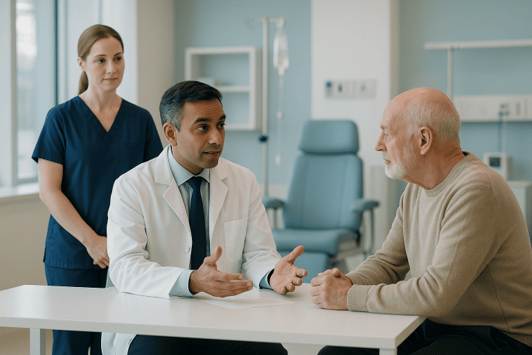
Melanoma Treatment
Melanoma is an aggressive skin cancer that requires early diagnosis, accurate staging and personalized treatment. Care may include surgery, immunotherapy, targeted therapy, radiation therapy or chemotherapy.
Quick answer
Melanoma is a cancer that begins in melanocytes, the pigment-producing cells of the skin. It usually appears as a new or changing mole and can spread to lymph nodes and organs if not removed early. Treatment depends on stage: early melanoma is usually treated with surgical excision, while advanced disease may involve immunotherapy, targeted therapy or radiation therapy alongside surgery.
Melanoma: What It Is and Why Early Recognition Matters
Melanoma is a cancer that begins in melanocytes, the cells that give skin its pigment. It usually develops on the skin as a new mole or a change in an existing one, and it matters more than most skin cancers because it has a greater tendency to spread beyond the original lesion when it is not removed early. Treatment depends on stage: many early melanomas are treated with surgery alone, while thicker or spreading disease calls for a coordinated plan that may combine surgery, immunotherapy, targeted therapy and radiation therapy.
Hearing the diagnosis is unsettling, and most people ask the same questions in the first days: has it spread, how quickly must treatment begin, will surgery be disfiguring, and are the newer medicines relevant to my case. This page answers those clinical questions plainly, and then explains how treatment is actually planned and delivered.
Melanoma behaves differently from one person to another. Some melanomas are detected very early and can often be treated effectively with a single operation. Others are thicker, ulcerated, located in complex areas, or have already reached lymph nodes or distant organs, and these require a treatment plan built across several specialties. That is why accurate staging and individualised decision-making sit at the centre of melanoma care: the name of the diagnosis alone does not determine the treatment.
The outlook for melanoma has changed considerably over the past decade. Advances in imaging, pathology, immunotherapy, molecular testing and multidisciplinary cancer care have altered what is possible for many patients, including some with advanced disease. The goal of modern care is to identify the exact stage and biology of the melanoma, choose treatment based on evidence-based protocols, and preserve quality of life as far as the disease allows.
At Acibadem, melanoma care is planned within an integrated oncology environment. Dermatology, surgical oncology, medical oncology, radiation oncology, nuclear medicine, radiology, pathology, and plastic and reconstructive surgery may all be involved, depending on the stage and location of the disease. This coordination matters because decisions often need to be made efficiently, with full understanding of the medical history, and without repeating tests unnecessarily.
What is melanoma?
Melanoma is a malignant tumour of melanocytes: the cells that produce melanin, the pigment responsible for skin, hair and eye colour. Because melanocytes exist wherever pigment exists, melanoma can arise not only on sun-exposed skin but also on the palms and soles, under the nails, in the eye, and on mucosal surfaces such as the nose, mouth and genital tract. Among all forms of melanoma cancer, cutaneous melanomamelanoma of the skinis by far the most frequent, which is why most public information focuses on moles and skin changes. The disease begins when melanocytes acquire genetic damage that allows them to grow without normal controls; left in place, the abnormal cells can grow downwards through the layers of the skin, reach lymphatic and blood vessels, and travel to lymph nodes and distant organs.
What causes melanoma?
The main cause of melanoma is damage to the DNA of melanocytes, most often from ultraviolet radiation. Sun exposure, particularly intermittent, intense exposure and blistering sunburns, especially in childhood, and the use of sunbeds are the best-established environmental factors. Personal risk is higher in people with fair skin that burns easily, red or blond hair, a large number of moles, atypical (dysplastic) moles, a personal history of melanoma or other skin cancer, a family history of melanoma, or a weakened immune system, for example after organ transplantation. Some inherited gene changes also raise risk in certain families.
Not every melanoma is explained by the sun. Acral melanomas on the palms, soles and nail units, mucosal melanomas and ocular melanomas arise in tissues with little or no ultraviolet exposure, and their causes are less clearly understood. This is worth stating plainly, because people sometimes dismiss a suspicious lesion on the sole of the foot or under a nail on the grounds that the area “never sees the sun”. Melanoma can occur there regardless.
What Does Melanoma Look Like?
Melanoma most often looks like a new pigmented spot, or an existing mole that is changing, growing, darkening, developing uneven colour, or acquiring an irregular edge. But it does not follow a single pattern. It can be flat or raised, brown, black, blue-grey, pink or red, and it can appear as a dark streak under a nail, a sore that will not heal, or a new lesion on skin that has never been sunburned. The single most useful signal is change: a spot that is visibly different from the person’s other moles, or different from what it was a few months ago, deserves professional examination.
What does skin cancer look like?
Skin cancer does not have one appearance, because there are several distinct types. Basal cell carcinoma often looks like a pearly or waxy bump, sometimes with visible small blood vessels, or a flat scar-like patch. Squamous cell carcinoma tends to be a firm red nodule or a rough, scaly, crusted lesion that may bleed. Melanoma is usually, though not always, a pigmented lesion with irregularity in shape or colour. The other skin cancer types are more common than melanoma but far less likely to spread; melanoma is the one where early recognition changes the picture most. Comparing a lesion against skin cancer pictures online can be a reasonable first step, but it has real limits: photographs show typical cases, and the melanomas most often missed are precisely the atypical ones. Galleries of melanoma pictures rarely feature the pink amelanotic variants or nail-unit lesions that are most likely to be mistaken for something harmless. A photograph cannot replace dermoscopy and, where needed, a biopsy.
What are 5 signs of melanoma?
The five classic warning signs of melanoma are summarised by dermatologists as the ABCDE features:
- A: Asymmetry: one half of the lesion does not match the other in shape.
- B: Border irregularity: edges that are ragged, notched, blurred or scalloped rather than smooth.
- C: Colour variation: more than one shade within the same lesion, browns, black, blue-grey, red, pink or even areas of white.
- D: Diameter: a lesion that is larger than most ordinary moles, or one that is clearly increasing in size.
- E: Evolution: any change over time in size, shape, colour, elevation, or new symptoms such as itching, crusting or bleeding.
A sixth, informal sign is often the most practical: the “ugly duckling”. Most of a person’s moles resemble each other; a lesion that stands out from its neighbours, darker, larger, differently textured, warrants attention even if it does not tick every ABCDE box. Equally, melanoma does not always follow these patterns. Nodular melanomas can be symmetrical, uniformly coloured and fast-growing, and amelanotic melanomas carry little or no pigment at all.
What does first stage melanoma look like?
First stage melanoma is a thin tumour confined to the skin, and it usually looks unremarkable: a flat or slightly raised pigmented spot, often smaller than the dramatic lesions shown in textbooks, with only subtle asymmetry or colour variation. Cancerous melanoma moles at this stage can be deceptively quiet, no pain, no bleeding, no obvious growth from week to week, which is exactly why they are found most reliably through deliberate skin checks rather than by chance. Under dermoscopy, a trained clinician can see structural patterns beneath the skin surface that separate an early melanoma from a benign mole far more reliably than the naked eye. The practical message is straightforward: early melanoma rarely announces itself, and waiting for a lesion to “look serious” means waiting for it to become thicker and harder to treat.
Melanoma Symptoms
Melanoma symptoms are often minimal or absent in the early stages, which is the disease’s most dangerous characteristic. When symptoms do occur in a skin lesion, they typically include itching, tenderness, bleeding, crusting, oozing, rapid growth, or a change in the surface or feel of a longstanding mole. A mole that begins to behave differently, becoming raised, fragile, or sensitive, has changed biologically, and change is the signal that matters.
In more advanced disease, symptoms depend on where the melanoma has spread. Patients may notice a swollen or firm lymph node in the neck, armpit or groin, new lumps in or under the skin, unexplained weight loss, persistent pain, headaches or neurological changes if the brain is involved, or breathing symptoms if the lungs are affected. None of these symptoms is specific to melanoma, and all of them have more common, benign explanations, but in a person with a current or previous melanoma diagnosis they are always worth reporting to the treating team.
Does melanoma itch?
Melanoma can itch, but most do not, and most itchy skin lesions are not melanoma. Itch arises when a growing tumour irritates nerve fibres in the skin, so a mole that has recently started itching, particularly one that is also changing in size or colour, deserves examination. The honest answer is that itch alone neither confirms nor excludes melanoma. Many melanomas cause no sensation whatsoever until they are advanced, which is why the visual features and the history of change carry far more diagnostic weight than any symptom.
Types of Melanoma and the Situations Treatment Addresses
Melanoma care addresses a spectrum of disease, from very early tumours confined to the upper layers of the skin to metastatic melanoma involving distant organs. It also spans several distinct subtypes. Superficial spreading melanoma grows outwards across the skin before growing down. Nodular melanoma grows vertically from the outset and can thicken quickly. Lentigo maligna melanoma develops slowly on chronically sun-damaged skin, typically on the face of older adults. Acral lentiginous melanoma arises on the palms, soles and nail units. Amelanotic melanoma, which can belong to any of these groups, carries little pigment and is the variant most often mistaken for something benign. Treatment is adapted to each stage and subtype.
Early-stage cutaneous melanoma is melanoma limited to the skin. It is usually treated with wide local excision, and the need for sentinel lymph node biopsy depends on tumour thickness and other microscopic risk features. The aim is complete removal and accurate risk assessment.
Intermediate or high-risk melanoma includes thicker tumours, ulcerated tumours, or melanomas with other microscopic features associated with a higher chance of recurrence. These patients are often discussed by a multidisciplinary tumour board to determine whether lymph node evaluation, adjuvant drug therapy, or closer surveillance is appropriate.
Regional melanoma involves nearby lymph nodes or in-transit metastases, deposits of melanoma in the skin or soft tissue between the original tumour and the regional lymph nodes. Management may include surgery, systemic therapy, radiation therapy, or localised treatments, depending on the extent of disease and whether it can be removed surgically.
Metastatic melanoma has spread to distant organs such as the lungs, liver, brain, bones, or distant skin and lymph node sites. Treatment usually centres on systemic therapy, especially immunotherapy, or targeted therapy when molecular testing identifies an actionable mutation. Radiation therapy and surgery may also play roles in selected situations, for example to control symptoms or treat isolated deposits.
Acral melanoma occurs on the palms, soles or under the nails. It is often diagnosed later than other melanomas because it can resemble trauma, fungal infection or benign pigmentation, and because these areas are rarely inspected. Treatment requires careful surgical planning to preserve hand and foot function wherever possible.
Mucosal melanoma arises in mucosal tissues such as the nasal passages, mouth, anorectal region or genital tract. It is uncommon and often more complex than skin melanoma, requiring site-specific surgical expertise and multidisciplinary planning from the outset.
Ocular melanoma involves structures of the eye and requires specialised ophthalmic oncology assessment. Its treatment pathway differs from skin melanoma and may involve eye-preserving therapies, surgery, radiation-based approaches and long-term systemic surveillance.
Recurrent melanoma may return near the original scar, in regional lymph nodes, or in distant organs. Recurrence requires a full reassessment, pathology, imaging, prior treatments and molecular profile, before the next evidence-based option is chosen. What worked as a plan at first diagnosis is not automatically the right plan at recurrence.
Who Needs Evaluation and How Melanoma Is Diagnosed
Anyone diagnosed with melanoma needs assessment by an experienced medical team, even when the lesion appears small: a point worth underlining, because thin melanomas can look trivial while still carrying defined risks. Some patients arrive with a biopsy that has already confirmed the diagnosis. Others seek evaluation because of a suspicious mole, a changing lesion, an abnormal spot flagged at a routine dermatology check, or a lymph node that feels wrong.
The diagnostic pathway usually follows a fixed sequence:
- Step 1: Clinical skin examination: the whole skin surface is examined, not only the lesion of concern, because a person with one suspicious lesion may have others.
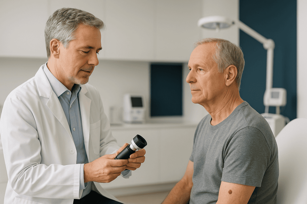
- Step 2: Dermoscopy: a handheld magnifying instrument lets the dermatologist see pigment networks and vascular structures beneath the skin surface, sharply improving the accuracy of the naked-eye assessment.
- Step 3: Biopsy: if melanoma is suspected, the lesion is removed or sampled. Where feasible, the preferred approach is to excise the whole lesion with a narrow margin, so the pathologist can assess its full depth.
- Step 4: Pathology: a pathologist examines the tissue microscopically. The report records the features that drive every subsequent decision, tumour thickness, ulceration, mitotic activity, margin status, and often the subtype.
- Step 5: Staging: depending on the pathology findings, further steps may include lymph node assessment, imaging, and molecular testing to define how far the disease extends.
Patients typically seek melanoma care in one of several situations:
- A suspicious lesion has been found and a biopsy is needed.
- A biopsy has confirmed melanoma and definitive treatment planning is required.
- Initial surgery was performed elsewhere and a second opinion is wanted on margins, staging or additional therapy.
- Sentinel lymph node biopsy or lymph node surgery is being considered.
- Melanoma has returned locally, regionally, or in distant organs.
- Advanced melanoma requires immunotherapy, targeted therapy, radiation therapy or combined treatment.
- The melanoma sits in a cosmetically or functionally sensitive area, face, scalp, hands, feet, nail unit, genital region or mucosal surfaces, where surgical planning and reconstruction need particular care.
Because melanoma treatment decisions affect both survival and quality of life, a structured evaluation matters before therapy begins. For patients who have already had a biopsy or surgery elsewhere, the original biopsy slides, pathology reports, imaging files, operative notes and medication records allow any experienced team to confirm the diagnosis and avoid repeating investigations unnecessarily.
How Melanoma Treatment Is Performed
Melanoma treatment is not a single procedure; it is the complete strategy used to remove, control or manage the disease based on its stage, location, molecular features and the patient’s overall health. It begins with confirmation of the diagnosis and careful staging, designed to answer a fixed set of questions: What type of melanoma is it? How deep is it? Has it reached lymph nodes or organs? Are there molecular features that open or close treatment options? And which approach offers the best balance of disease control, safety, appearance, function and the patient’s personal circumstances?
Preparation and Diagnostic Review
Before treatment, the medical team reviews the biopsy report, prior photographs if available, personal and family history, sun exposure history, previous skin cancers, immune status, current medications and other medical conditions. A full skin examination looks for additional suspicious lesions, and the regional lymph nodes are examined clinically.
When a patient arrives with an outside diagnosis, pathology review is often recommended, particularly if the melanoma is thick, unusual, recurrent, or located in a complex site. Melanoma pathology is genuinely difficult, and an expert second reading of the original slides can confirm the diagnosis and clarify the features that determine what happens next. This is one of the least visible but most consequential steps in the whole pathway.
Imaging is not required for every early melanoma. For higher-risk or symptomatic patients, ultrasound, computed tomography, magnetic resonance imaging, or PET-CT may be used to assess lymph nodes and look for spread. For evaluating the brain in advanced melanoma, MRI is preferred because it shows brain tissue in fine detail. Requesting scans that the stage does not justify adds anxiety and cost without adding safety; a good team explains why imaging is or is not indicated in your specific case.
Molecular testing may be performed in higher-risk or advanced melanoma to identify mutations, most importantly in growth-signalling genes, that can make targeted therapy an option. This testing does not replace staging; it adds a layer of information that can be decisive for treatment selection in stage III and IV disease.
Surgery for Melanoma
Surgery is the cornerstone of treatment for most localised melanomas. The standard operation is wide local excision: the surgeon removes the biopsy scar or residual lesion together with a measured margin of surrounding normal-looking tissue. The recommended margin depends on the tumour’s thickness and location, and its purpose is to remove microscopic disease that may extend beyond what is visible on the skin. For thin melanomas this is often a modest, outpatient procedure.
When the melanoma sits where appearance or function is critical, the face, eyelid region, ear, fingers, toes or sole of the foot, surgical planning involves reconstructive thinking from the start. Plastic and reconstructive surgeons may help close the wound, restore contour and preserve movement, either during the same operation or in a staged approach. Oncological completeness always comes first, but it does not have to come at an avoidable cosmetic or functional price.
Sentinel lymph node biopsy may be recommended for melanomas above certain thickness and risk thresholds. Mapping techniques, a tracer injected around the melanoma site, identify the first lymph node or nodes that drain the area. These sentinel nodes are removed through a small incision and examined microscopically for melanoma cells. The result is primarily a staging tool: it tells the team whether the disease has begun to travel, and it directly shapes recommendations about additional therapy and the intensity of surveillance.
If lymph nodes are clinically enlarged or confirmed to contain melanoma, treatment may combine surgery to remove involved nodes, systemic therapy, and in selected cases radiation therapy. In modern stage III pathways, the sequencing of drug therapy and surgery is itself a deliberate decision, in some situations systemic treatment is given before an operation rather than after it, and this is exactly the kind of question a multidisciplinary tumour board exists to settle.
Immunotherapy, Targeted Therapy, Radiation Therapy and Chemotherapy
Immunotherapy is now central to the treatment of many patients with advanced or high-risk melanoma. These medicines work by releasing the brakes on the immune system so that immune cells can recognise and attack melanoma cells. They may be used after surgery to reduce the risk of recurrence in selected high-risk patients, before surgery in some situations, or as the main treatment for metastatic disease. Because immunotherapy activates the immune system throughout the body, it can cause inflammation-related side effects affecting the skin, bowel, liver, lungs, thyroid or other glands. These effects are manageable when caught early, which is why structured monitoring, not just prescribing, is part of the treatment itself.
Targeted therapy is an option when the melanoma carries specific mutations, most commonly in genes involved in cell growth signalling. These medicines block the abnormal signals driving the cancer’s growth and are usually taken as oral tablets. They can produce meaningful disease control in patients whose tumours carry the relevant mutation, though side effects and the development of resistance over time both require ongoing attention from the treating team.
Radiation therapy uses precisely planned beams to treat disease in a defined area. In melanoma it may be used after surgery where the risk of local or regional recurrence is elevated, to relieve symptoms from metastases, to manage certain lymph node regions, or to treat brain metastases with highly focused stereotactic techniques. Modern planning systems shape the dose around the target while limiting exposure to nearby healthy tissue.
Chemotherapy plays a smaller role in melanoma than it once did, because immunotherapy and targeted therapy are generally preferred when suitable. It has not disappeared entirely: chemotherapy may still be considered in specific circumstances, particularly when other options are not appropriate or have already been exhausted. All of these systemic decisions belong within a broader oncology treatment framework, where the sequence and combination of therapies are chosen deliberately rather than one drug at a time.
How Long Does Melanoma Treatment Take?
The length of melanoma treatment varies enormously with stage. A patient with an early melanoma may complete a wide local excision as an outpatient and recover over several weeks. A patient needing sentinel lymph node biopsy spends additional time in preoperative mapping, surgery and pathology review. Patients receiving immunotherapy, targeted therapy or radiation therapy follow schedules that run over months or longer, with regular monitoring and periodic imaging built in. There is no honest single answer: the pathology and stage set the timetable.
Planning usually begins with a full review of the existing medical records, so that dermatology assessment, imaging, pathology review, surgical and oncology consultations, and tumour board discussion where indicated can be arranged in a logical sequence rather than piecemeal. Where long-term systemic therapy is needed, the team explains how monitoring is typically structured, the rhythm of blood tests, clinic visits and periodic imaging, so that patients and families know what the months ahead will actually involve and can plan around it.
Recovery After Melanoma Treatment
Recovery depends on what was done. After a local excision, patients typically manage straightforward wound care, temporary activity restrictions, and a follow-up visit for suture removal and healing assessment. Where a larger reconstruction was required, wound instructions are more detailed and healing takes longer. After sentinel lymph node biopsy or lymph node surgery, there may be soreness, swelling, drain care in some cases, and specific guidance to reduce the risk of lymphoedema in the affected limb.
Patients on systemic therapy need regular monitoring for both treatment response and side effects: blood tests, physical examinations, symptom review and periodic imaging. Radiation therapy side effects depend on the treated area and may include fatigue, skin changes or site-specific symptoms. The care team provides clear instructions on which changes to report, fever, worsening pain, shortness of breath, diarrhoea, neurological symptoms, new swelling, and reporting early is consistently better than waiting to see whether a symptom settles.
Is Melanoma Curable?
Melanoma that is found early is often removed completely with surgery, and many people treated at an early stage never see the disease return. Clinicians tend to speak in terms of complete removal, remission and long-term surveillance rather than making absolute promises, because melanoma can recur years after treatment and a small ongoing risk justifies continued skin checks. For thicker tumours and disease that has reached lymph nodes, the risk of recurrence is higher, which is why adjuvant drug therapy and closer follow-up enter the picture. In metastatic melanoma, immunotherapy and targeted therapy have made durable, long-lasting disease control a realistic goal for some patients, a genuine change from the era before these medicines, while for others the disease remains difficult to control. The honest summary: stage at diagnosis is the single biggest factor, and it is the one factor a patient can influence by acting on a suspicious lesion promptly.
Is melanoma a fatal cancer?
Melanoma can be fatal, particularly when it has spread to distant organs before diagnosis, but it is not uniformly so, outcome depends primarily on how advanced the disease is when treatment begins. Thin melanomas confined to the skin behave very differently from metastatic disease, and treating a melanoma while it is still thin is precisely what screening, skin awareness and early biopsy are designed to achieve. Modern systemic therapies have also improved what is possible in advanced disease, though they do not help every patient equally. Fear of the diagnosis should never delay evaluation; delay is the one response that reliably makes the situation worse.
Can you live a long life after having melanoma?
Yes, many people treated for melanoma, especially early-stage melanoma, go on to live long lives. What changes after treatment is not so much daily life as the relationship with one’s own skin: survivors carry a higher-than-average risk of developing a second melanoma or other skin cancer, so lifelong sun protection, regular self-examination and scheduled dermatology reviews become permanent habits. Follow-up appointments also monitor for recurrence at the original site, in the regional lymph nodes, and, for higher-stage disease, with periodic imaging. A structured surveillance plan is not a sign that the team expects trouble; it is what allows any trouble to be caught while it is still small.
Why Acting Early Matters
Melanoma is often highly treatable when found early, but it becomes more difficult to control once it grows deeper into the skin or spreads to lymph nodes and organs. Acting early preserves the opportunity for complete surgical removal and accurate staging before the disease becomes complex.
Delaying evaluation of a suspicious lesion allows the melanoma to increase in thickness, and thickness is one of the most important factors used to estimate risk and guide treatment. A delay can also change whether sentinel lymph node biopsy is recommended, and it increases the likelihood that additional therapy beyond surgery will be needed.
Delay is especially risky for melanomas that do not look typical. Amelanotic melanomas, which carry little or no pigment, are mistaken for benign pink bumps. Nail melanomas are attributed to trauma. Acral melanomas on the sole of the foot are treated as warts or calluses for months. Mucosal melanomas cause symptoms that resemble common inflammatory conditions. The pattern in every case is the same: a persistent or changing lesion that keeps being explained away. Persistent or changing lesions deserve medical evaluation, whatever they superficially resemble.
For patients already diagnosed, timely staging matters just as much as timely biopsy. Starting the wrong treatment before the stage or molecular profile is confirmed can complicate every future decision. The goal is a pathway that is careful but efficient, avoiding both unnecessary delay and rushed treatment based on incomplete information.
Benefits of Melanoma Treatment
The benefits of melanoma treatment depend on the stage of disease and the therapies used, but the aims are consistent: accurate control of the cancer, preservation of function and appearance, and a surveillance plan that catches any recurrence early.
| Benefit | What It Means for You |
|---|---|
| Accurate diagnosis and staging | Pathology review, imaging where needed, lymph node assessment and molecular testing define the true extent of disease before any treatment decision is made. |
| Complete removal of localised melanoma | For early melanoma, surgery aims to remove the cancer with appropriate margins while planning the closure to support healing, function and appearance. |
| Personalised treatment selection | Therapy is adapted to tumour thickness, lymph node status, mutation profile, prior treatments, medical history and your own priorities. |
| Access to modern systemic therapies | Immunotherapy and targeted therapy can be considered for high-risk or advanced melanoma when clinically appropriate. |
| Coordinated specialist input | Complex cases are reviewed by the relevant specialists together, so surgery, drug therapy, radiation therapy, reconstruction and follow-up are aligned rather than sequential improvisations. |
| Structured follow-up | Ongoing skin checks, imaging when indicated, and symptom monitoring detect recurrence or new skin cancers as early as possible. |
Recovery Timeline After Melanoma Treatment
Recovery varies with the stage of melanoma and the treatment performed, but the general pattern of care after diagnosis follows a recognisable shape.
| Time Period | What Patients Can Expect |
|---|---|
| Day 1 | After a biopsy or excision, wound care instructions and activity guidance are given. If systemic therapy is starting, baseline blood tests and side effect education are reviewed. |
| First Week | Mild discomfort, swelling, bruising or tightness around the surgical site can occur. Patients are advised to watch for signs of infection and avoid strain on the treated area. |
| First Month | Pathology results are reviewed, sutures may be removed, and any additional treatment decisions are finalised. Patients who had lymph node procedures receive more detailed recovery guidance. |
| During Active Therapy | Patients on immunotherapy, targeted therapy or radiation therapy attend scheduled monitoring visits. Side effects should be reported early, even when they seem mild. |
| Longer Term | Follow-up focuses on skin surveillance, lymph node examination, imaging when appropriate, management of any late effects, sun protection, and education about the signs of recurrence. |
What Influences Outcomes and a Good Result
Melanoma outcomes are shaped by factors assessed at diagnosis and staging: tumour thickness, ulceration, mitotic activity, lymph node involvement, spread to distant organs, the melanoma subtype, and the patient’s overall health. Response to systemic therapy also varies between individuals, and no clinician can promise a specific result in advance.
Early stage at diagnosis is strongly associated with better outcomes. A thin melanoma that is completely removed generally carries a more favourable outlook than a thick melanoma with nodal or distant spread. Even within the same stage, individual risk differs, which is precisely why the fine detail of the pathology report, and multidisciplinary review of it, matter so much.
The quality of the first biopsy and the first operation matters. An appropriate biopsy technique lets the pathologist measure depth accurately; a poor one can make accurate staging impossible. Correct surgical margins reduce the chance of local persistence. Sentinel lymph node biopsy, when indicated, provides staging information that may change the entire treatment plan. For melanomas in complex locations, combined experience in cancer surgery and reconstruction affects function, healing and appearance.
For advanced melanoma, molecular testing and careful treatment sequencing influence results. Some patients are better candidates for immunotherapy; others may benefit from targeted therapy when the relevant mutations are present. In selected cases, systemic therapy before surgery can shrink disease or reveal how it responds. Each of these options has benefits, risks and timing considerations, and choosing between them is genuine specialist work rather than a menu selection.
General health plays its part. Autoimmune disease, organ transplant history, chronic infections, liver or kidney disease and certain existing medications can all affect which treatments are suitable, decisions that belong to the treating doctor with the full picture in front of them. Patients on immunotherapy need close monitoring because immune-related side effects can be serious if not recognised early; patients on targeted therapy need attention to fever, skin effects, heart function in selected cases, liver tests and eye symptoms.
A good result in melanoma care is not defined only by removing a lesion. It includes accurate staging, appropriate therapy, safe management of side effects, thoughtful reconstruction where needed, and a surveillance plan the patient can realistically follow. It also includes clear communication, patients need to understand what was found, what was done, what risks remain, and what should prompt contact with the treating team.
Melanoma Care at Acibadem
Patients facing melanoma usually need more than an appointment with a single physician. They need a reliable diagnostic pathway, access to several specialties working from the same information, clear explanations they can genuinely understand, and a treatment plan that can be shared with every doctor involved in their ongoing care. Acibadem’s melanoma pathway is organised around this kind of coordinated care rather than around individual departments.
Multidisciplinary evaluation matters more in melanoma than in many diagnoses because the required expertise shifts with the stage. A patient with an early lesion primarily needs dermatology, pathology and surgery. A patient with stage III disease needs surgical oncology, medical oncology, nuclear medicine, radiology and radiation oncology input. A melanoma on the face or hand adds reconstructive expertise. Metastatic disease adds molecular testing, systemic therapy planning, brain imaging and supportive care. Tumour boards exist to align these decisions with evidence-based treatment protocols rather than leaving them to whichever specialist a patient happens to see first.
Diagnostic capability underpins the whole process. Dermoscopic evaluation supports the assessment of suspicious lesions. Detailed pathology defines tumour depth and risk features. Cross-sectional, metabolic and ultrasound imaging are used where the stage justifies them. Radiation planning systems shape treatment around defined targets, and surgical mapping supports sentinel lymph node biopsy in the patients who need it. None of this replaces clinical judgement: it equips it.
Supportive care runs alongside the medical pathway rather than after it. Side effect management, wound care education, nutrition guidance during systemic therapy, psychological support and clear written treatment summaries all belong to melanoma care in practice, because a plan a patient cannot tolerate or follow is not a good plan. Clear documentation at each stage also means that every clinician involved later, in surveillance, in managing late effects, in assessing a new skin lesion years on, can see exactly what was found and what was done.
Second opinions are a legitimate part of melanoma care, not a discourtesy to the first team. Some patients want confirmation of the original pathology reading; others want to understand whether immunotherapy, targeted therapy or surgery is the right next step, or whether the sequence should change. A second opinion carries the most value when the melanoma is high-risk, recurrent, metastatic, rare in subtype, or located in a challenging anatomical area: the situations where reasonable experts can genuinely differ and where the details decide.
Preparing for Melanoma Treatment Planning
A melanoma diagnosis deserves urgency, but it deserves clarity more. The most useful next step for any patient, wherever treatment eventually happens, is to establish the exact stage of the disease and to understand which options apply to that stage. For some, this means a focused surgical procedure followed by skin surveillance. For others, it means a longer plan combining surgery with immunotherapy, targeted therapy or radiation therapy.
Certain documents shape the quality of any specialist consultation, and gathering them early saves weeks: the original biopsy and pathology reports, the pathology slides themselves where available, all imaging studies with their reports, operative notes from any previous surgery, and a current medication list. A complete record set allows a new team to confirm the diagnosis, avoid repeating investigations, and move directly to the decisions that matter.
It also helps to know which questions the consultation should answer. What is the exact stage, and what pathology features drive it? Were the surgical margins adequate? Is sentinel lymph node biopsy indicated, and what would its result change? Is molecular testing worthwhile at this stage? What does the surveillance schedule look like afterwards, and which parts of it can be carried out at home? Melanoma care is time-sensitive, but the strongest outcomes come from plans built on complete information, accurate staging, multidisciplinary review, and a strategy matched to the individual rather than to the diagnosis alone.
Preparation
- Preparation usually includes dermatologic examination, biopsy review, imaging for staging and laboratory tests. Your oncology team reviews medications, allergies and previous treatments before creating a personalized plan. If surgery is planned, fasting and anesthesia instructions may be required.
Aftercare
- Aftercare may include wound care, follow-up imaging, skin checks and monitoring for treatment side effects. Patients should protect skin from UV exposure and report new or changing moles promptly. Long-term surveillance is important because melanoma can recur or spread.
Frequently Asked Questions
What mainly affects the cost of melanoma treatment?
The main factors are diagnostic tests, pathology and molecular testing, imaging, surgical complexity, hospital stay, and whether immunotherapy, targeted therapy, radiation therapy or chemotherapy is needed. The final plan must be based on specialist review.
How can I get a personalised quote for melanoma care in Turkey?
You can request a free consultation by sharing your pathology report, biopsy details, imaging results, previous treatment records and current photos if relevant. The medical team can then recommend the next steps and prepare a personalised estimate.
Does a melanoma treatment package include all therapies?
Packages vary by patient and hospital. Some may include consultation, selected diagnostics, surgery and coordination services, while medicines, advanced imaging, radiation therapy or extended follow-up may be quoted separately.
Will I need to travel more than once?
This depends on the treatment plan. Some patients travel for diagnosis review and surgery, while others need ongoing systemic therapy, radiation therapy or follow-up imaging that may require longer stays or coordinated shared care at home.
Is the lowest quote always the best option?
Not necessarily. Patients should consider the completeness of staging, pathology quality, specialist experience, hospital accreditation, medication access, follow-up planning, language support and what is included in the quote. This information is general and is not medical or financial advice.
Medically reviewed by the Acıbadem International Medical Board August 31, 2026
See our medical review board →
Update history
- PublishedJune 8, 2026
- Medical review approvedAugust 31, 2026
- Last content updateSeptember 8, 2026
References3
- Melanoma Treatment (PDQ) - Patient Version cancer.gov
- Melanoma skin cancer nhs.uk
- Melanoma medlineplus.gov
Trusted care for international patients
From the Acibadem Blog
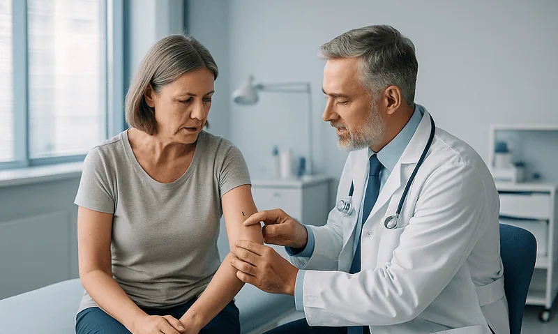
How Fast Melanoma Spreads: Growth Rates, Stages and Why Early Detection Matters
Melanoma spreads at very different speeds. Many melanomas grow slowly for months or even years within the top layer of skin, but once cells…

Targeted Therapy or Immunotherapy for Melanoma: How Mutation Testing Guides the Choice
For melanoma, mutation testing checks the tumor for changes in the BRAF gene. If no BRAF V600 mutation is found, targeted BRAF and MEK…

What a Sentinel Lymph Node Biopsy Tells Your Team About Melanoma Spread
A sentinel lymph node biopsy for melanoma removes the first one or two lymph nodes that drain the skin where the melanoma grew, so…