Eyelid Ptosis
Eyelid Ptosis is drooping of the upper eyelid that may affect vision. Learn symptoms, causes, diagnosis and treatment options.
Quick answer
Eyelid ptosis is a drooping of the upper eyelid that can affect vision and appearance, usually caused by weakness of the lifting muscle, nerve problems, aging, or congenital factors. At Acibadem in Turkey, evaluation focuses on the cause and severity, and treatment may include monitoring, managing underlying conditions, or surgery to tighten or reposition the eyelid muscles.
What is eyelid ptosis?
Eyelid ptosis (pronounced “TOE-sis”) is the medical term for a drooping upper eyelid. In this condition, the edge of the upper eyelid sits lower than normal, and in more noticeable cases it can partially or even completely cover the pupil, the dark opening in the center of the eye that lets light in. Ptosis can affect one eye or both eyes, and the amount of drooping can range from barely visible to severe enough to block vision.
Many people first ask “what is eyelid ptosis?” after noticing a tired or sleepy appearance in photographs, or after a family member points out that one eye looks smaller than the other. It is important to understand that ptosis is not simply loose or wrinkled skin on the eyelid; it involves the position of the eyelid margin itself. Excess skin that hangs over the lid, called dermatochalasis, is a separate condition, although the two can occur together, especially in older adults.
Eyelid ptosis can affect people of any age. When it is present at birth or appears in the first year of life, doctors call it congenital ptosis. When it develops later in life, it is called acquired ptosis. Acquired ptosis becomes more common with age, because the tissues that lift the eyelid can gradually stretch or weaken over time. However, ptosis can also appear suddenly at any age, and a sudden droop can occasionally signal a more serious underlying problem, which is why medical evaluation is important.
Symptoms of eyelid ptosis
The most obvious sign is the drooping eyelid itself, but eyelid ptosis symptoms often go beyond appearance. Common symptoms include:
- A visibly lower upper eyelid on one or both sides, which may make the eyes look uneven or sleepy
- Reduced field of vision, especially in the upper part of what you see, when the lid covers part of the pupil
- Raising the eyebrows or forehead constantly to lift the lids, which can lead to forehead strain and headaches
- Tilting the head back (a “chin-up” posture) to see underneath the drooping lid
- Eye fatigue or heaviness, particularly late in the day or when reading
- Difficulty keeping the eye fully open when tired
How symptoms appear often depends on the type and stage of the condition. In mild ptosis, the lid may sit only slightly lower than usual, and the main concern is often cosmetic or a subtle feeling of heaviness. In moderate to severe ptosis, the lid can cover part or all of the pupil, and vision in that eye becomes clearly restricted. Many people with age-related ptosis notice that the droop worsens gradually over years and is more pronounced in the evening, when the eyelid muscles are tired.
In children with congenital ptosis, the symptoms can be different and, in some ways, more urgent. A young child’s visual system is still developing, and a lid that blocks the pupil can interfere with normal vision development. This can lead to amblyopia, often called “lazy eye,” in which the brain does not learn to use that eye properly. Children with ptosis may also develop astigmatism (an irregular curve of the eye’s surface that blurs vision) because the drooping lid presses on the eye. For this reason, ptosis in children should always be evaluated by an eye doctor, even when it looks mild.
A sudden onset of ptosis, especially when it appears together with double vision, a change in pupil size, eye pain, or weakness elsewhere in the body, is not a typical pattern of ordinary age-related drooping and should be assessed urgently, as explained later in this article.
Causes and risk factors
Understanding eyelid ptosis causes helps explain why treatment differs from person to person. The upper eyelid is lifted mainly by a muscle called the levator muscle, with help from a smaller muscle known as Müller’s muscle. Ptosis occurs when something interferes with these muscles, the nerves that control them, or the tendon-like tissue that connects the levator muscle to the lid.
Common causes include:
- Age-related (involutional) ptosis: The most common cause in adults. Over time, the levator muscle’s attachment to the eyelid stretches or slips, so the lid sits lower even though the muscle itself works normally.
- Congenital ptosis: Present from birth, usually because the levator muscle did not develop fully. It often affects one eye and may run in families.
- Nerve-related (neurogenic) causes: Problems with the nerves controlling the eyelid muscles, such as third cranial nerve palsy (a problem with the nerve that moves the eye and lifts the lid) or Horner syndrome (a disruption of certain nerve pathways that also causes a smaller pupil on the affected side).
- Muscle (myogenic) conditions: Disorders that weaken muscles generally, such as myasthenia gravis, an autoimmune condition in which the connection between nerves and muscles is impaired. Ptosis from myasthenia gravis often varies during the day and may worsen with fatigue.
- Mechanical causes: Extra weight or bulk on the lid, such as a stye, cyst, tumor, or significant swelling, can physically push the lid down.
- Trauma or prior surgery: Injury to the eyelid, or in some cases eye surgery such as cataract surgery, can stretch or damage the lifting structures.
- Long-term contact lens wear: Many years of inserting and removing rigid contact lenses may contribute to stretching of the levator attachment in some people.
Risk factors therefore include older age, a family history of congenital ptosis, previous eye or eyelid surgery, eye injury, long-term contact lens use, and certain neurological or muscular conditions. Diabetes and other conditions affecting blood vessels can, in some cases, contribute to nerve-related ptosis.
Diagnosis
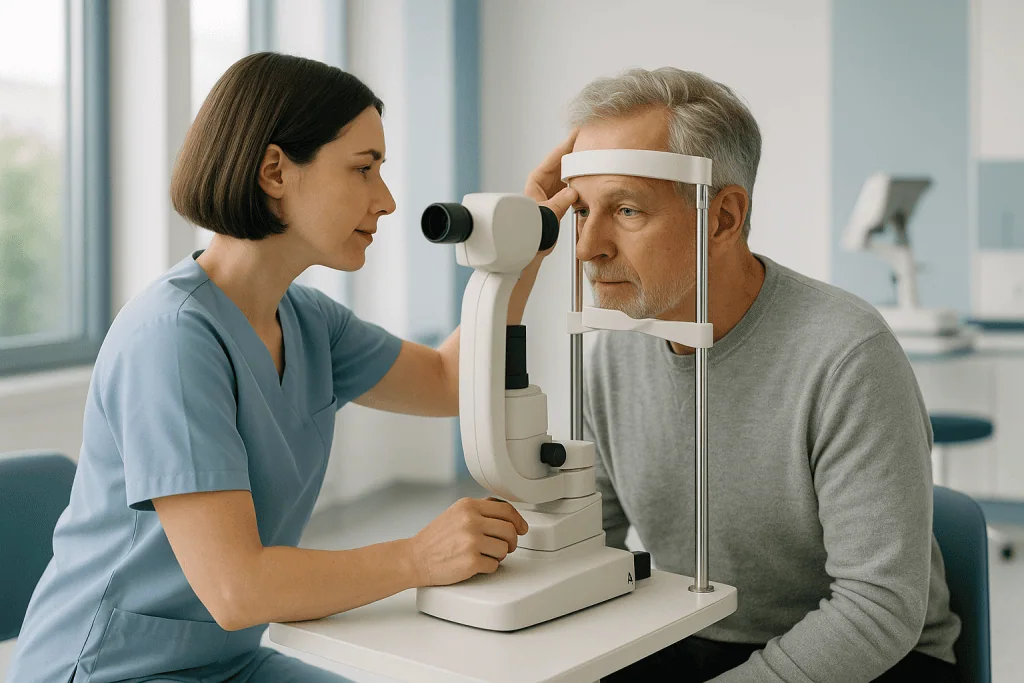
Eyelid ptosis diagnosis begins with a careful history and a detailed eye examination, usually performed by an ophthalmologist, a physician who specializes in eye and eyelid conditions. The doctor will typically ask when the drooping started, whether it changes during the day, whether it affects one or both eyes, and whether there are other symptoms such as double vision, difficulty swallowing, or muscle weakness. Old photographs can be surprisingly useful, because they help show whether the droop is new or long-standing.
During the examination, the doctor takes several standard measurements:
- Margin reflex distance (MRD): The distance between the center of the pupil’s light reflection and the edge of the upper eyelid. A smaller distance indicates more drooping.
- Palpebral fissure height: The vertical opening between the upper and lower eyelids.
- Levator function: How far the eyelid can move when the patient looks from down to up while the forehead muscle is held still. This measurement is important because it strongly influences which surgical technique, if any, is most appropriate.
The doctor will also examine the pupils, eye movements, and the surface of the eye, and will usually check vision in each eye. In adults, a visual field test, a test that maps how much of your surroundings you can see, may be performed with the eyelid in its natural position and again with the lid taped up. This helps document how much the ptosis is interfering with sight, which is relevant when surgery is being considered for functional (vision-related) reasons.
Additional tests are ordered only when the examination suggests an underlying cause beyond simple stretching of the eyelid tissues. For example, if myasthenia gravis is suspected, blood tests for specific antibodies and other specialized tests may be arranged. If a nerve problem such as third nerve palsy or Horner syndrome is suspected, particularly with sudden onset, pupil changes, or double vision, the doctor may order imaging of the brain, orbit (eye socket), neck, or chest, such as MRI or CT scans, to look for the cause. In children, the evaluation also includes checking for amblyopia and astigmatism so that vision development can be protected.
Treatment options
Eyelid ptosis treatment depends on the cause, the severity of the drooping, how much it affects vision, and, in children, the risk to visual development. There is no single approach that fits everyone, and the first step is always identifying and addressing any underlying condition.
Watchful waiting
Mild ptosis that does not block vision and does not bother the patient often does not require any treatment. In these cases, the doctor may simply recommend periodic monitoring to check whether the drooping is progressing. In children with mild congenital ptosis, regular eye examinations are important to make sure vision is developing normally, and treatment can be deferred as long as the lid is not covering the pupil and no amblyopia is developing.
Treating the underlying cause
When ptosis results from another condition, treating that condition comes first. For example, ptosis caused by myasthenia gravis is usually managed with medications for that disease rather than eyelid surgery, and the lid position often improves as the condition is controlled. Ptosis caused by a stye, cyst, or swelling typically improves when that problem is treated. Some cases of nerve-related ptosis improve on their own over months as the nerve recovers, so doctors may recommend waiting before considering surgery.
Medication
For certain adults with mild to moderate acquired ptosis, prescription eye drops that temporarily stimulate Müller’s muscle can lift the eyelid by a small amount for several hours. These drops do not correct the underlying anatomy and must be used regularly to maintain the effect; they are not suitable for everyone, and your doctor can advise whether they are an option in your case.
Surgery
Surgery is the main long-term treatment for ptosis that interferes with vision or causes significant symptoms. The choice of technique depends largely on how well the levator muscle works:
- Levator advancement or resection: When the levator muscle functions reasonably well, the surgeon tightens or reattaches it to lift the lid. This is the most common operation for age-related ptosis and is often performed under local anesthesia in adults.
- Müller’s muscle surgery: For milder ptosis with good muscle function, a procedure that shortens Müller’s muscle from the inside of the lid may be used.
- Frontalis sling: When levator function is poor, as in many cases of congenital ptosis, the surgeon connects the eyelid to the forehead (frontalis) muscle using a small sling of tissue or medical material, so raising the eyebrow lifts the lid.
Like all surgery, ptosis repair carries risks, including under-correction, over-correction, asymmetry between the two eyes, dry eye or difficulty fully closing the eye, bleeding, and infection. In some cases a second adjustment procedure is needed. Recovery usually involves bruising and swelling for a period of weeks, and the final lid position may take some time to settle. Your surgeon will explain the expected course based on your specific situation.
Ptosis is generally evaluated and managed within an ophthalmology service, often by oculoplastic specialists, ophthalmologists with additional training in eyelid surgery. At Acibadem, this condition is managed by the ophthalmology department, where assessment, non-surgical management, and surgical repair can be coordinated depending on the individual case.
Supportive measures
For people who cannot or prefer not to have surgery, simple supportive options exist. Special glasses fitted with a small bar, sometimes called a ptosis crutch, can hold the lid up mechanically, and lubricating drops may relieve eye discomfort. These measures do not correct the ptosis but can improve daily comfort and function in selected cases.
Living with eyelid ptosis and outlook
The outlook for eyelid ptosis is generally good, but it varies with the cause. Age-related ptosis tends to progress slowly and does not damage the eye itself; the main effects are on vision through the upper field, appearance, and comfort. When surgery is performed for this type of ptosis, most people experience a meaningful improvement in lid position and field of vision, although results vary from person to person and no outcome can be guaranteed.
Congenital ptosis, when monitored and treated appropriately, usually allows normal visual development, though children may need glasses, patching for amblyopia, or more than one operation as they grow. Ptosis caused by neurological or muscular disease follows the course of the underlying condition: it may fluctuate, improve with treatment of that condition, or persist.
Day to day, people living with untreated or partially treated ptosis often find practical adjustments helpful, such as positioning reading material or screens slightly lower to reduce the need to look upward, ensuring good lighting, and taking breaks when the eyes feel heavy. Because chronic brow-raising can cause forehead tension and headaches, some people find symptoms improve after the lid position is corrected. If you drive, it is worth discussing with your doctor whether the drooping affects your visual field enough to matter for road safety.
It is also normal for ptosis to affect self-image, particularly when it is noticeable in one eye. Discussing both the functional and appearance-related aspects openly with your doctor helps ensure the management plan addresses what matters most to you.
Frequently asked questions
What is eyelid ptosis in simple terms?
Eyelid ptosis is a drooping of the upper eyelid, so that the lid edge sits lower than it should and may partly cover the pupil. It can affect one or both eyes and can be present from birth or develop later in life. It is different from loose eyelid skin, although the two can occur together, and an eye doctor can tell them apart during an examination.
Can eyelid ptosis heal on its own?
It depends on the cause. Age-related ptosis, caused by stretching of the lid-lifting tissues, does not usually improve on its own and often progresses slowly. However, some ptosis caused by nerve problems, temporary swelling, or certain medical conditions can improve over weeks to months, either spontaneously or as the underlying condition is treated. A doctor can help determine which situation applies to you.
How serious is eyelid ptosis?
In many cases, ptosis is not dangerous in itself: the main concerns are blocked vision, eye strain, and appearance. However, a sudden droop can occasionally be a warning sign of a serious neurological problem, especially if it appears with double vision, an unequal pupil, headache, or weakness. In children, untreated severe ptosis can interfere with vision development. This is why any new or changing ptosis deserves a proper medical evaluation.
What causes a drooping eyelid to develop suddenly?
Sudden ptosis may result from nerve problems such as third nerve palsy or Horner syndrome, from conditions like myasthenia gravis, or from injury or swelling of the lid. Because some of these causes need urgent attention, a sudden droop, particularly with double vision, pupil changes, or pain, should be assessed promptly rather than watched at home.
Is surgery the only treatment for eyelid ptosis?
No. Mild ptosis may simply be monitored, ptosis caused by another condition is treated by addressing that condition, and certain adults may be candidates for prescription eye drops that temporarily lift the lid. Supportive options such as ptosis-crutch glasses also exist. Surgery is the main long-term option when the drooping significantly affects vision or quality of life, and the best approach depends on the cause and on how well the eyelid muscle works.
What is recovery like after ptosis surgery?
Recovery experiences vary, but bruising and swelling of the eyelid for a period of weeks are common, and the final lid position may take time to settle. Doctors typically advise avoiding rubbing the eye and strenuous activity for a time, and lubricating drops or ointment may be recommended, since the eye sometimes does not close completely at first. Your surgeon will give instructions specific to the procedure performed, and follow-up visits are used to check healing and lid position.
Can children outgrow congenital ptosis?
Congenital ptosis usually does not disappear on its own, because it typically involves underdevelopment of the muscle that lifts the lid. Mild cases may only need monitoring, but regular eye examinations are important to detect amblyopia or astigmatism early. If the lid threatens vision development, doctors may recommend surgery in childhood; the timing depends on how severe the droop is and how the child’s vision is developing.
When to see a doctor
Any new, worsening, or bothersome eyelid droop is a reasonable reason to arrange an eye examination, and children with any degree of ptosis should always be evaluated. However, certain warning signs suggest a potentially serious cause and call for urgent medical attention:
- Sudden onset of a drooping eyelid, over hours or days rather than months or years
- Double vision or difficulty moving the eye normally
- Unequal pupilsone pupil noticeably larger or smaller than the other
- Severe or sudden headache, eye pain, or pain around the eye accompanying the droop
- Weakness of other muscles, such as trouble swallowing, speaking, breathing, or holding the head up, or drooping that clearly worsens with fatigue
- Ptosis after a head or eye injury
- In a child, a lid covering the pupil, a constant chin-up head posture, or any sign that vision is affected
If a sudden eyelid droop occurs together with double vision, a dilated pupil, or severe headache, seek emergency care immediately, as these can point to conditions that require rapid diagnosis and treatment. For gradual, long-standing drooping without warning signs, a routine appointment with an ophthalmologist is the appropriate next step, so the cause can be confirmed and suitable options discussed.
From the Acibadem Blog
Horner Syndrome: One Droopy Eyelid, Three Nerve Clues
Horner syndrome is a rare condition marked by a drooping upper eyelid, a smaller pupil, and reduced sweating on one side of the face.…
Medically reviewed by the Acıbadem International Medical Board September 3, 2026
See our medical review board →
Update history
- PublishedJune 8, 2026
- Medical review approvedSeptember 3, 2026
- Last content updateSeptember 3, 2026