Meningioma
Meningioma is usually a slow-growing tumor of the brain or spinal cord lining. Learn symptoms, causes, diagnosis and treatment options.

Quick answer
Meningioma is a usually slow-growing tumor that arises from the membranes surrounding the brain or spinal cord, and treatment depends on its size, location, growth, and symptoms. At Acibadem in Turkey, evaluation may include neurological assessment and advanced imaging, with management ranging from monitoring to surgery and, when appropriate, radiotherapy.
What is meningioma?
A meningioma is a tumor that grows from the meninges, the thin layers of tissue that cover and protect the brain and spinal cord. Although meningiomas are often called brain tumors, they do not actually grow from brain tissue itself. Instead, they arise from the protective membranes and can press on the brain, the spinal cord, nerves, or nearby blood vessels as they enlarge.
Most meningiomas are benign, meaning they are not cancerous and grow slowly. A smaller number are classified as atypical, which means they grow faster and are more likely to return after treatment, and a small minority are malignant (cancerous). Doctors grade meningiomas from grade 1 (benign and slow-growing) to grade 3 (malignant and faster-growing) based on how the tumor cells look under a microscope.
Meningiomas are among the most common tumors that form inside the skull in adults. They are diagnosed more often in women than in men, and the risk increases with age, with most cases found in people over 40. Some meningiomas are discovered by chance during a brain scan performed for an unrelated reason, because small, slow-growing tumors may cause no symptoms at all for many years.
Understanding what is meningioma and how it behaves helps patients and families make sense of the treatment choices their care team may offer. Because many meningiomas grow very slowly, not every tumor needs immediate treatment, and in some cases careful monitoring is a reasonable medical approach.
Symptoms of meningioma
Meningioma symptoms depend mainly on where the tumor is located, how large it is, and how quickly it grows. Because most meningiomas grow slowly, symptoms often develop gradually over months or even years. In many cases, small tumors cause no symptoms at all and are found incidentally on imaging done for another reason.
Common meningioma symptoms may include:
- Headaches that gradually become more frequent or severe, often worse in the morning
- Seizures, which can be the first sign of a meningioma in someone with no history of epilepsy
- Vision changes, such as blurred vision, double vision, or partial loss of vision
- Hearing loss or ringing in the ears (tinnitus)
- Weakness or numbness in an arm or leg
- Memory problems or difficulty concentrating
- Changes in personality or behavior that family members may notice first
- Loss of smell, particularly with tumors near the base of the front of the skull
- Difficulty with speech or language
- Balance problems or unsteadiness when walking
The location of the tumor strongly influences which symptoms appear. A meningioma pressing on the optic nerve (the nerve that carries visual information from the eye to the brain) may cause vision problems, while one near the areas that control movement may cause weakness on one side of the body. Meningiomas that grow along the spinal cord can cause back pain, weakness, or numbness in the limbs.
Higher-grade (atypical or malignant) meningiomas tend to cause symptoms sooner and more severely, because they grow faster and are more likely to swell the surrounding brain tissue. However, symptoms alone cannot tell doctors the grade of a tumor; that requires imaging and, in most cases, examination of tumor tissue.
It is important to remember that all of these symptoms can be caused by many conditions other than a meningioma. Only a medical evaluation can determine the cause.
Causes and risk factors
In most cases, doctors cannot identify a specific cause for an individual meningioma. Research suggests that these tumors develop when certain cells in the meninges acquire genetic changes that allow them to multiply abnormally, but what triggers those changes is usually unknown. Still, several factors are known to increase risk. Recognized meningioma causes and risk factors include:
- Radiation exposure. Previous radiation therapy to the head, especially in childhood, is the most clearly established environmental risk factor. Tumors related to radiation may appear many years after the exposure.
- Female sex and hormones. Meningiomas are more common in women, and some tumors carry receptors for female hormones such as progesterone. Researchers continue to study possible links between hormone exposure and meningioma growth, and some tumors have been observed to change in size during pregnancy.
- Increasing age. Meningiomas become more common with age and are most often diagnosed in middle-aged and older adults.
- Inherited conditions. Neurofibromatosis type 2 (a genetic condition that causes tumors to grow on nerves) significantly increases the risk of developing meningiomas, sometimes more than one.
- Obesity. Some studies have found an association between higher body weight and meningioma risk, although the reason for this link is not fully understood.
Having one or more risk factors does not mean a person will develop a meningioma, and many people diagnosed with the condition have no identifiable risk factors at all. There is no proven way to prevent meningiomas.
Diagnosis of meningioma
Meningioma diagnosis usually begins when a doctor evaluates symptoms such as persistent headaches, seizures, or neurological changes, or when a tumor is discovered by chance on a brain scan. The diagnostic process typically includes several steps.
Neurological examination
A doctor first performs a neurological examination, which tests vision, hearing, balance, coordination, strength, reflexes, and thinking. This helps identify which part of the nervous system may be affected and guides further testing.
Imaging tests
Imaging is the cornerstone of diagnosis. The most important tests are:
- Magnetic resonance imaging (MRI). MRI uses magnetic fields, not radiation, to create detailed pictures of the brain and spinal cord. An MRI with contrast dye is the preferred test, because meningiomas typically take up the dye in a characteristic pattern along the meninges. MRI also shows the tumor’s size, location, and relationship to nearby structures.
- Computed tomography (CT). A CT scan uses X-rays to create cross-sectional images. It may be used when MRI is not possible, and it is helpful for showing calcification (calcium deposits) within a tumor or changes in the nearby skull bone, which are common with meningiomas.
In many cases, the appearance of the tumor on MRI is so typical that doctors can be reasonably confident it is a meningioma based on imaging alone, particularly for small tumors that are being monitored rather than treated.
Biopsy and tumor grading
The only way to confirm the diagnosis with certainty and determine the tumor’s grade is to examine tumor tissue under a microscope. In most cases, this tissue is obtained during surgery to remove the tumor rather than through a separate biopsy. A pathologist (a doctor who studies tissue samples) then assigns a grade from 1 to 3, which helps guide decisions about further treatment and follow-up. In some centers, additional molecular tests on the tumor tissue may provide more information about how the tumor is likely to behave.
At large hospital groups such as Acibadem, meningioma care is typically coordinated by the neurosurgery department, working together with neurology, radiation oncology, and radiology specialists.
Treatment options for meningioma
Meningioma treatment depends on the tumor’s size, location, and grade, how quickly it appears to be growing, the symptoms it causes, and the patient’s age and overall health. Because many meningiomas grow slowly, treatment is not always needed right away. The main approaches are described below.
Active monitoring (watchful waiting)
For small meningiomas that cause no symptoms, doctors often recommend observation with regular MRI scans rather than immediate treatment. Scans are typically repeated at set intervals to check whether the tumor is growing. If the tumor remains stable and causes no problems, monitoring may continue for years. If it grows or begins to cause symptoms, treatment can then be considered. This approach spares many patients the risks of surgery or radiation for a tumor that might never cause harm.
Surgery
Surgery is the main treatment for meningiomas that are growing or causing symptoms. The goal is to remove as much of the tumor as safely possible, along with the portion of the meninges from which it grew. Complete removal often provides long-term control, and for many grade 1 tumors no further treatment is needed afterward.
However, complete removal is not always possible. Some meningiomas grow close to critical structures, such as major blood vessels, important nerves, or the brainstem, and removing every part of the tumor could cause serious harm. In these situations, surgeons may remove most of the tumor and leave a small portion behind, which can then be monitored or treated with radiation. Like all brain and spinal surgery, meningioma surgery carries risks, including infection, bleeding, and neurological problems, and your surgical team will discuss these risks in detail before any operation.
Radiation therapy
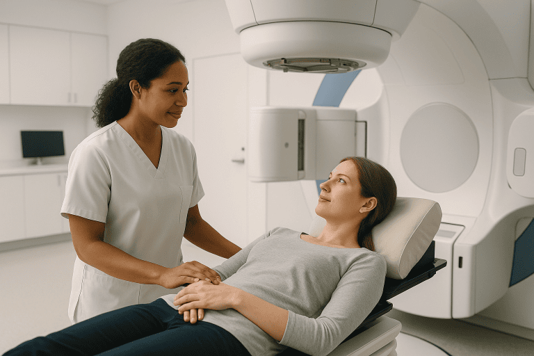
Radiation therapy uses precisely targeted beams of energy to damage tumor cells and stop them from growing. It may be recommended:
- After surgery, when part of the tumor could not be removed or when the tumor is atypical or malignant
- Instead of surgery, when an operation would be too risky because of the tumor’s location or the patient’s health
- When a tumor returns after previous treatment
One commonly used form is stereotactic radiosurgery, which, despite its name, is not surgery. It delivers a highly focused dose of radiation to the tumor in one or a few sessions while sparing surrounding healthy tissue. Conventional radiation therapy, given in smaller doses over several weeks, may be used for larger tumors or those near sensitive structures. The choice between these approaches depends on the individual case.
Medications
There is currently no medication that reliably shrinks or cures meningiomas. Drug treatment is generally reserved for tumors that continue to grow despite surgery and radiation, and it may be offered within clinical trials. Medications are, however, often used to manage symptoms: anti-seizure drugs can control seizures, and corticosteroids (medicines that reduce swelling) may temporarily relieve symptoms caused by swelling around the tumor.
Follow-up care
Whatever treatment is chosen, long-term follow-up with periodic MRI scans is a standard part of care, because meningiomas can return even after apparently complete removal. Higher-grade tumors require closer monitoring.
Living with meningioma and outlook
The outlook for people with meningioma is generally favorable, particularly for grade 1 tumors that can be fully removed. Many patients return to their usual activities after treatment and live full lives. That said, every case is different, and no doctor can guarantee a specific outcome.
Several factors influence prognosis, including the tumor’s grade, its location, whether it could be completely removed, and the patient’s age and overall health. Atypical and malignant meningiomas are more likely to come back and often require additional treatment and closer surveillance. Even benign tumors can recur, which is why long-term imaging follow-up is important.
Living with a meningioma, or recovering from its treatment, can involve practical and emotional challenges. Some people experience fatigue, difficulty concentrating, or mood changes after surgery or radiation, and recovery times vary widely. Rehabilitation services such as physical therapy, occupational therapy, or speech therapy can help when the tumor or its treatment has affected movement, daily activities, or communication. People being monitored without treatment sometimes describe anxiety around scan appointments; discussing these feelings with the care team, a counselor, or a patient support group can help.
Practical steps that many patients find helpful include keeping a record of symptoms, attending all scheduled follow-up scans, taking anti-seizure medication exactly as prescribed if it has been recommended, and asking the care team about driving restrictions, which may apply after a seizure or brain surgery depending on local regulations.
Frequently asked questions
What is meningioma in simple terms?
A meningioma is a tumor that grows from the meninges, the protective layers of tissue covering the brain and spinal cord. It is not a tumor of the brain tissue itself, but it can press on the brain or nerves as it grows. Most meningiomas are benign, meaning they are not cancerous and tend to grow slowly, although a smaller number grow faster and behave more aggressively.
Is a meningioma cancer?
In most cases, no. The majority of meningiomas are grade 1, which means they are benign and slow-growing. However, some meningiomas are atypical (grade 2) and a small percentage are malignant (grade 3), meaning they are cancerous and grow more quickly. The grade can only be confirmed by examining tumor tissue under a microscope, which is usually done after surgical removal.
How serious is a meningioma?
The seriousness varies widely. Many small meningiomas cause no symptoms and may never need treatment beyond regular monitoring. Others can cause significant problems, such as seizures, vision loss, or weakness, depending on their size and location. Even a benign tumor can be serious if it presses on critical parts of the brain or spinal cord. Your doctor can explain what your specific tumor’s size, location, and growth pattern mean for you.
Can a meningioma go away on its own?
Meningiomas do not typically disappear on their own. Some remain stable for many years without growing, and in older adults a small tumor may never cause problems during their lifetime. In rare situations, a tumor may appear to shrink slightly, but this is not the usual course. If a meningioma needs to be removed or controlled, surgery or radiation therapy is generally required.
What are the first symptoms of a meningioma?
Early meningioma symptoms are often subtle because these tumors usually grow slowly. Gradually worsening headaches, a first-ever seizure in adulthood, slowly progressing vision or hearing changes, mild weakness or numbness, and changes in memory or personality are among the more common early signs. Many meningiomas cause no symptoms at all and are found incidentally on a scan done for another reason.
What is the recovery like after meningioma surgery?
Recovery varies depending on the tumor’s size and location, the extent of the surgery, and the patient’s overall health. Many people spend several days in the hospital and then continue recovering at home over weeks to months. Fatigue is common at first, and some patients need rehabilitation to regain strength, coordination, or speech. Your surgical team will give you individualized guidance about activity, medications, and follow-up scans.
Can a meningioma come back after treatment?
Yes, recurrence is possible, which is why long-term follow-up imaging is part of standard care. The risk of a meningioma returning depends largely on its grade and whether it was completely removed. Grade 1 tumors that are fully removed come back less often, while atypical and malignant tumors have a higher chance of recurring and may need additional treatment such as radiation therapy.
When to see a doctor
If you have been diagnosed with a meningioma, follow the monitoring and follow-up schedule your care team recommends, and report any new or changing symptoms between appointments. If you have not been diagnosed but experience persistent or worsening neurological symptoms, a medical evaluation is important, because early assessment allows more treatment options.
Seek urgent medical attention if you or someone near you experiences any of the following red-flag warning signs:
- A seizure, especially a first-ever seizure in an adult
- A sudden, severe headache unlike any experienced before, or a headache accompanied by vomiting, confusion, or drowsiness
- Sudden weakness or numbness in the face, arm, or leg, particularly on one side of the body
- Sudden vision loss, double vision, or other rapid changes in eyesight
- Sudden difficulty speaking or understanding speech
- Sudden loss of balance, severe dizziness, or inability to walk normally
- Increasing drowsiness, confusion, or unresponsiveness
- After brain surgery: fever, worsening headache, redness or fluid leaking from the wound, or new neurological symptoms
These symptoms can indicate a medical emergency, whether or not they are related to a meningioma, and should never be ignored. For non-urgent concerns, such as gradually worsening headaches, subtle memory changes, or slowly progressing hearing or vision problems, arrange a medical appointment so the cause can be properly evaluated. Conditions such as meningioma are typically assessed and managed by neurology and neurosurgery specialists, including the relevant departments at hospital groups such as Acibadem.
Medically reviewed by the Acıbadem International Medical Board September 2, 2026
See our medical review board →
Update history
- PublishedJune 8, 2026
- Medical review approvedSeptember 2, 2026
- Last content updateSeptember 2, 2026