18 Week Ultrasound: A Complete Medical Overview
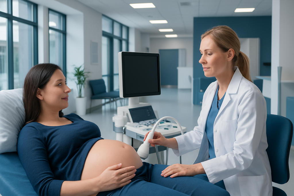
The 18 week ultrasound is often part of the mid-pregnancy anatomy scan. It evaluates fetal growth, major organs, limbs, placenta, amniotic fluid, and often the cervix.
Key Takeaways
- The 18 week ultrasound is often part of the mid-pregnancy anatomy scan.
- It evaluates fetal growth, major organs, limbs, placenta, amniotic fluid, and often the cervix.
- Most scans are reassuring, but some findings may need repeat imaging or specialist review.
- The scan cannot detect every condition, and image quality can vary with fetal position and other factors.
- Preparation is usually simple, and the exam is painless and noninvasive.
An 18 week ultrasound is a detailed second-trimester scan used to assess fetal development, examine major organs and body structures, and review the placenta, cervix, and amniotic fluid. It is a routine part of prenatal care for many pregnancies and helps guide the next steps if any findings need closer follow-up.
Overview: what an 18 week ultrasound is
An 18 week ultrasound is a detailed imaging examination performed during the second trimester of pregnancy. In many care settings, it forms part of the routine mid-pregnancy anatomy scan, usually done between about 18 and 22 weeks. The main purpose is to look carefully at the baby’s growth and body structures, while also checking the placenta, amniotic fluid, and sometimes the cervix.
This scan uses sound waves, not radiation. A trained sonographer or doctor moves a probe over the abdomen to create images of the pregnancy on a screen. The examination is generally painless, although slight pressure on the abdomen may be felt while the sonographer tries to obtain clear views.
For many expectant parents, this visit is memorable because the baby’s features and movements can often be seen more clearly than in earlier scans. Medically, however, the value of the 18 week ultrasound lies in its systematic review of anatomy and pregnancy structures. It can provide reassurance when development appears typical and help identify concerns that may benefit from monitoring, further tests, or consultation with a specialist.
What the scan checks
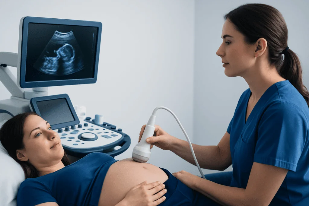
The 18 week ultrasound is more detailed than an early dating scan. It usually includes measurements of the baby’s head, abdomen, and thigh bone to assess growth. The heart is reviewed for basic structure and rhythm, and the sonographer also examines the brain, face, spine, stomach, kidneys, bladder, abdominal wall, arms, legs, hands, and feet as far as visibility allows.
The scan also assesses the pregnancy environment. This includes the location of the placenta, the amount of amniotic fluid, and the appearance of the umbilical cord. In many cases, the cervix is measured because cervical length can give useful information about the risk of early cervical shortening in some pregnancies.
If the baby’s position allows, fetal sex may sometimes be seen, but this is separate from the medical purpose of the test and depends on local practice and parental preference. It is also important to understand that an ultrasound is very useful but not perfect. Some conditions are subtle, develop later, or cannot be seen clearly because of fetal position, maternal body habitus, abdominal scarring, or limited image windows.
- Fetal growth and estimated gestational development
- Brain, spine, face, heart, abdomen, kidneys, and bladder
- Arms, legs, hands, and feet
- Placenta location and appearance
- Amniotic fluid volume
- Cervical length when appropriate
Why this timing matters in pregnancy

The timing of the 18 week ultrasound is medically useful because the baby is developed enough for many anatomical structures to be visible, while still early enough for the healthcare team to plan follow-up if needed. This balance makes the second trimester an ideal period for a detailed survey of anatomy.
At this stage, the scan can help confirm whether growth is in line with the pregnancy dates and whether there are any findings that may require a repeat scan, fetal echocardiography, genetic counseling, or maternal-fetal medicine review. If placenta location is low or close to the cervix, for example, later imaging may be advised because the placenta can appear to move upward as the uterus grows.
For people with higher-risk pregnancies, this appointment may also fit into a broader care plan. A clinician may recommend additional surveillance for concerns such as bleeding, a family history of congenital conditions, abnormal screening results, or a pregnancy involving more than one baby. In these settings, the 18 week ultrasound complements other parts of prenatal assessment rather than replacing them.
How to prepare and what happens during the appointment
Preparation is usually straightforward. The clinic may give specific instructions about arriving with a moderately full bladder, especially if views of the cervix or lower uterus are needed early in the examination. Wearing comfortable two-piece clothing can make the scan easier, since the abdomen will need to be exposed.
During the appointment, gel is placed on the abdomen and a handheld transducer is moved across the skin. The sonographer captures multiple still images and measurements, often pausing to obtain the right angle. Because the baby may change position, the scan can be brief in some cases and longer in others. Sometimes the patient is asked to change position, empty the bladder, or walk briefly to help the baby move.
In some situations, a transvaginal ultrasound may be recommended to assess the cervix more accurately or to clarify the position of the placenta. This is a common and safe technique that uses a slim probe placed in the vagina and often provides clearer views of structures close to the cervix.
After the scan, the findings are reviewed by the reporting clinician according to local practice. Results may be discussed immediately or after a formal report is completed. If any area could not be seen well enough, a repeat ultrasound may be suggested simply to complete the anatomical survey.
Understanding the results and possible findings
Most 18 week ultrasound results are reassuring. A normal report generally means that the structures visible on the day of the scan appeared typical for that stage of pregnancy and that growth measurements were within an expected range. Even so, no ultrasound can rule out every condition, and some issues become apparent only later in pregnancy or after birth.
Sometimes the report notes a finding that is minor, uncertain, or likely to resolve, such as a view that was incomplete because of fetal position. In other cases, the scan may identify a structural difference or a marker that raises the need for further evaluation. This does not automatically mean there is a serious problem, but it does mean the finding should be interpreted in context by a qualified obstetric team.
Examples of next steps can include a repeat anatomy scan, a focused ultrasound imaging assessment, additional blood tests, consultation with maternal-fetal medicine specialists, or a targeted fetal heart study if the cardiac views were unclear or concerning. If bleeding, placental location, or cervical concerns are part of the picture, clinicians may also review symptoms linked with placenta previa or monitor for signs associated with preterm birth.
Limits of the scan and when extra tests may be needed
One of the most important points about an 18 week ultrasound is that it is a screening and assessment tool, not a guarantee. It can detect many major structural problems, but it cannot identify all genetic conditions, developmental differences, or later-appearing complications. A normal scan is very helpful, yet it does not remove the need for routine prenatal care.
Additional tests may be considered if the anatomy scan is incomplete, if a structural concern is seen, or if there are other pregnancy risk factors. Depending on the situation, a clinician may advise repeat scanning, noninvasive prenatal screening review, diagnostic testing such as amniocentesis, or a specialist consultation. When heart anatomy needs closer review, referral for fetal echocardiography may be appropriate.
In some pregnancies, the 18 week ultrasound is also one part of monitoring conditions that may affect growth or the placenta. Follow-up plans are individualized. The safest approach is for the patient to ask what was seen clearly, what was not fully assessed, and whether any repeat visit is recommended.
When to seek medical care
The 18 week ultrasound is usually a routine check rather than an emergency test. However, medical advice should be sought promptly if pregnancy symptoms raise concern, whether or not a scan has already been performed. An ultrasound result should always be considered alongside symptoms and clinical examination.
A pregnant person should contact a doctor, midwife, or urgent maternity service if there is vaginal bleeding, leaking fluid, severe or persistent abdominal pain, fever, fainting, severe headache, vision changes, or a sudden marked reduction in previously noticed fetal movement later in pregnancy. Local guidance may differ on when fetal movement patterns become reliable, so personalized advice matters.
If the scan report suggests follow-up, keeping that appointment is important even when there are no symptoms. Early review can clarify uncertain findings, track placental position, reassess cervical length, or evaluate growth. Where needed, care may involve a multidisciplinary team; Acibadem International’s specialists in obstetrics, radiology, and fetal assessment at JCI-accredited hospitals diagnose and manage pregnancy concerns for international patients, including advanced perinatology evaluation and support from obstetrics and gynecology teams.
Frequently asked questions
Is the 18 week ultrasound the same as the anatomy scan?
Often, yes. In many clinics, the 18 week ultrasound is part of the detailed mid-pregnancy anatomy scan performed between about 18 and 22 weeks. The exact timing depends on local practice and the needs of the pregnancy.
Can an 18 week ultrasound show if the baby is healthy?
It can provide important information about fetal growth and visible anatomy, and many results are reassuring. However, no ultrasound can confirm overall health with complete certainty or detect every condition. It is best understood as a detailed assessment that works alongside the rest of prenatal care.
Can all birth defects be seen at 18 weeks?
No. Many structural differences can be detected, but some are too subtle, develop later, or cannot be seen clearly at the time of the scan. A normal result is encouraging, but it does not rule out every congenital or genetic condition.
What happens if the sonographer cannot see everything clearly?
This is common and often happens because of fetal position, body habitus, or the location of the placenta. A repeat scan may be arranged to complete the missing views. It does not necessarily mean there is a problem.
Is the 18 week ultrasound safe for the baby?
Diagnostic pregnancy ultrasound is widely used and is considered safe when performed appropriately by trained professionals. It uses sound waves rather than ionizing radiation. The scan is noninvasive and is a standard part of prenatal assessment in many settings.
Do patients need a full bladder for the scan?
Sometimes, but not always. Some clinics ask patients to arrive with a moderately full bladder, while others do not. It is best to follow the instructions given by the imaging center or obstetric team.
This article is for general information only and is not a substitute for professional medical advice. Please consult a qualified doctor about your individual situation.
Ultrasound in Turkey — costs, top hospitals & a free quote
JCI-accredited · board-certified surgeons · reply within 24h
Add us as a Preferred Source to see more of our trusted health content across Google Search, AI Overviews and Discover.

Check-up & Preventive Medicine
Comprehensive health screenings designed for early detection and prevention.
1 specialists in this unitMore from the Health Library

Seed Ticks: A Complete Medical Overview

Basaglar: A Complete Medical Overview

Thieves Oil: A Complete Medical Overview

Saxagliptin — Explained by Medical Evidence, Not Myths

Weighted Blanket: An Evidence-Based Guide for Patients

Bow Tie Pasta — Explained by Medical Evidence, Not Myths
Gynecology & Obstetrics Specialists at Acibadem

Assoc. Prof. Dr. Emine Karabük
Gynecology & Obstetrics
Assoc. Prof. Dr. Emre Özgü
Gynecology & Obstetrics
Assoc. Prof. Dr. Engin Çelik
Gynecology & Obstetrics
Prof. Dr. Engin Oral (m)
Gynecology & Obstetrics