

Ascites Ultrasound: Preparation, Procedure and Results
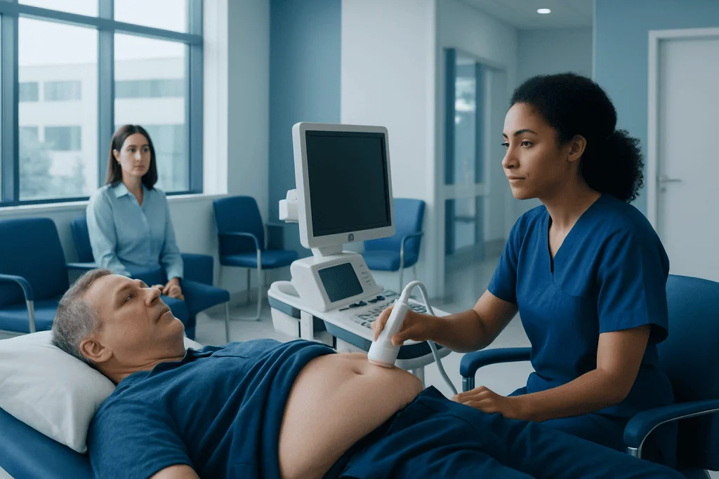
Ascites ultrasound can detect even small amounts of abdominal fluid that may not be evident on physical examination. The test is non-invasive, does not use radiation and usually takes about 15 to 30 minutes.
Key Takeaways
- Ascites ultrasound can detect even small amounts of abdominal fluid that may not be evident on physical examination.
- The test is non-invasive, does not use radiation and usually takes about 15 to 30 minutes.
- Ultrasound findings help assess where fluid is located, whether it is simple or complex, and whether drainage may be appropriate.
- The scan cannot determine the cause of ascites on its own; blood tests, fluid analysis and other imaging may be needed.
- Urgent medical assessment is important for new abdominal swelling, severe pain, fever, confusion, vomiting blood or breathing difficulty.
An ascites ultrasound is a painless imaging examination that uses sound waves to identify fluid within the abdomen and estimate its distribution. It can help clinicians confirm ascites, look for related findings and safely plan procedures such as fluid drainage when needed.
Overview: What Is an Ascites Ultrasound?
An ascites ultrasound is an abdominal scan that uses high-frequency sound waves to look for fluid in the peritoneal cavity, the space around the abdominal organs. It is a safe, radiation-free way to confirm whether abdominal enlargement is related to ascites, identify where the fluid has collected and provide useful information for further evaluation.
Ascites is a medical term for excess fluid in the abdomen. It may develop with liver disease, heart failure, kidney disease, cancer, infection, inflammation or other conditions. Ultrasound does not usually establish the underlying cause by itself, but it is often an important first imaging test because it can identify fluid accurately and support decisions about tests, follow-up and treatment.
The examination may be requested when a person has abdominal swelling, weight gain, discomfort, early fullness after meals, leg swelling or shortness of breath. It may also be used to monitor known ascites over time or to guide a procedure called paracentesis, in which a clinician removes a sample of fluid or relieves pressure by draining fluid.
How It Works and Who May Need the Scan
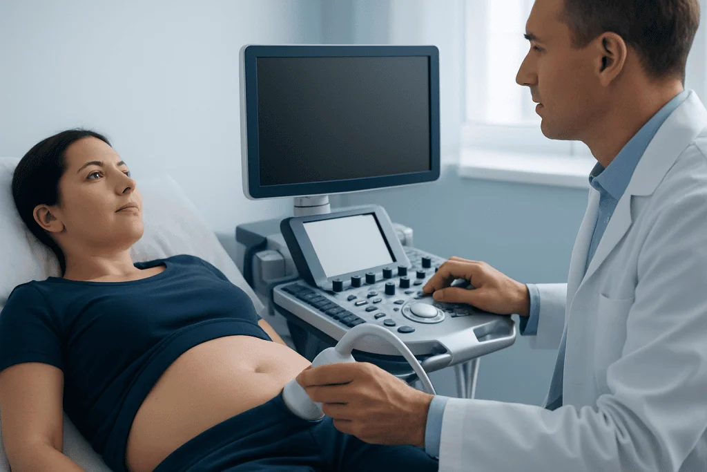
Ultrasound works by sending sound waves from a handheld device called a transducer into the body. The waves reflect differently from fluid and tissues, allowing the scanner to produce real-time images on a monitor. Free fluid usually appears dark or black on ultrasound, while organs, bowel and internal structures have different patterns of echoes.
The ascites ultrasound definition is therefore straightforward: it is an ultrasound examination performed to detect, characterize and map fluid in the abdominal cavity. It may be useful for people with suspected new ascites, known cirrhosis or other chronic liver conditions, unexplained abdominal distension, abdominal cancer, pancreatitis, heart or kidney disease, or symptoms that suggest fluid accumulation.
There is no meaningful radiation exposure, so ultrasound can generally be repeated when medically necessary. It is also frequently used in people for whom computed tomography may be less suitable, although a clinician may still recommend CT or MRI when more detailed assessment of abdominal organs, tumors or complications is needed.
Preparation and Step-by-Step Procedure
Preparation for an ascites ultrasound is usually simple. The imaging center may ask the person to avoid eating for several hours beforehand, particularly if the scan will also evaluate the liver, gallbladder or pancreas. Fasting can reduce bowel gas and improve image quality. However, preparation instructions vary, so patients should follow the instructions from their own care team and continue prescribed medicines unless told otherwise.
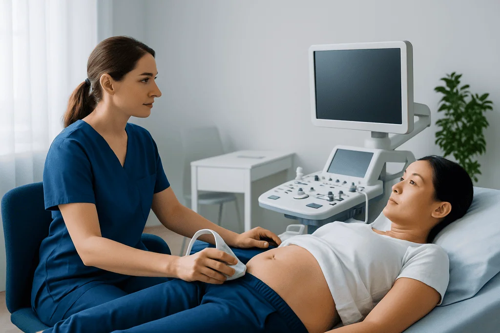
During the examination, the person usually lies on their back and may be asked to turn onto one side. A sonographer applies warm gel to the abdomen and moves the transducer over several areas. Gentle pressure may be used to improve the view, but the scan should not be painful. The person can usually breathe normally, although brief breath-holding may occasionally be requested.
An ascites ultrasound protocol commonly includes scanning dependent areas where fluid tends to collect, such as around the liver and spleen, the lower abdomen and pelvis, and spaces between bowel loops. The ascites ultrasound probe is typically a low-frequency curved transducer that can image deeper abdominal structures; a higher-frequency probe may be used to assess more superficial areas or guide a needle procedure.
An ascites check ultrasound protocol may also include measurements of fluid pockets and an assessment for features such as septations, debris, thickened peritoneum or masses. If drainage is planned, the operator identifies the safest accessible pocket of fluid, avoiding bowel, blood vessels and other organs.
What Are the Ultrasound Findings of Ascites?
On ultrasound, uncomplicated ascites most often appears as an anechoic, or dark, collection of free fluid surrounding abdominal organs or separating loops of bowel. Small volumes may first be seen in dependent spaces, while larger volumes can extend throughout the abdomen and pelvis. The sonographer may document the location and approximate amount of visible fluid.
Fluid is not always simple in appearance. Internal echoes, strands, septations or loculated pockets can indicate complex fluid, although these findings are not specific to one diagnosis. Blood, infection, inflammation and high-protein fluid may sometimes produce a more complex ultrasound appearance. A radiologist or treating clinician interprets these findings together with symptoms, examination results and laboratory tests.
The scan may also identify related features, such as a nodular liver contour, enlarged spleen, dilated veins, abdominal masses, enlarged lymph nodes or changes in the kidneys. These features can help direct further evaluation but are not a substitute for a full diagnostic assessment. When clinicians need to determine why the fluid has developed, they may recommend blood tests and analysis of a fluid sample obtained through paracentesis.
How Is Ascites Graded on an Ultrasound?
There is no single universal ultrasound grading system used in every hospital. Clinicians commonly describe ascites as mild, moderate or large based on how much fluid is seen and how widely it is distributed. Reports may also describe fluid as trace, small, moderate or large, rather than assigning a precise total volume.
In clinical practice, mild ascites may only be visible on imaging and may be limited to small dependent pockets. Moderate ascites is usually more readily visible around abdominal organs and in the pelvis. Large or tense ascites occupies much of the abdominal cavity and may cause obvious distension, discomfort, reduced appetite, reflux or shortness of breath.
Ultrasound can identify and map fluid well, but estimating the exact number of liters from imaging alone is difficult. Fluid volume may change with body position, timing, treatment and the distribution of fluid between abdominal spaces. For this reason, clinicians focus on symptoms, examination findings, changes over time and the clinical reason for measuring the fluid, rather than relying only on a volume estimate.
Is 5 Liters of Ascites a Lot?
Yes. Five liters of ascitic fluid is generally considered a large volume. In many adults, this amount can produce noticeable abdominal swelling and may contribute to pressure, difficulty eating normal portions, reduced mobility, shortness of breath or swelling in the legs. The impact varies according to a person’s body size, the speed at which fluid accumulated and any underlying illness.
A large fluid volume does not by itself explain why ascites is present or determine the best treatment. Some people may need careful adjustment of diet and medicines, while others may need diagnostic or therapeutic paracentesis. If a large-volume drainage procedure is being considered, clinicians assess blood pressure, kidney function, liver function and the likely cause of ascites to plan care safely.
Ultrasound is particularly valuable before paracentesis because it helps locate a suitable pocket of fluid and reduces the chance of needle injury to surrounding structures. Depending on the clinical situation, a specialist may recommend ultrasound-guided paracentesis to obtain fluid for laboratory testing or to relieve symptoms.
Benefits, Risks, Recovery and Next Steps
The principal benefits of an ascites ultrasound are that it is non-invasive, widely available, free from ionizing radiation and able to show fluid in real time. It can detect small amounts of ascites, guide needle placement and be repeated to monitor change. Results may be available promptly, although the final interpretation is usually provided in a formal imaging report.
Diagnostic ultrasound itself has no known significant risks when performed appropriately. The gel may feel cool, and pressure from the transducer can cause temporary discomfort if the abdomen is already tender. Risks are different when ultrasound is used to guide paracentesis, because needle drainage can occasionally cause bleeding, infection, leakage, low blood pressure or injury to nearby structures; these complications are uncommon when the procedure is performed by an experienced clinical team.
There is no recovery period after a routine scan. Most people can return to usual activities, food and medicines immediately unless they have been given other instructions. If the scan was part of a drainage procedure, the care team will provide specific aftercare advice and explain when to seek help.
Results should be discussed with the clinician who ordered the test. Further assessment may include liver and kidney blood tests, heart assessment, CT or MRI, or analysis of ascitic fluid. Acibadem International’s multidisciplinary specialists and JCI-accredited hospitals can assess ascites and coordinate imaging, gastroenterology, hepatology, oncology or interventional care for international patients when appropriate.
When to Seek Medical Care
New or increasing abdominal swelling should be assessed by a qualified clinician, particularly when it develops over days to weeks or is accompanied by weight gain, ankle swelling, reduced appetite or abdominal discomfort. Early review can help identify the cause and determine whether imaging, blood tests or fluid sampling is needed.
Urgent medical care is needed for severe or worsening abdominal pain, fever, chills, confusion, fainting, marked sleepiness, vomiting blood, black stools, new severe shortness of breath or rapidly increasing abdominal size. These symptoms can indicate complications that require prompt evaluation.
People with chronic liver disease or previously diagnosed ascites should follow their clinician’s monitoring plan. They should not start or stop diuretics, restrict fluids excessively or attempt to drain abdominal fluid outside a medical setting. A healthcare professional can advise on safe nutrition, salt intake, medicines and follow-up according to the underlying condition.
Frequently asked questions
How long does an ascites ultrasound take?
A routine ascites ultrasound often takes about 15 to 30 minutes. The time may be longer if the examination also evaluates abdominal organs in detail or if ultrasound guidance is needed for a procedure. The person can usually leave soon after a diagnostic scan.
Do people need to fast before an ascites ultrasound?
Some centers ask patients to fast for several hours, especially when the liver, gallbladder or pancreas will be assessed at the same time. Others may not require fasting for a focused fluid check. The imaging provider’s preparation instructions should be followed.
Can ultrasound detect a small amount of ascites?
Yes. Ultrasound is sensitive for detecting small pockets of free abdominal fluid, including fluid that may not be apparent during a physical examination. It can also show where the fluid is located and whether a safely accessible pocket is present for drainage.
Can an ultrasound tell what caused ascites?
Ultrasound may reveal findings that suggest an underlying liver, heart, kidney or abdominal condition, but it usually cannot confirm the cause of ascites alone. Clinicians often combine the scan with medical history, examination, blood tests and, when appropriate, ascitic fluid analysis.
Is an ascites ultrasound painful?
The ultrasound scan itself is usually painless. Mild pressure from the probe may feel uncomfortable if the abdomen is tender or very distended, but patients should tell the sonographer if they experience pain. If fluid drainage is performed, local anesthetic is typically used at the needle site.
What happens if fluid is found on an ascites ultrasound?
The next step depends on the amount of fluid, symptoms and medical history. A clinician may arrange laboratory tests, additional imaging, follow-up scanning or paracentesis to test or remove fluid. Treatment is directed at the underlying cause as well as symptom relief when needed.
References
- American College of Radiology
- American Association for the Study of Liver Diseases
- National Institute of Diabetes and Digestive and Kidney Diseases
- Radiological Society of North America
This article is for general information only and is not a substitute for professional medical advice. Please consult a qualified doctor about your individual situation.
Ultrasound in Turkey — costs, top hospitals & a free quote
JCI-accredited · board-certified surgeons · reply within 24h
Add us as a Preferred Source to see more of our trusted health content across Google Search, AI Overviews and Discover.

Check-up & Preventive Medicine
Comprehensive health screenings designed for early detection and prevention.
2 specialists in this unitMore from the Health Library



Related Specialists

Prof. Dr. Ahmet Arnaz
Cardiovascular Surgery
Assoc. Prof. Dr. Mustafa Çorum
Physical Medicine & Rehabilitation
Prof. Dr. Celaletdin Camcı
Medical Oncology
Dr. Şirin Efe
Clinical Laboratory