AVM Radiation: How It Works, Results and What to Expect

AVM radiation is a non-invasive treatment that can be useful for small or difficult-to-reach brain AVMs. The treatment effect is gradual, so follow-up imaging is essential until the AVM is confirmed closed.
Key Takeaways
- AVM radiation is a non-invasive treatment that can be useful for small or difficult-to-reach brain AVMs.
- The treatment effect is gradual, so follow-up imaging is essential until the AVM is confirmed closed.
- An AVM can still bleed during the interval before it is fully obliterated.
- Treatment decisions depend on AVM size, location, drainage pattern, symptoms, prior bleeding and overall health.
- Care is usually planned by a multidisciplinary team including neurosurgery, radiation oncology and neuroradiology.
AVM radiation, usually delivered as stereotactic radiosurgery, directs highly focused radiation at a brain arteriovenous malformation (AVM). It does not remove the AVM immediately; instead, it causes the abnormal vessels to thicken and close gradually over months to years.
Overview: What Is AVM Radiation?
AVM radiation is a focused radiation treatment for a brain arteriovenous malformation (AVM), a tangle of abnormal blood vessels that connects arteries directly to veins. Most AVM radiation treatment is delivered through stereotactic radiosurgery, despite the name: it is not an operation involving an incision. Multiple radiation beams are precisely aimed so that their combined effect is concentrated within the AVM while limiting exposure to surrounding brain tissue.
The purpose is to trigger gradual changes in the abnormal vessel walls. Over time, the vessels may narrow, scar and close, a result called obliteration. This approach may be considered when an AVM is small enough to target accurately, is located in an area where open surgery carries substantial risk, or remains after other treatment. Stereotactic radiosurgery is planned individually and is not the best approach for every AVM.
Radiation is one option within a broader care plan. Depending on the AVM, doctors may recommend observation with monitoring, microsurgical removal, endovascular embolization, radiosurgery, or a combination of these approaches. The safest plan balances the natural risks of the AVM against the potential risks and benefits of treatment.
What Does Radiation Do to an AVM?
Radiation damages cells lining the abnormal AVM blood vessels in a controlled, targeted way. The body responds with inflammation and healing changes that make the vessel walls progressively thicker. Blood flow through the malformation then decreases, and the vessels may eventually seal off.
This process is slow. AVM after radiation is not expected to disappear on the day of treatment or within a few weeks. Obliteration commonly takes several years, although the timing varies with the AVM’s size, blood-flow pattern, radiation plan and individual response. Imaging follow-up, often with MRI and sometimes catheter angiography, determines whether closure has occurred.
The avm radiation dose is calculated carefully by the radiation oncology and neurosurgery team. A higher dose may improve the chance of closure in some situations, but it can also increase the likelihood of injury to nearby brain tissue. The team therefore selects a plan that aims for effective treatment while protecting important structures such as areas involved in movement, speech, vision or memory.
Who May Be a Candidate for AVM Radiation?

AVM radiation therapy is most often considered for small to medium-sized AVMs, particularly those located deep in the brain or near areas that control essential functions. It may also be an option for a remaining portion of an AVM after embolization or surgery. A person’s age, symptoms, history of bleeding, seizures, imaging findings and personal treatment goals all matter.
Before recommending treatment, specialists assess the AVM with detailed brain imaging. MRI or CT may show its relationship to brain tissue, while cerebral angiography maps the blood vessels and blood flow in greater detail. These studies help the team estimate the likelihood of treatment benefit and potential effects on surrounding tissue. Brain arteriovenous malformation evaluation should be performed by clinicians experienced in cerebrovascular conditions.
Large AVMs may be harder to treat safely in one radiosurgery session. In selected cases, a staged radiation approach or another strategy may be discussed. For some unruptured AVMs, careful observation may be appropriate; this decision should be made after a thorough discussion of the individual AVM’s features and the uncertainty that can exist around long-term risk.
How Is AVM Radiation Performed?
Planning begins with a consultation involving specialists such as a neurosurgeon, radiation oncologist and neuroradiologist. The team reviews symptoms, medical history and imaging, then explains the expected benefits, limitations and potential complications. Certain medicines may need to be reviewed before the procedure, but patients should not stop anti-seizure, blood pressure or other prescribed medicines unless their clinician advises them to do so.
On the treatment day, imaging is used to define the AVM target with high precision. A fitted head frame or a custom mask may be used to keep the head still. Depending on the treatment system and individual circumstances, imaging may include MRI, CT and angiography. The planning process can take longer than the actual radiation delivery.
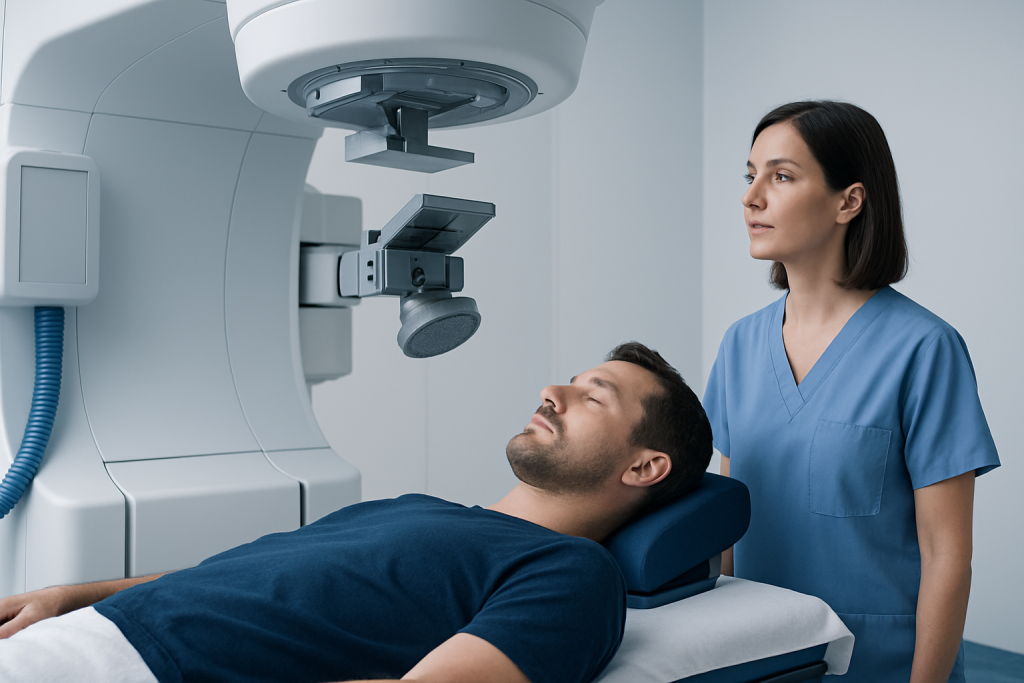
During treatment, the patient lies still on a treatment table while the machine delivers radiation from multiple angles. The procedure itself is painless. It is commonly completed in one outpatient session, although some larger lesions may be treated in stages. The patient is monitored by the clinical team throughout and can communicate with staff.
Afterward, most people go home the same day with instructions tailored to their situation. The care team arranges follow-up scans and explains which symptoms require urgent assessment. Because AVM closure is delayed, ongoing monitoring is a central part of avm radiation oncology care.
Benefits, Risks and Recovery After AVM Radiation
The principal potential benefit of AVM radiation is eventual closure of the abnormal vessels without open brain surgery. It may offer a treatment route for AVMs that are not safely accessible through microsurgery. Successful obliteration can reduce the future risk of bleeding from that AVM, but it cannot reverse every effect of a previous brain hemorrhage or guarantee that all neurological symptoms will resolve.
Many patients have little or no immediate recovery time beyond temporary tiredness, headache or scalp tenderness. A person may usually resume light daily activities within a short period, according to their clinician’s advice. However, biological healing continues for a long time. Follow-up imaging is often performed at intervals over several years, with catheter angiography sometimes used to confirm complete obliteration.
Possible side effects include headache, nausea, fatigue, temporary worsening of existing symptoms or swelling around the treated area. Less common but more serious risks include radiation-related brain injury, neurological changes, seizures, cyst formation or bleeding before the AVM closes. The likelihood depends on the AVM’s location and treatment details. New symptoms should be reported promptly rather than assumed to be a normal effect of recovery.
People should keep scheduled appointments and follow the team’s individual instructions about activity, work, driving and seizure safety. Endovascular embolization or brain surgery may be discussed in some treatment plans, either instead of radiation or in combination with it.
How Serious Is a Brain AVM?
A brain AVM can be serious because its fragile, high-flow blood vessels may bleed. A hemorrhage can cause sudden severe headache, weakness, numbness, trouble speaking, seizures, loss of consciousness or other stroke-like symptoms. The possible impact varies widely, ranging from no lasting symptoms to significant disability or, in severe cases, death.
Not every AVM causes symptoms or behaves the same way. Some are discovered after a bleed or seizure, while others are found incidentally during imaging for a different reason. Features such as prior rupture, AVM size, location, the pattern of venous drainage and associated aneurysms help specialists evaluate risk. An individualized assessment is more useful than judging severity by a single feature alone.
It is understandable to feel anxious after an AVM diagnosis. A cerebrovascular team can explain the imaging findings in clear terms and review whether monitoring or treatment is appropriate. A second opinion may also be helpful when choices are complex or when the AVM lies in an especially sensitive area of the brain.
What to Avoid With Brain AVM?
There is no universal list of activities that every person with a brain AVM must avoid. Recommendations depend on whether the AVM has bled, whether seizures have occurred, planned treatment and other medical conditions. Patients should ask their specialist for individualized advice before making major changes to exercise, work or travel.
It is generally sensible to avoid smoking and recreational drugs, particularly stimulants such as cocaine or amphetamines, because they can adversely affect blood pressure and blood vessels. Alcohol should be discussed with the treating clinician, especially for anyone with seizures or medicines that interact with alcohol. Blood pressure should be managed according to a healthcare professional’s advice.
People with seizures may need precautions related to driving, swimming alone, heights, machinery and bathing, based on local laws and their clinician’s recommendations. They should take prescribed anti-seizure medicine consistently and report changes in seizure pattern. Patients should also tell every clinician about the AVM before starting medicines or supplements, particularly drugs that may affect bleeding.
When to Seek Medical Care
Emergency medical care is needed for a sudden, severe headache unlike usual headaches; new weakness or numbness; facial drooping; difficulty speaking or understanding speech; vision loss; confusion; fainting; repeated vomiting; or a seizure lasting longer than five minutes or followed by failure to regain usual awareness. These symptoms can indicate bleeding or another urgent neurological problem.
Patients receiving avm radiation should contact their treating team promptly for a new or worsening headache, seizure, change in strength, coordination, speech, vision, thinking or persistent nausea. It is important not to wait for the next routine scan if concerning symptoms develop.
Acibadem International’s multidisciplinary specialists and JCI-accredited hospitals evaluate and treat cerebrovascular conditions for international patients, coordinating neurosurgery, neuroradiology and radiation oncology when appropriate.
Frequently asked questions
How long does it take for AVM radiation to work?
AVM radiation works gradually rather than immediately. The abnormal vessels may take months to years to narrow and close, and follow-up imaging is needed to confirm whether the AVM has been obliterated. The timeline varies according to the AVM and the treatment plan.
Can a brain AVM bleed after radiation treatment?
Yes. Until the AVM is fully closed, bleeding can still occur. This is why patients need ongoing surveillance and should seek emergency care for symptoms such as a sudden severe headache, weakness, speech difficulty, collapse or a seizure.
Is AVM radiation painful?
Radiation delivery itself is painless. Some people may experience temporary headache, fatigue, nausea or scalp discomfort afterward, depending on the positioning method and treatment details. The care team can advise on expected side effects and symptom relief.
What is the survival rate for someone with an AVM after surgery?
There is no single survival rate that applies to everyone after AVM surgery. Outcomes depend on whether the AVM has ruptured, its size and location, the person’s neurological condition, age and other health factors, as well as surgical complexity. A neurosurgeon can discuss individualized expectations based on the specific AVM.
Does AVM radiation cure seizures or headaches?
Some people experience improvement in seizures or headaches after successful AVM treatment, but this is not guaranteed. Symptoms may have more than one cause, and existing brain injury from a prior bleed may continue to affect neurological function. Ongoing seizure care may still be needed.
How is success after AVM radiation confirmed?
MRI is commonly used for follow-up over time, but catheter cerebral angiography is often used to confirm complete AVM obliteration. The treating team determines the timing and type of imaging based on the AVM and the response to treatment. Patients should continue follow-up until their clinicians confirm that monitoring can be reduced or stopped.
References
- American Stroke Association
- National Institute of Neurological Disorders and Stroke
- American Association of Neurological Surgeons
- International Society of Stereotactic Radiosurgery
This article is for general information only and is not a substitute for professional medical advice. Please consult a qualified doctor about your individual situation.
Explore treatments in Turkey — costs, top hospitals & a free quote
JCI-accredited · board-certified surgeons · reply within 24h
Add us as a Preferred Source to see more of our trusted health content across Google Search, AI Overviews and Discover.
Check your numbers in seconds
BMI, calories, due date, blood pressure and 30+ more clinical calculators — free, instant, doctor-reviewed ranges.