Barrett’s Screening: How It Works, Results and What to Expect

Barrett's screening generally involves an upper endoscopy with biopsies of any suspicious tissue. Screening is most often considered for people with long-term reflux symptoms plus additional risk factors.
Key Takeaways
- Barrett's screening generally involves an upper endoscopy with biopsies of any suspicious tissue.
- Screening is most often considered for people with long-term reflux symptoms plus additional risk factors.
- Most people receive sedation, go home the same day, and resume usual activities within about 24 hours.
- A diagnosis of Barrett’s esophagus does not mean cancer is present, but it may require periodic monitoring.
- Biopsy results determine whether there are no precancerous changes, dysplasia, or a need for further assessment or treatment.
Barrett's screening is usually performed with an upper endoscopy, a short procedure that allows a gastroenterologist to examine the esophagus and take small tissue samples if needed. It is not recommended for everyone with heartburn, but it can help identify Barrett’s esophagus and guide appropriate follow-up for people with higher risk factors.
Overview: What Barrett's Screening Checks For
Barrett’s screening looks for Barrett’s esophagus, a change in the lining of the lower esophagus that can occur after long-term exposure to stomach acid and, sometimes, bile reflux. The usual test is an upper endoscopy, also called esophagogastroduodenoscopy (EGD). A flexible camera is passed through the mouth to inspect the esophagus, stomach, and first part of the small intestine while the person is sedated.
During the procedure, the specialist may take tiny samples of tissue, called biopsies. A pathologist examines these samples under a microscope to confirm whether Barrett’s tissue is present and whether there are abnormal precancerous changes, known as dysplasia. Screening does not diagnose cancer in most people; instead, it helps identify who may benefit from monitoring or early treatment.
Barrett’s esophagus is associated with gastroesophageal reflux disease (GERD), but many people with reflux never develop it. Likewise, some people with Barrett’s esophagus have few or no noticeable reflux symptoms. Decisions about screening should therefore be individualized with a qualified clinician.
Who May Be a Candidate for Barrett's Screening?

Routine screening is not needed for every person with occasional heartburn. Clinicians commonly consider a one-time screening endoscopy for people with chronic reflux symptoms, particularly when symptoms have been present weekly for several years and other risk factors are also present.
Factors that may increase the likelihood of Barrett’s esophagus include age over 50, male sex, White race, obesity around the abdomen, tobacco use, and a close family history of Barrett’s esophagus or esophageal adenocarcinoma. These factors do not mean a person will have Barrett’s esophagus or develop cancer. They help a clinician judge whether the potential benefit of testing outweighs its small risks and inconvenience.
Screening may also be discussed for a person with reflux and warning features such as swallowing difficulty, unintentional weight loss, gastrointestinal bleeding, persistent vomiting, or iron-deficiency anemia. In these situations, endoscopy may be recommended to investigate symptoms rather than solely to screen for Barrett’s esophagus.
- Chronic, frequent reflux or regurgitation
- Several additional risk factors for Barrett’s esophagus
- A first-degree relative with Barrett’s esophagus or esophageal cancer
- Symptoms that could suggest narrowing, bleeding, or another upper digestive condition
How the Procedure Works: Preparation, Endoscopy and Biopsies
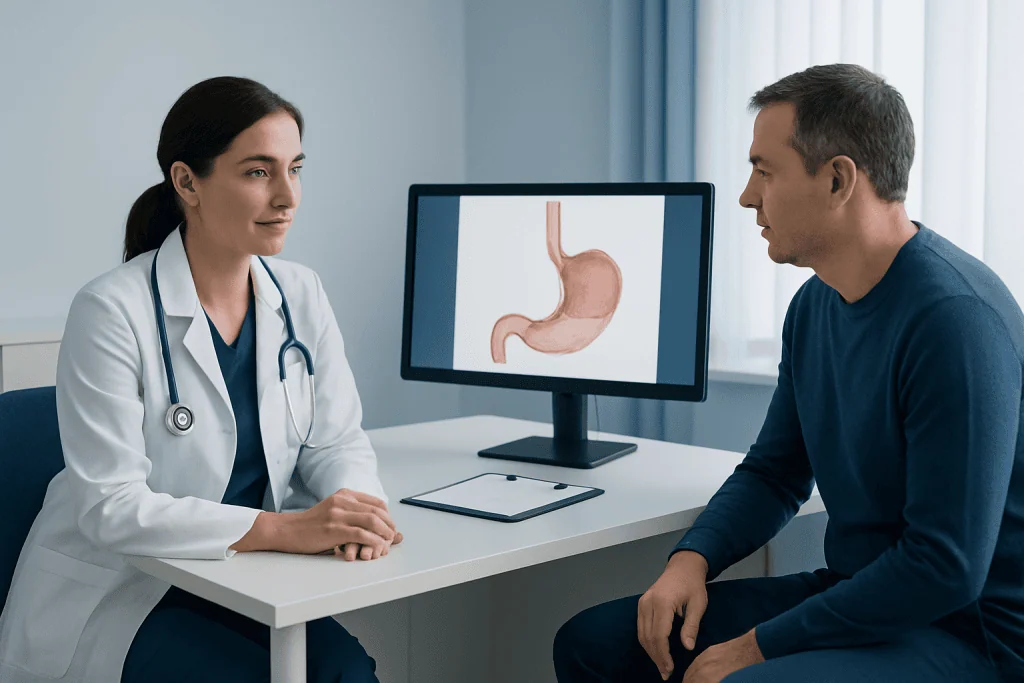
Before Barrett’s screening, the care team reviews medical history, allergies, medications, and conditions such as diabetes, sleep apnea, heart disease, or bleeding disorders. A person is usually asked not to eat or drink for a specified period before the examination. The clinician may advise temporary adjustments to medicines that affect blood clotting or blood sugar; these changes should only be made under medical guidance.
At the endoscopy unit, an intravenous line is usually placed for sedation. Most people receive medication that makes them sleepy and relaxed, so they typically remember little or none of the procedure. A mouth guard protects the teeth, and the clinician gently guides the endoscope through the mouth and into the esophagus. The examination itself often takes less than 30 minutes, although preparation and recovery make the visit longer.
If the lower esophagus has an appearance that suggests Barrett’s tissue, the endoscopist takes multiple biopsies in a systematic pattern. These samples are very small and generally cannot be felt. Advanced imaging methods may be used in selected cases to inspect the lining more closely. If abnormal tissue is identified, a specialist may discuss endoscopic evaluation and treatment options after the pathology report is available.
Recovery, Benefits and Possible Risks
After the procedure, the person rests in a recovery area until the sedative effects lessen. A mild sore throat, bloating, or gassiness can occur for a short time because air is used to view the digestive tract. Most people can eat and drink when advised by the care team and return home the same day with a responsible adult.
Driving, operating machinery, drinking alcohol, signing important documents, and making major decisions should be avoided until the following day or until the sedation team says it is safe. Many people return to regular activities within 24 hours. If biopsies were taken, results are commonly available within several days to a couple of weeks, depending on laboratory processes.
The main benefit of screening is that it may detect Barrett’s esophagus and dysplasia at a stage when follow-up or treatment can be planned carefully. Upper endoscopy is generally safe, but uncommon risks include reactions to sedation, bleeding after biopsy, aspiration, infection, and a tear in the digestive tract. Severe chest pain, fever, worsening abdominal pain, vomiting blood, black stools, shortness of breath, or trouble swallowing after endoscopy warrants urgent medical attention.
Understanding Results and Follow-Up Planning
A normal result means that Barrett’s esophagus was not found. If the esophagus is inflamed from reflux, the clinician may recommend lifestyle measures or medication and decide whether further assessment is needed. A diagnosis of Barrett’s esophagus is confirmed by both the endoscopic appearance and biopsy findings showing intestinal-type cells in the esophageal lining.
Pathology may report Barrett’s esophagus without dysplasia, indefinite for dysplasia, low-grade dysplasia, or high-grade dysplasia. “Indefinite” can occur when inflammation makes cells difficult to interpret. In that situation, reflux treatment may be optimized and biopsies repeated after healing, sometimes with review by an expert gastrointestinal pathologist.
For confirmed dysplasia, a gastroenterology team may recommend expert repeat assessment or endoscopic therapy to remove or destroy abnormal tissue. Options can include endoscopic resection of visible lesions and ablation techniques. Ongoing management also addresses reflux, often with acid-suppressing medicines and individualized lifestyle guidance. Acibadem International’s multidisciplinary specialists and JCI-accredited hospitals provide assessment and treatment planning for international patients with Barrett’s esophagus.
How Long Does It Take for Barrett's Esophagus to Turn Cancerous?
Barrett’s esophagus often does not turn into cancer. When cancer develops, the process usually occurs gradually over years through a sequence of cellular changes, rather than over days or weeks. The chance of progression varies substantially between individuals and is influenced most strongly by whether dysplasia is present.
People with Barrett’s esophagus without dysplasia generally have a low annual risk of developing esophageal adenocarcinoma. The risk is higher when low-grade dysplasia is confirmed and higher still with high-grade dysplasia. Because pathology interpretation can affect management, dysplasia is often reviewed by a pathologist with expertise in gastrointestinal disease.
Regular surveillance and, when appropriate, endoscopic treatment are intended to find important changes early. A clinician can explain the meaning of an individual pathology result, account for personal risk factors, and recommend a follow-up plan that is neither unnecessarily frequent nor delayed.
What Are the Four Stages of Barrett's Esophagus?
Barrett’s esophagus is not usually described in “stages” in the same way as cancer. However, people often use this phrase to refer to four commonly discussed pathology categories: Barrett’s esophagus without dysplasia, indefinite for dysplasia, low-grade dysplasia, and high-grade dysplasia.
Without dysplasia means Barrett’s cells are present but do not show precancerous changes. Indefinite for dysplasia means inflammation or other changes make the sample difficult to classify confidently. Low-grade dysplasia indicates early abnormal cellular changes, while high-grade dysplasia indicates more advanced abnormalities that have a greater likelihood of being associated with or progressing to cancer.
These categories guide the next steps, which may include surveillance endoscopy, repeat biopsies after inflammation is controlled, or endoscopic therapy. Cancer is a separate diagnosis and requires further staging tests and a personalized treatment discussion if it is found.
How Often Should Someone With Barrett's Esophagus Have an Endoscopy?
Endoscopy frequency depends on the biopsy result, the length and appearance of Barrett’s tissue, whether dysplasia is present, and prior treatment. For Barrett’s esophagus without dysplasia, surveillance is often performed at intervals of several years rather than every year. The exact interval should be set by the treating gastroenterologist using current guidelines and the person’s clinical circumstances.
When a result is indefinite for dysplasia, clinicians may first improve control of reflux-related inflammation and repeat endoscopy within a shorter period. Confirmed low-grade dysplasia may lead to a discussion of endoscopic eradication therapy or closer surveillance. High-grade dysplasia generally needs prompt expert evaluation and treatment planning rather than routine observation alone.
After successful endoscopic treatment for dysplasia, surveillance remains important because Barrett’s tissue or dysplasia can recur. Keeping scheduled appointments and reporting new swallowing problems, persistent chest discomfort, bleeding, or unexplained weight loss helps the care team respond appropriately.
Is Barrett's Overdiagnosed?
Barrett’s esophagus can be overdiagnosed if an irregular junction between the stomach and esophagus is labeled as Barrett’s without adequate biopsy evidence or when very short, uncertain changes are interpreted inconsistently. For this reason, diagnosis relies on careful endoscopic assessment and pathology findings, not on appearance alone.
At the same time, underdiagnosis can occur because many people with risk factors do not have endoscopy and some people have little or no reflux discomfort. Appropriate screening focuses on people most likely to benefit instead of testing everyone with occasional heartburn.
Expert review is particularly valuable when dysplasia is reported, because inflammation can mimic abnormal changes and because treatment decisions may depend on a precise diagnosis. Asking whether the biopsy was reviewed by an experienced gastrointestinal pathologist can be reasonable when results will significantly affect management.
When to Seek Medical Care
A person should arrange a medical review for persistent reflux symptoms, especially if they occur frequently, disrupt sleep, require regular over-the-counter medication, or have continued for years. A clinician can assess likely causes, discuss whether testing is appropriate, and help develop a safe plan to control symptoms.
Prompt medical assessment is important for difficulty or pain with swallowing, food getting stuck, vomiting blood, black or tar-like stools, unexplained weight loss, ongoing vomiting, or symptoms of anemia such as unusual fatigue or shortness of breath. These symptoms have several possible causes and should not be assumed to be Barrett’s esophagus.
Maintaining a healthy weight where possible, avoiding tobacco, limiting alcohol if advised, and identifying foods or habits that worsen reflux may reduce symptoms. People should not stop prescribed acid-suppressing treatment or change surveillance schedules without discussing it with their doctor.
Frequently asked questions
Is Barrett's screening painful?
Most people receive sedation for upper endoscopy and do not feel pain during the examination. They may notice temporary throat irritation, bloating, or sleepiness afterward. The care team monitors comfort and safety throughout the visit.
Can Barrett's screening be done without an endoscopy?
Standard evaluation usually uses upper endoscopy because it allows the clinician to see the esophageal lining and collect biopsies. Less invasive tests may be available in some settings for selected people, but an endoscopy may still be needed to confirm abnormal findings and assess dysplasia.
Do proton pump inhibitors prevent Barrett's esophagus?
Proton pump inhibitors reduce stomach acid and are commonly used to treat GERD and help heal reflux-related inflammation. They may be part of care for people with Barrett’s esophagus, but they do not eliminate the need for individualized screening or surveillance decisions. A clinician should advise on the most appropriate medicine and duration.
What does it mean if biopsies show no dysplasia?
No dysplasia means Barrett’s tissue is present but the sampled cells do not show precancerous abnormalities. This is reassuring, although periodic surveillance may still be recommended because risk is low rather than zero. The interval depends on the individual findings and guideline-based clinical assessment.
Can Barrett's esophagus go away?
Reflux symptoms and inflammation can improve with lifestyle measures and medication, but established Barrett’s tissue may persist. In people with dysplasia, endoscopic treatment can remove or ablate abnormal Barrett’s tissue. Follow-up remains important even after successful treatment.
Should everyone with GERD have Barrett's screening?
No. Most people with GERD do not need screening endoscopy solely because they have heartburn. Screening is usually considered when chronic reflux occurs alongside additional risk factors, or when alarm symptoms require investigation. A gastroenterologist can help determine whether testing is appropriate.
References
- American College of Gastroenterology
- American Gastroenterological Association
- National Institute of Diabetes and Digestive and Kidney Diseases
- National Cancer Institute
- Mayo Clinic
This article is for general information only and is not a substitute for professional medical advice. Please consult a qualified doctor about your individual situation.
Persistent digestive symptoms? Get evaluated in Turkey
JCI-accredited · board-certified surgeons · reply within 24h
Add us as a Preferred Source to see more of our trusted health content across Google Search, AI Overviews and Discover.