Bedsores Pressure Ulcers Pictures and Early Skin Changes
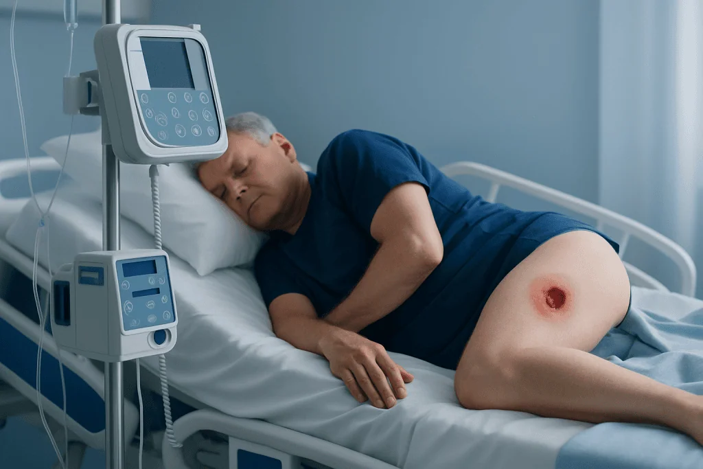
Pressure ulcers may begin as persistent discoloration, warmth, firmness, tenderness, or pain before the skin opens. They commonly develop over the heels, tailbone, hips, ankles, elbows, shoulder blades, and back of the head.
Key Takeaways
- Pressure ulcers may begin as persistent discoloration, warmth, firmness, tenderness, or pain before the skin opens.
- They commonly develop over the heels, tailbone, hips, ankles, elbows, shoulder blades, and back of the head.
- Regular repositioning, pressure-relieving surfaces, skin care, nutrition, and mobility support can reduce risk.
- Any open sore, non-blanching discoloration, drainage, fever, or worsening pain needs timely clinical assessment.
- Deep tissue injury can be present even when the skin surface is unbroken, especially in darker skin tones.
Bedsores, also called pressure ulcers or pressure injuries, are areas of skin and underlying tissue damage caused by prolonged pressure, often over bony parts of the body. Pictures can support recognition of possible early changes, but skin tone, wound depth, and other conditions can make visual self-diagnosis unreliable.
What bedsores pressure ulcers pictures can and cannot show
Bedsores pressure ulcers pictures may help a person recognize possible warning signs, such as an area of skin that remains red, purple, blue, or darker than the surrounding skin after pressure is relieved. They are also called pressure injuries or pressure sores. These wounds occur when sustained pressure, sometimes combined with friction or shear, reduces blood flow to the skin and tissues beneath it.
Images should be used as a guide rather than a way to diagnose or stage a wound at home. The appearance of pressure damage varies with skin tone, location, circulation, swelling, moisture, and the depth of tissue injury. A wound may look small on the surface while extending more deeply, and some early injuries remain covered by intact skin.
In lighter skin tones, an early pressure injury may appear as persistent redness that does not briefly fade when gentle finger pressure is applied. In darker skin tones, it may look darker, purple, bluish, gray, ashen, or unusually shiny compared with nearby skin. It may also feel warmer or cooler, firmer, softer, swollen, or more painful than surrounding skin.
How pressure ulcers develop and where they occur
Pressure ulcers develop when body weight presses soft tissue between a bony area and a bed, chair, cast, medical device, or other firm surface for too long. Pressure compresses small blood vessels and limits oxygen delivery. If it continues, cells can be damaged and tissue can break down.
Friction can occur when skin rubs against bedding or clothing, while shear happens when the skin stays in place but deeper tissues slide, such as when a person slides down in bed. Moisture from sweating, urine, stool, or wound drainage can weaken the skin and make injury more likely.
Common sites depend on a person’s position. For someone lying on their back, these include the tailbone, buttocks, heels, elbows, shoulder blades, and back of the head. For someone lying on their side, the hips, ankles, knees, and ears may be at risk. Wheelchair users may develop pressure injuries over the tailbone, buttocks, hips, or backs of the thighs.
- Heels are especially vulnerable because there is little tissue between the skin and bone.
- Pressure can also occur beneath masks, tubing, braces, casts, or prosthetic devices.
- People who cannot feel discomfort or change position independently need regular skin checks and support.
What the stages and visual signs mean

Clinicians describe pressure injuries by stage after examining the wound. Staging helps communicate the depth of visible tissue damage and guide care; it does not indicate how quickly a wound will heal. Not every pressure injury can be staged, particularly when the wound base is covered by dead tissue.
Stage 1 pressure injury has intact skin with persistent discoloration. It may be painful, itchy, warmer or cooler than nearby skin, or feel firmer or softer. Stage 2 involves partial-thickness skin loss, which may appear as a shallow open sore, a blister, or a pink or red moist wound bed. It should not be assumed that every blister or skin tear is a pressure injury, so assessment is important.
Stage 3 is full-thickness skin loss, where fat tissue may be visible and the wound may look deeper or crater-like. Stage 4 involves extensive tissue loss with exposed or directly palpable deeper structures such as fascia, muscle, tendon, cartilage, or bone. These wounds require expert care.
An unstageable pressure injury has full-thickness tissue loss, but its depth cannot be confirmed because the base is obscured by slough or eschar. Deep tissue pressure injury may present as intact or non-intact skin with a persistent deep red, maroon, purple, or dark discoloration, sometimes with a blood-filled blister. It can evolve quickly and warrants prompt clinical review.
Who is at risk and why early detection matters
Anyone with limited mobility can develop a pressure ulcer, but risk is higher for people who spend long periods in bed or seated, including those recovering from surgery, serious illness, injury, or stroke. Reduced sensation, paralysis, dementia, sedation, and severe weakness can make it difficult to notice discomfort or shift position.
Other contributors include poor blood circulation, diabetes, dehydration, anemia, fever, swelling, incontinence, fragile skin, low body weight, and inadequate protein or calorie intake. Medical devices and ill-fitting equipment can create concentrated pressure as well.
Early action is important because pressure injuries can worsen if pressure continues. An intact area of abnormal color may improve when pressure is promptly relieved and the contributing factors are addressed. Once the skin breaks down, healing commonly takes longer and the risk of infection rises.
Caregivers can help by checking skin daily, especially over bony areas and beneath devices. Good lighting is useful, and changes should be compared with the nearby skin rather than relying only on whether an area looks red. New pain, unusual tenderness, warmth, coolness, firmness, or swelling can be meaningful even before an obvious wound appears.
Diagnosis and treatment options
A healthcare professional diagnoses a pressure injury through a skin and wound assessment. They will consider the location, wound appearance, depth, drainage, odor, pain, circulation, mobility, nutrition, continence, medical conditions, and use of medical devices. They may also determine whether another condition, such as a skin tear, moisture-associated skin damage, arterial ulcer, venous ulcer, or diabetic foot ulcer, is more likely.
The first treatment step is to remove or reduce pressure from the affected area. This may involve a repositioning plan, heel elevation, a pressure-redistributing mattress or cushion, seating assessment, and review of braces, casts, or other devices. The plan should be individualized; people should not use ring-shaped cushions, which can concentrate pressure around the area.
Wound care may include gentle cleansing, dressings selected for the wound’s moisture level and depth, protection of surrounding skin, and management of incontinence. Dead tissue sometimes needs removal by an appropriately trained clinician. Antibiotics are not routinely needed for every pressure ulcer, but may be prescribed if there is a diagnosed infection.
Nutrition and hydration support healing, particularly adequate protein and calories when clinically appropriate. Complex, deep, infected, or slow-healing wounds may need care from a multidisciplinary wound team. Acibadem International’s multidisciplinary specialists and JCI-accredited hospitals provide assessment and treatment for international patients with complex wound-care needs.
Prevention and daily self-care
Preventing pressure ulcers is mainly about reducing prolonged pressure and keeping skin healthy. People who can move independently should shift their weight frequently and avoid remaining in one position for a long time. Those who need assistance should have an individualized repositioning schedule developed with a nurse, doctor, physiotherapist, or occupational therapist.
Use supportive mattresses, cushions, and heel-protection strategies recommended by a clinician. Keep sheets smooth, dry, and free of crumbs or objects. When moving someone in bed, use appropriate handling techniques or equipment to reduce dragging and shear. A physiotherapist can advise on safe mobility, transfers, and positioning.
Cleanse skin promptly after episodes of incontinence using gentle products, pat dry rather than rubbing, and consider a barrier product if recommended by a healthcare professional. Moisturize dry skin, but avoid massaging directly over bony areas or skin that is discolored, painful, or at risk, as massage may cause further tissue damage.
Eating a balanced diet and drinking enough fluids, where medically suitable, support skin health. A clinician or dietitian can assess people who have poor appetite, unintentional weight loss, swallowing difficulties, or increased nutritional needs. Do not attempt to cut away dead tissue, pop blisters, or apply harsh antiseptics to a suspected pressure ulcer without medical advice.
When to seek medical care
Seek medical advice promptly for skin discoloration that does not improve after pressure is relieved, especially if it is painful, warm, cool, swollen, firm, or blistered. A clinician should also examine any open wound, drainage, foul odor, black or yellow tissue, or wound that appears to be getting larger or deeper.
Urgent medical assessment is needed if a person with a pressure ulcer develops fever, chills, confusion, increasing weakness, rapidly spreading redness, worsening pain, pus-like drainage, or red streaking around the wound. These symptoms can indicate infection or another serious problem and should not be managed with home remedies alone.
People with diabetes, poor circulation, reduced sensation, immune suppression, or a history of difficult-to-heal wounds should contact their healthcare team early when a new skin change appears. Early review can help identify pressure damage before it becomes more extensive.
A photo can be useful for tracking a change over time when taken with consent and stored securely, but it should not delay assessment. If photographs are shared with a healthcare professional, include the date and note symptoms such as pain, drainage, fever, and changes in mobility or pressure exposure.
Frequently asked questions
What do early bedsores look like?
Early bedsores often appear as skin that is persistently discolored compared with the surrounding area. The area may be red in lighter skin or purple, blue, darker, gray, or ashen in darker skin. It may also be painful, itchy, warm, cool, swollen, firm, or soft before the skin opens.
Can bedsores look like bruises?
Yes. A deep tissue pressure injury can look purple, maroon, dark red, or bruise-like, particularly in darker skin tones. However, bruising and pressure injury can have different causes, so a new unexplained discolored area should be assessed by a healthcare professional.
How quickly can a pressure ulcer develop?
The timing varies depending on the amount of pressure, movement, skin condition, circulation, nutrition, moisture, and overall health. Tissue damage can develop sooner than expected in people who are seriously ill or unable to reposition themselves. Regular inspection and pressure relief are therefore important.
Should a pressure ulcer be covered or left open to air?
The right approach depends on the wound and should be decided by a healthcare professional. Many pressure ulcers benefit from a suitable dressing that protects the wound, manages moisture, and supports healing. Leaving a wound open to air is not automatically safer or more effective.
Can pressure ulcers heal completely?
Many pressure ulcers can heal, especially when pressure is relieved early and underlying factors are addressed. Healing time varies widely with wound depth, blood flow, nutrition, mobility, infection, and other health conditions. Deep wounds may require specialist treatment and ongoing follow-up.
How often should someone at risk be repositioned?
There is no single schedule that fits everyone. Repositioning should be individualized according to mobility, skin condition, support surfaces, comfort, and clinical risk. A nurse, doctor, physiotherapist, or occupational therapist can recommend a safe plan for the person's needs.
References
- National Pressure Injury Advisory Panel
- National Health Service
- Mayo Clinic
- MedlinePlus
- World Health Organization
This article is for general information only and is not a substitute for professional medical advice. Please consult a qualified doctor about your individual situation.
Persistent digestive symptoms? Get evaluated in Turkey
JCI-accredited · board-certified surgeons · reply within 24h
Add us as a Preferred Source to see more of our trusted health content across Google Search, AI Overviews and Discover.

More from the Health Library

Bicep: What Patients Need to Know

Claw Toe: An Evidence-Based Guide for Patients

Ciliary Body — Explained by Medical Evidence, Not Myths

Rhinovirus: An Evidence-Based Guide for Patients

Multivitamins Make Me Feel Like I Have a Cold: A Complete Medical Overview