Benign Mole: An Evidence-Based Guide for Patients

Most moles are harmless and remain stable for years. A mole that changes in size, shape, color, surface, or sensation should be medically assessed.
Key Takeaways
- Most moles are harmless and remain stable for years.
- A mole that changes in size, shape, color, surface, or sensation should be medically assessed.
- Dermatologists examine moles using the skin examination, dermoscopy, and sometimes a biopsy.
- Removal may be recommended when a mole is suspicious, repeatedly irritated, or bothersome for cosmetic reasons.
- Sun protection and regular skin awareness support early identification of concerning changes.
A benign mole is a common collection of pigment-producing skin cells that is not cancerous and usually does not need treatment. Because melanoma can sometimes resemble an ordinary mole, any new, changing, bleeding, or unusual-looking spot should be checked by a qualified clinician.
What Is a Benign Mole?
A benign mole, medically called a melanocytic nevus, is a noncancerous growth formed by melanocytes. Melanocytes are the cells that make melanin, the pigment that gives skin, hair, and eyes their color. Moles may be flat or raised and can range from tan or brown to pink, red, or skin-colored.
Many people develop moles during childhood and adolescence, although new moles can appear into early adulthood. A person may have only a few or many moles, and their appearance can vary across different areas of the body. Most benign moles are harmless and require no treatment.
However, it is not possible to confirm that every changing spot is benign by appearance alone. Skin cancer, including melanoma, may occasionally look like a mole or develop within an existing mole. Professional assessment is therefore important when a spot is new, evolving, or noticeably different from a person’s other marks.
How Benign Moles Usually Look and Feel
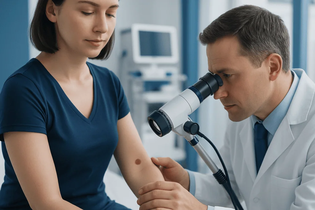
Typical benign moles often have a round or oval shape, a relatively even color, and a clear border. They may be smooth, slightly raised, or dome-shaped. Some contain hairs, which is usually not harmful on its own. Moles can occur almost anywhere, including the scalp, face, trunk, arms, legs, and genital area.
There is no single “normal” mole appearance for every person. Skin tone, genetics, sun exposure, age, and hormonal changes can affect how moles look. For example, moles can darken temporarily during pregnancy or puberty, although a significant or persistent change still deserves medical review.
A useful personal observation method is the “ugly duckling” sign: a mole that clearly looks different from a person’s other moles may merit attention. This approach complements, rather than replaces, the ABCDE guide and a clinical skin examination.
- Asymmetry: one half does not resemble the other.
- Border: edges are irregular, blurred, or notched.
- Color: more than one color or a new uneven color pattern is present.
- Diameter: a growing spot, especially one larger than about 6 mm, should be reviewed.
- Evolving: any change in appearance, sensation, or behavior is important.
Why Moles Develop and Who May Have More

Moles develop when melanocytes grow in clusters rather than being evenly distributed throughout the skin. The exact reasons differ between individuals, but inherited traits have a major role. Some moles are present at birth, while most are acquired over time.
Ultraviolet radiation from sunlight and tanning devices may contribute to the development of acquired moles and can damage skin cells. People with lighter skin, a tendency to freckle or burn, a family history of melanoma, or many moles may have a higher need for regular skin surveillance. Having many moles does not mean a person will develop skin cancer, but it can make self-checks and professional monitoring more important.
Hormonal changes may also influence pigmentation, which is why some moles become darker or more noticeable during adolescence or pregnancy. A clinician can help distinguish expected pigment changes from features that need closer assessment.
How a Mole Is Checked and Diagnosed
A clinician usually begins by asking when the mole appeared, whether it has changed, and whether there is itching, pain, crusting, bleeding, or a personal or family history of skin cancer. The skin is then examined, often including other areas that may be difficult for a person to inspect themselves.
Dermatologists may use dermoscopy, a handheld device that magnifies the skin and uses light to reveal pigment structures not visible to the naked eye. Dermoscopy can improve assessment of pigmented lesions, but it does not replace clinical judgment or laboratory testing when there is uncertainty.
If a mole appears suspicious, the most reliable way to determine whether it is benign is a biopsy. All or part of the lesion is removed under local anesthetic and examined by a pathologist. This procedure is generally brief, and the pathology result guides any further care. Photographs or digital mole mapping may also be used to monitor selected moles over time.
Treatment and Mole Removal Options
A confirmed benign mole usually does not need to be removed. Observation may be appropriate when the mole is stable and does not cause symptoms or repeated irritation. It is important not to attempt home removal with acids, cutting tools, or over-the-counter products, as these can cause burns, infection, scarring, and delayed diagnosis of a potentially serious lesion.
A clinician may recommend removal if a mole has concerning features, is frequently rubbed by clothing or shaving, catches on jewelry, or causes significant cosmetic concern. The approach depends on the mole’s type, size, depth, and location. Common medical techniques include shave removal, surgical excision, or a biopsy designed to remove the full lesion.
After removal, the area may need simple wound care and protection from sun exposure while healing. Some procedures leave a scar, and the clinician can discuss realistic expectations before treatment. When a lesion requires specialist removal and tissue analysis, dermatology assessment and treatment can help ensure the choice of procedure is appropriate.
Skin Awareness, Sun Protection and Self-Care
Regular skin awareness is a practical way to notice changes early. Many people find it helpful to check their skin about once a month in good light, using a mirror for the back, scalp, buttocks, and other hard-to-see areas. A partner or family member may help inspect areas that are difficult to view.
Taking dated photographs can be useful for observing a mole over time, particularly for people with many moles. Images should not be used to self-diagnose or to delay an appointment if a lesion seems concerning. Changes that occur over weeks or months are particularly important to report.
Sun protection supports overall skin health and helps reduce ultraviolet damage. This includes seeking shade when practical, wearing protective clothing and a wide-brimmed hat, using broad-spectrum sunscreen as directed, and avoiding tanning beds. Sunscreen is valuable but should not be used to stay in intense sun for longer periods.
When to Seek Medical Care
A person should arrange a medical assessment for a new mole in adulthood or a mole that is changing in size, shape, color, elevation, or texture. Review is also advisable for a spot that repeatedly itches, hurts, crusts, bleeds, or does not heal. These symptoms do not automatically mean cancer, but they should not be ignored.
Prompt assessment is especially important if a lesion has marked asymmetry, irregular borders, multiple colors, rapid growth, or looks unlike the person’s other moles. People with a previous melanoma, a strong family history of melanoma, many atypical moles, or reduced immune function may benefit from individualized advice on skin-check intervals.
Acibadem International’s multidisciplinary specialists and JCI-accredited hospitals can assess suspicious skin lesions and arrange appropriate diagnosis and treatment for international patients. A dermatologist can provide reassurance for a harmless mole or recommend timely next steps if further investigation is needed.
Frequently asked questions
Are benign moles dangerous?
Benign moles are noncancerous and are usually not dangerous. Most remain unchanged and do not require treatment. A new or changing mole should still be examined because some skin cancers can resemble an ordinary mole.
Can a benign mole become cancerous?
Most benign moles do not become cancerous. Melanoma can arise in or near a mole in some cases, but it more often develops on previously normal-looking skin. Monitoring for change and seeking assessment promptly are the safest steps.
What changes in a mole should be checked?
Changes in size, shape, border, color, thickness, or surface should be checked by a clinician. Itching, tenderness, bleeding, crusting, or failure to heal also warrant assessment. A mole that looks distinctly different from the rest should be reviewed.
Is it safe to remove a mole for cosmetic reasons?
Mole removal can be safe when it is performed by a qualified healthcare professional after appropriate assessment. The clinician may recommend sending the removed tissue for laboratory examination. Home removal methods are not safe and can lead to scarring, infection, or missed diagnosis.
Does hair growing from a mole mean it is benign?
Hair growing from a mole is commonly seen and does not by itself indicate cancer. However, it also cannot confirm that a mole is benign. Any mole with concerning changes should be assessed regardless of whether hair is present.
How often should moles be checked?
Many people can perform a simple self-check about once a month to become familiar with their skin. The frequency of professional examinations depends on personal risk factors, including many moles, a family history of melanoma, or a history of skin cancer. A dermatologist can recommend an individual schedule.
References
- American Academy of Dermatology
- American Cancer Society
- National Cancer Institute
- NHS
- World Health Organization
This article is for general information only and is not a substitute for professional medical advice. Please consult a qualified doctor about your individual situation.
Hair transplant in Turkey — costs, graft counts & a free assessment
JCI-accredited · board-certified surgeons · reply within 24h
Add us as a Preferred Source to see more of our trusted health content across Google Search, AI Overviews and Discover.