

Bladder Ultrasound: Preparation, Procedure and Results

A bladder ultrasound does not use radiation and is usually painless. Patients are often asked to drink water and avoid urinating before the examination.
Key Takeaways
- A bladder ultrasound does not use radiation and is usually painless.
- Patients are often asked to drink water and avoid urinating before the examination.
- The scan may be performed before and after urination to measure post-void residual urine.
- Results can help investigate urinary symptoms, infections, stones, obstruction or bladder changes, but may need other tests for a diagnosis.
- Severe pain, inability to pass urine, visible blood in urine or fever with urinary symptoms should be assessed promptly.
A bladder ultrasound is a safe, non-invasive imaging test that uses sound waves to create pictures of the bladder and measure how much urine it holds or retains after urination. Preparation often involves arriving with a comfortably full bladder, but the exact instructions depend on why the scan has been requested.
Bladder Ultrasound Overview
A bladder ultrasound is an imaging examination that uses high-frequency sound waves to produce images of the urinary bladder. A handheld device called a transducer is moved over the lower abdomen. The reflected sound waves are processed into images, allowing the clinician to assess the bladder’s shape, wall appearance and the volume of urine inside it.
The test is commonly requested for symptoms such as difficulty urinating, frequent urination, urinary leakage, recurrent urinary tract infections, pelvic discomfort or a feeling that the bladder does not empty completely. It may also be included in an ultrasound assessment of the kidneys and urinary tract.
One important use is measuring the amount of urine left in the bladder after a person urinates. This is called the post-void residual. A higher-than-expected residual can occur for several reasons, including an obstruction to urine flow, reduced bladder muscle function or certain neurological conditions. The result is interpreted alongside symptoms, examination findings and other tests.
How a Bladder Ultrasound Works and Who May Need One
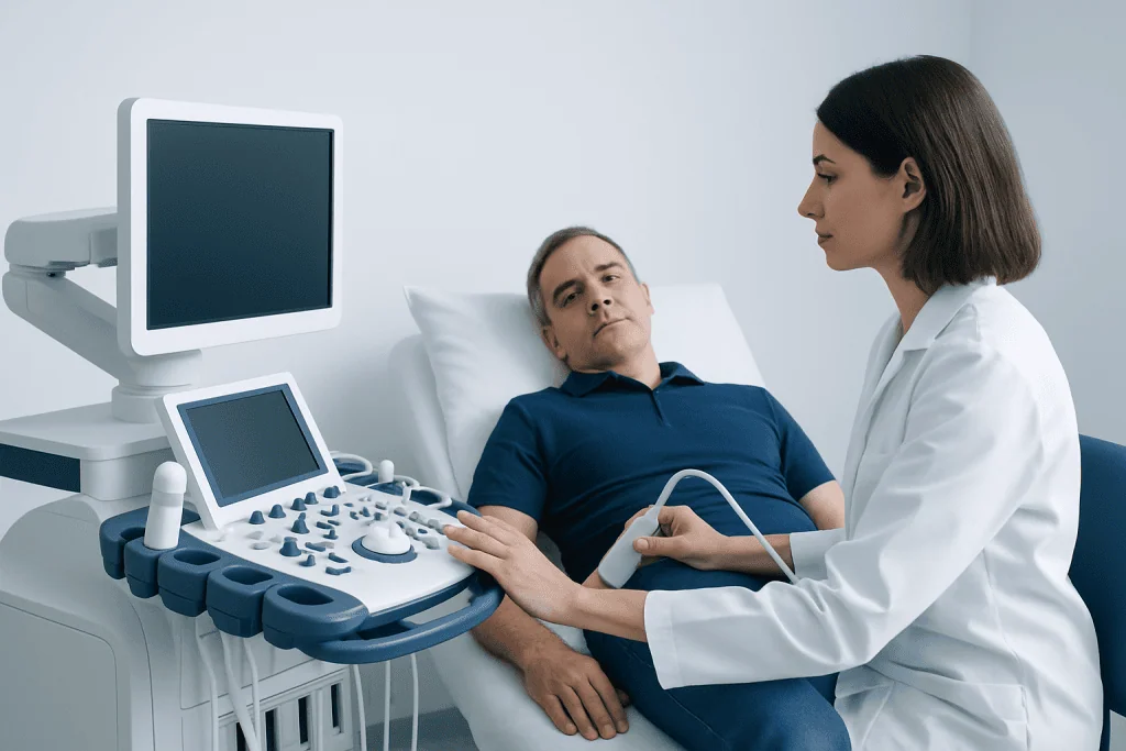
Ultrasound works by sending sound waves through the skin into the body. Unlike X-rays and CT scans, it does not use ionising radiation. Fluid in the bladder provides a clear acoustic window, which is why a full bladder is often helpful for the first part of the examination.
A clinician may recommend a bladder ultrasound for adults or children with persistent urinary symptoms. It can help identify bladder distension, retained urine, some bladder stones, structural changes or features that need further assessment. It may also be used to monitor bladder emptying in people with an enlarged prostate, pelvic organ prolapse, diabetes, spinal conditions or neurological disorders.
Ultrasound is useful, but it has limits. It cannot reliably rule out every cause of urinary symptoms, and small lesions inside the bladder may require direct inspection with cystoscopy or other imaging. If the concern is a suspected urinary stone or urinary blockage, assessment may include kidney stone evaluation or additional urinary tract imaging, depending on the clinical situation.
Preparing for a Bladder Ultrasound

Preparation instructions vary by hospital, the type of ultrasound ordered and whether the kidneys, prostate, uterus or other pelvic organs are also being examined. Patients should follow the instructions provided by their imaging department, as these take priority over general guidance.
For a bladder-focused scan, people are commonly asked to arrive with a comfortably full bladder. This may mean drinking water in the hour before the appointment and avoiding urination until the initial images are complete. A very painful urge to urinate is not necessary and should not be endured; the imaging team can often adapt the examination.
Comfortable, loose clothing can make the appointment easier. Patients should tell the team about recent pelvic surgery, a urinary catheter, pregnancy, mobility limitations or difficulty holding urine. These details can affect the approach and help the sonographer provide practical support.
How much water should I drink before an ultrasound of my bladder?
Many imaging departments advise adults to drink several glasses of plain water, often around 500 to 1,000 mL, before a bladder or pelvic ultrasound. The amount and timing differ between facilities and depend on body size, age, kidney or heart health, and whether other organs are being scanned. Patients should use the amount given in their appointment instructions rather than forcing extra fluids.
People with conditions requiring fluid restriction, such as some forms of heart failure or kidney disease, should ask their doctor or imaging department for personalised instructions. The aim is a comfortably filled bladder, not discomfort or excessive fluid intake.
Can I eat anything 2 hours before my ultrasound?
For an ultrasound that examines only the bladder, eating is often allowed. However, some combined abdominal or pelvic ultrasound examinations require fasting because food and bowel gas can affect images of organs such as the gallbladder, liver or pancreas.
Patients should check their appointment letter or contact the imaging department if instructions are unclear. They should not stop prescribed medicines unless a clinician specifically advises them to do so.
What to Expect During the Procedure
A bladder ultrasound is usually performed by a sonographer, radiologist or trained clinician. The patient may be asked to lie on an examination couch with the lower abdomen exposed. Gel is applied to the skin; it may feel cool, but it helps the transducer make good contact and improves image quality.
The clinician moves the transducer over the lower abdomen and may apply light pressure to obtain views from different angles. The scan itself is generally painless and commonly takes only a short time. If the bladder is very full, the patient may feel uncomfortable because of the urge to urinate rather than the ultrasound probe.
For a post-void residual measurement, the first scan may be done with the bladder full. The patient is then asked to urinate, usually in a nearby toilet, and returns promptly for a second scan. Comparing the images before and after urination helps estimate how completely the bladder empties.
There are no injections, incisions or anaesthesia for a standard external bladder ultrasound. In some care settings, a portable bladder scanner may be used at the bedside to estimate bladder volume quickly, for example after surgery or when urinary retention is suspected.
What happens if I can’t hold my pee before an ultrasound?
If a person urinates before the scan or cannot comfortably hold urine, they should tell the imaging team. The examination may still be possible, particularly if it is intended to measure urine remaining after voiding, or the team may ask the person to drink water and wait until the bladder refills.
There is no need to feel embarrassed. Holding urine can be difficult for people with urgency, incontinence, pregnancy, pain or certain medical conditions. The team can advise on the safest and most practical next step.
Is it best to empty your bladder before an ultrasound?
For the initial part of many bladder and pelvic ultrasounds, it is usually better not to empty the bladder because a full bladder can improve the images. In contrast, a post-void residual examination specifically requires the person to empty their bladder after the first scan so the remaining urine can be measured.
The best approach therefore depends on the reason for the examination. Patients should follow the instructions from their imaging department and ask on arrival if they are uncertain.
Results, Benefits and Possible Limitations
A radiologist or the requesting clinician reviews the images and places the findings in clinical context. The report may describe bladder volume, bladder wall appearance, visible debris or stones, whether there is a mass-like finding, and the estimated amount of urine remaining after urination. A result may be available the same day in some settings, while a formal report can take longer.
A post-void residual result is not interpreted in isolation. Urine remaining in the bladder can vary with hydration, time since the urge to urinate, medication use and the testing environment. If the result is unexpected, the clinician may repeat the measurement or arrange urine tests, kidney imaging, urodynamic testing, cystoscopy or specialist review.
The main benefits of bladder ultrasound are that it is quick, non-invasive and free from radiation exposure. It can provide useful information without requiring a catheter in many situations. However, bowel gas, body shape, an insufficiently filled bladder and movement can reduce image quality, and ultrasound cannot confirm every bladder condition.
Urinary symptoms may be related to conditions such as infection, benign prostate enlargement, pelvic floor dysfunction or bladder overactivity. Depending on the findings, a clinician may discuss further testing and treatment options, including assessment for urinary tract infection where appropriate.
Recovery, Risks and Next Steps
There is no recovery period after a standard bladder ultrasound. Patients can normally return to usual activities immediately, eat and drink as normal, and take prescribed medicines. If they had been asked to keep the bladder full, they can use the toilet as soon as the relevant images have been obtained.
External ultrasound is considered very safe. The gel can occasionally cause minor skin irritation in people with sensitive skin, but significant complications are not expected. Any discomfort is usually brief and related to holding urine or gentle pressure on a tender lower abdomen.
If ultrasound suggests incomplete bladder emptying or another concern, the next step depends on the cause and severity. Management may range from reviewing medicines and treating an infection to urology assessment for obstruction or bladder dysfunction. When symptoms are ongoing, clinicians may recommend specialised testing such as urodynamic testing to understand how the bladder stores and releases urine.
Acibadem International’s multidisciplinary specialists and JCI-accredited hospitals can assess urinary concerns and coordinate diagnostic imaging and urology care for international patients.
When to Seek Medical Care
A planned bladder ultrasound is suitable for many non-urgent urinary concerns, but some symptoms need more timely medical assessment. A person should contact a qualified clinician if urinary symptoms persist, recur, disrupt daily life or are accompanied by pelvic pain, unexplained weight loss or repeated infections.
Urgent medical care is important for an inability to urinate with increasing lower abdominal pain or swelling, visible blood in the urine, fever or chills with urinary symptoms, severe flank or back pain, or confusion in an older or medically vulnerable person. These symptoms can have several causes and should not be self-diagnosed.
People who are pregnant, immunocompromised, have known kidney disease or have recently had urinary tract surgery should seek clinical advice promptly when new urinary symptoms develop. A doctor can decide whether a bladder ultrasound, urine testing or another form of assessment is most appropriate.
Frequently asked questions
Does a bladder ultrasound hurt?
A standard bladder ultrasound is generally painless. The transducer is moved over the lower abdomen with gel, and only gentle pressure is usually needed. Discomfort may occur if the bladder is very full or the lower abdomen is already tender.
How long does a bladder ultrasound take?
The scanning portion commonly takes a short time, often around 10 to 20 minutes. It can take longer if the examination includes the kidneys or other pelvic organs, or if images are needed before and after urination. Allow additional time for registration and preparation.
Can a bladder ultrasound detect bladder cancer?
An ultrasound can sometimes show an abnormal area in or around the bladder, but it cannot diagnose or exclude bladder cancer on its own. If there is a suspicious finding or blood in the urine, a doctor may recommend cystoscopy, urine tests, CT imaging or other investigations. Early assessment is important when visible blood is present.
What is a post-void residual bladder ultrasound?
A post-void residual ultrasound measures the amount of urine remaining in the bladder after a person urinates. It is used when there is concern that the bladder may not be emptying fully. The result helps guide further evaluation but is interpreted alongside symptoms and other clinical information.
Do I need to stop my medicines before a bladder ultrasound?
Most people can continue their regular medicines before a bladder ultrasound. Appointment instructions may differ if other abdominal tests are being performed at the same time. Patients should ask their doctor or imaging department before changing any prescribed medicine.
Can a bladder ultrasound show a urinary tract infection?
Ultrasound does not directly diagnose most urinary tract infections. It may identify related findings, such as incomplete bladder emptying or stones, that can contribute to recurrent infections. Urine testing and clinical assessment are usually needed to confirm an infection.
References
- American College of Radiology
- Radiological Society of North America
- National Institute of Diabetes and Digestive and Kidney Diseases
- National Health Service
- Urology Care Foundation
This article is for general information only and is not a substitute for professional medical advice. Please consult a qualified doctor about your individual situation.
Ultrasound in Turkey — costs, top hospitals & a free quote
JCI-accredited · board-certified surgeons · reply within 24h

Check-up & Preventive Medicine
Comprehensive health screenings designed for early detection and prevention.
3 specialists in this unitMore from the Health Library



Related Specialists

Assoc. Prof. Dr. Mehmet Sürmeli
Otorhinolaryngology
Dr. Birol Sarkut
Emergency Service
Dt. Aybike Karaüzüm
Dentistry
Asst. Prof. Dr. Alper Özdilek
Otorhinolaryngology