Bone Mets Xray: Preparation, Procedure and Results

A bone mets xray uses a small amount of radiation to create images of a painful or at-risk bone. X-rays are useful for detecting fractures and established bone destruction but may not reveal early metastatic deposits.
Key Takeaways
- A bone mets xray uses a small amount of radiation to create images of a painful or at-risk bone.
- X-rays are useful for detecting fractures and established bone destruction but may not reveal early metastatic deposits.
- MRI, CT, PET/CT, and bone scans can provide additional information when metastases are suspected.
- Persistent focal bone pain, new weakness, or symptoms of spinal cord compression require prompt medical assessment.
- Treatment is individualized and may include systemic cancer therapy, radiotherapy, surgery, pain management, and bone-strengthening medicines.
A bone mets xray is a quick, painless imaging test that may identify bone damage, fractures, or areas suspicious for metastatic cancer. However, standard X-rays can miss early bone metastases, so clinicians often combine them with MRI, CT, PET/CT, or a nuclear medicine bone scan when more detail is needed.
Bone Mets Xray: What It Can Show
A bone mets xray is a conventional X-ray examination used to assess whether cancer may have spread to a bone. It is most often ordered when a person with a current or previous cancer has new, localized bone pain, tenderness, a suspected fracture, or an abnormality found on another scan. The test can help clinicians see changes in bone shape, density, and strength.
Bone metastases occur when cancer cells from a primary tumor travel to bone. They are more common with several cancers, including breast, prostate, lung, kidney, and thyroid cancers, although they can occur with many types of cancer. A bone mets xray does not confirm the primary cancer type on its own; results are interpreted alongside medical history, symptoms, blood tests, and other imaging.
On X-ray, metastases may appear as areas where bone has been broken down, areas of increased bone formation, or mixed changes. The test is especially helpful for evaluating structural complications, such as a fracture or a bone that may be weakened enough to fracture. Normal X-ray findings do not always exclude early bone involvement.
How the X-Ray Works and Who May Need It
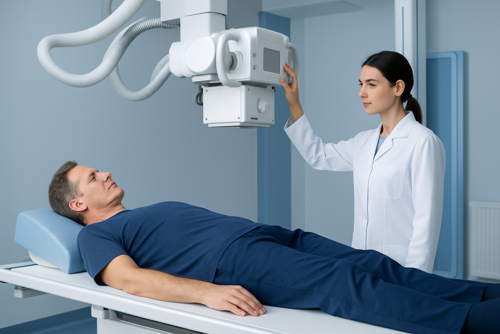
X-rays use a controlled, low dose of ionizing radiation to make images of dense body structures. Bone absorbs more X-rays than soft tissue, which creates the contrast needed to assess the outer bone surface and internal bone pattern. A radiologist reviews the images and sends a report to the requesting clinician.
A clinician may recommend a bone mets xray for new pain in one specific area, pain that worsens with movement or weight-bearing, an unexplained fracture, or follow-up of a known bone lesion. It may also be used after an abnormal bone scan, CT, MRI, or PET/CT to examine a particular location more closely.
Pregnancy should be discussed before any X-ray, even though imaging may still be appropriate when medically necessary. The radiology team can take steps to minimize exposure. People should also tell the team if they have severe pain or difficulty positioning, as the examination can usually be adapted to improve comfort.
Preparing for a Bone Mets Xray and What Happens During the Procedure

Most people do not need special preparation for a bone X-ray. They can usually eat, drink, and take regular medicines as directed by their healthcare professional. It is helpful to wear comfortable clothing and to remove jewelry, belts, or other metal items near the body part being scanned, as metal can obscure the image.
During the procedure, a radiographer positions the affected body part on the X-ray table or against an upright image detector. Depending on the location, the person may sit, stand, or lie down. Several views are typically taken from different angles, and the radiographer may ask the person to stay still or briefly hold their breath.
The image capture itself takes only moments, while positioning may take longer if the area is painful. The examination commonly takes about 10 to 20 minutes. It does not involve needles, contrast dye, or anesthesia. Afterward, people can generally return to normal activities immediately.
The radiation exposure from a diagnostic X-ray is kept as low as reasonably achievable. The benefit of finding a fracture, assessing a painful lesion, or guiding further testing usually outweighs the small radiation risk when the test is clinically indicated.
Understanding Bone Mets Xray Results
A report may describe a lesion as lytic, sclerotic, mixed, or indeterminate. Lytic lesions are areas of bone loss that can make bone weaker. Sclerotic lesions are denser-than-usual areas of bone formation. Some cancers more often produce one pattern than another, but imaging appearance alone does not always establish the cause.
Radiologists also look for cortical destruction, changes in the bone marrow region that are visible on X-ray, periosteal reaction, joint involvement, and fractures. If a long bone is substantially weakened, the report may note concern for an impending fracture. In the spine, alignment and vertebral compression fractures may also be assessed.
An abnormal result may lead to additional imaging, blood tests, or occasionally a biopsy. If metastatic disease is known or confirmed, the oncology team considers the imaging with the person’s overall cancer status to plan care. Depending on the situation, options may include radiotherapy for painful or high-risk bone lesions, systemic anticancer treatment, orthopedic stabilization, and supportive care.
It is important not to interpret wording in an imaging report without clinical context. Terms such as “suspicious,” “cannot exclude,” or “recommend correlation” do not provide a final diagnosis by themselves; they indicate that further assessment may be useful.
What Is the Best Test for Bone Metastases?
There is no single best test for bone metastases in every situation. The most appropriate examination depends on the suspected cancer type, symptoms, body area involved, whether the goal is staging or investigating one painful site, and what previous imaging has shown. A plain X-ray remains valuable for checking bone stability and fractures, but it is not the most sensitive test for early disease.
MRI is particularly useful for examining bone marrow, the spine, nerves, and possible spinal cord compression. In bone mets radiology, MRI can detect marrow changes before enough bone destruction or formation has occurred to be visible on an X-ray. CT provides detailed images of bone structure and can help assess lesion size, fracture risk, and suitability for procedures.
A nuclear medicine bone scan can survey much of the skeleton and may show areas of increased bone activity. PET/CT may identify metabolically active cancer and provide information about both bone and soft-tissue disease. Each test has limitations, including false-positive and false-negative findings, so clinicians choose and combine tests thoughtfully.
If a lesion remains uncertain or the diagnosis would change treatment, a biopsy may be recommended. Biopsy allows a pathologist to examine tissue and can help distinguish metastatic cancer from infection, a benign bone condition, or a new primary bone tumor.
What Does Metastatic Cancer Look Like on a Bone Scan?
On a conventional nuclear medicine bone scan, metastatic cancer often appears as one or more areas of increased tracer uptake, sometimes called “hot spots.” These areas reflect increased bone remodeling or repair activity. They may be found in the spine, pelvis, ribs, skull, or long bones, but location alone cannot confirm metastasis.
A hot spot is not specific to cancer. Arthritis, prior injuries, healing fractures, infection, and other benign conditions can also increase tracer uptake. For this reason, a bone scan is interpreted together with symptoms, the person’s cancer history, and other imaging such as X-ray, CT, MRI, or PET/CT.
Some lesions may be less apparent on a bone scan, particularly if they cause little bone-forming response. This is one reason why MRI, CT, PET/CT, or more than one imaging method may be necessary. The care team will explain what a particular scan pattern means in the individual clinical setting.
How Fast Do Bone Mets Progress?
The rate at which bone metastases progress varies widely. It depends on the primary cancer, its biological characteristics, the extent of disease, response to treatment, and the person’s overall health. Some bone lesions can remain stable for long periods with treatment, while others may change more quickly.
Imaging performed at intervals helps clinicians monitor known lesions and identify complications. A new or worsening symptom should not wait for a scheduled scan, particularly if there is severe pain, reduced ability to bear weight, or new neurological symptoms. Earlier assessment can help prevent complications such as fracture or pressure on the spinal cord.
It is understandable to seek a timeline, but imaging cannot reliably predict the course for every individual. An oncologist can provide the most meaningful estimate based on the cancer type, current treatment, scan findings, and response over time.
What Do Early Bone Metastases Feel Like?
Early bone metastases may cause no symptoms at all. When symptoms occur, the most common is persistent, localized bone pain that may gradually become more noticeable, occur at rest, or disturb sleep. However, bone pain is common and has many non-cancer causes, including arthritis, muscle strain, and injury.
Other possible signs include increasing tenderness over a bone, reduced mobility, or pain with weight-bearing. If a lesion weakens bone, a fracture can occur with minimal trauma. Metastases affecting the spine can sometimes cause back or neck pain along with numbness, tingling, weakness, or changes in bladder or bowel control.
Symptoms alone cannot diagnose bone metastases. Anyone with a cancer history and persistent new bone pain should contact their healthcare team. A timely examination and appropriate imaging can clarify the cause and guide symptom relief.
Benefits, Limitations, Recovery and When to Seek Medical Care
The main benefits of a bone mets xray are that it is fast, widely available, painless, and effective for detecting many established bone changes and fractures. There is no recovery period, sedation, or aftercare beyond returning to usual activities. If positioning causes discomfort, this typically settles once the examination is over.
The primary limitation is sensitivity: early bone metastases can be invisible on X-ray because a substantial change in bone mineral content may be needed before abnormalities are seen. This is why a normal X-ray may be followed by MRI, CT, PET/CT, or a bone scan when symptoms or clinical concern persist.
Medical care should be sought promptly for severe or rapidly increasing bone pain, pain after a minor fall or movement, inability to bear weight, new weakness or numbness, difficulty walking, or new loss of bladder or bowel control. These symptoms can have several causes, but urgent evaluation is important, especially for people with cancer or a history of cancer.
Care for bone metastases often involves oncologists, radiologists, radiation oncologists, orthopedic specialists, pain clinicians, and rehabilitation professionals. Acibadem International’s multidisciplinary specialists and JCI-accredited hospitals diagnose and treat cancer-related bone concerns for international patients, with investigations and treatment plans tailored to clinical needs.
Frequently asked questions
Can an X-ray detect bone metastases?
An X-ray can detect some bone metastases, particularly when they have already caused visible bone destruction, increased bone density, or a fracture. It may miss early lesions, so MRI, CT, PET/CT, or a bone scan may be needed when suspicion remains.
Is a bone mets xray painful?
The X-ray itself is painless and does not involve injections. Positioning an already painful area can be uncomfortable, but the radiography team can make adjustments and work as gently as possible.
How long does it take to get bone X-ray results?
The images are usually available immediately after the examination, but a radiologist needs time to review them and prepare a report. The timing of communication varies by care setting and urgency, and the requesting clinician explains what the findings mean.
Can a normal X-ray rule out bone metastases?
No. A normal X-ray does not reliably rule out early bone metastases because some changes begin in bone marrow or are too subtle to be seen with conventional radiography. Further imaging may be appropriate if symptoms or other test results remain concerning.
Do bone metastases always cause pain?
No. Bone metastases can be symptom-free, especially early on, and may first be detected during staging or follow-up imaging. When pain occurs, it is often localized and persistent, but pain alone does not prove metastatic disease.
What symptoms need urgent assessment with possible bone metastases?
Urgent assessment is needed for sudden severe bone pain, inability to use a limb or bear weight, new leg weakness, numbness, trouble walking, or loss of bladder or bowel control. These symptoms may indicate a fracture or spinal nerve compression and should be evaluated without delay.
References
- National Cancer Institute
- American Cancer Society
- RadiologyInfo.org
- European Society for Medical Oncology
- National Health Service
This article is for general information only and is not a substitute for professional medical advice. Please consult a qualified doctor about your individual situation.
Joint and spine care in Turkey — expert assessment & treatment
JCI-accredited · board-certified surgeons · reply within 24h
Add us as a Preferred Source to see more of our trusted health content across Google Search, AI Overviews and Discover.