Bone Scan for Joint and Spine Problems: What to Expect
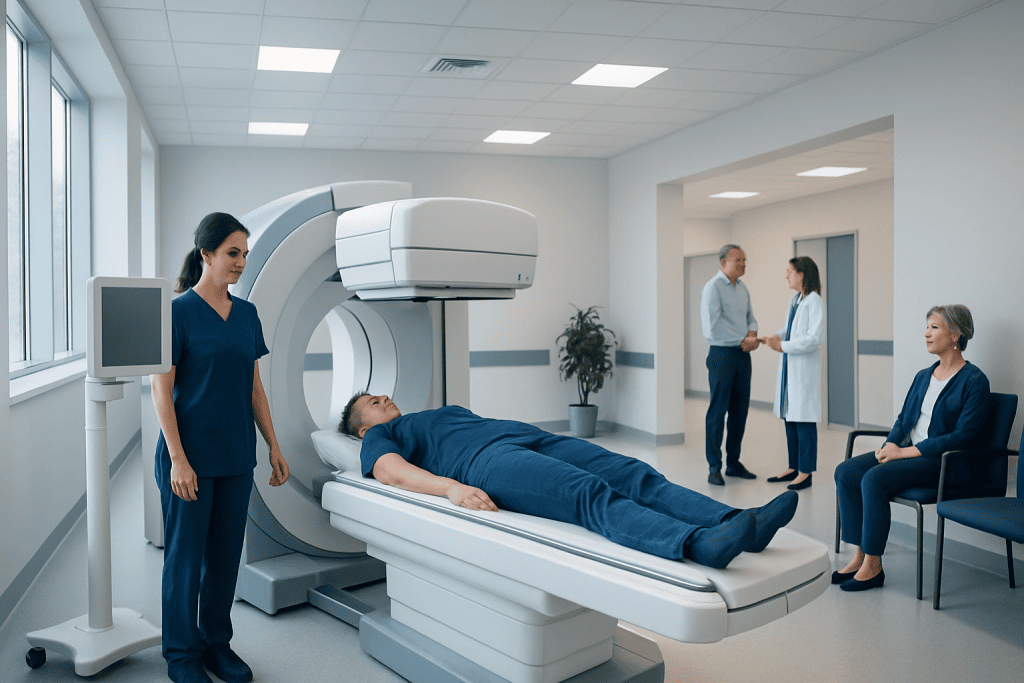
A bone scan shows how active the bones are, not just their shape. It can help identify fractures, arthritis, infection, inflammation, and some bone tumors.
Key Takeaways
- A bone scan shows how active the bones are, not just their shape.
- It can help identify fractures, arthritis, infection, inflammation, and some bone tumors.
- The test usually involves a small injection, a waiting period, and then images taken with a special camera.
- Bone scans are generally safe, and the amount of radiation used is low.
- Results are interpreted together with symptoms, examination findings, and other imaging tests.
A bone scan for joint and spine problems is a nuclear medicine imaging test that shows areas of increased or decreased bone activity. It can help doctors investigate joint pain, back pain, fractures, infection, arthritis, and other bone-related conditions when more detail is needed.
Overview
A bone scan is an imaging test that uses a small amount of radioactive tracer to look at bone metabolism, which means how active the bones are. Unlike a standard X-ray, which mainly shows bone structure, a bone scan can highlight areas where the bone is repairing itself or reacting to disease. This makes it especially useful when joint or spine symptoms are difficult to explain with routine imaging alone.
For people with ongoing joint pain, unexplained back pain, suspected stress fractures, or concern for infection or inflammation, a bone scan may provide helpful information. It can show “hot spots,” where bone activity is increased, or less commonly “cold spots,” where bone activity is reduced. These patterns do not give a diagnosis by themselves, but they help doctors narrow down possible causes.
Bone scans are commonly performed in the field of nuclear medicine. They are often used alongside other tests such as X-rays, CT, MRI, blood tests, and physical examination findings. In many cases, the value of the scan comes from seeing changes that are not yet obvious on other imaging studies.
Why a Bone Scan May Be Recommended
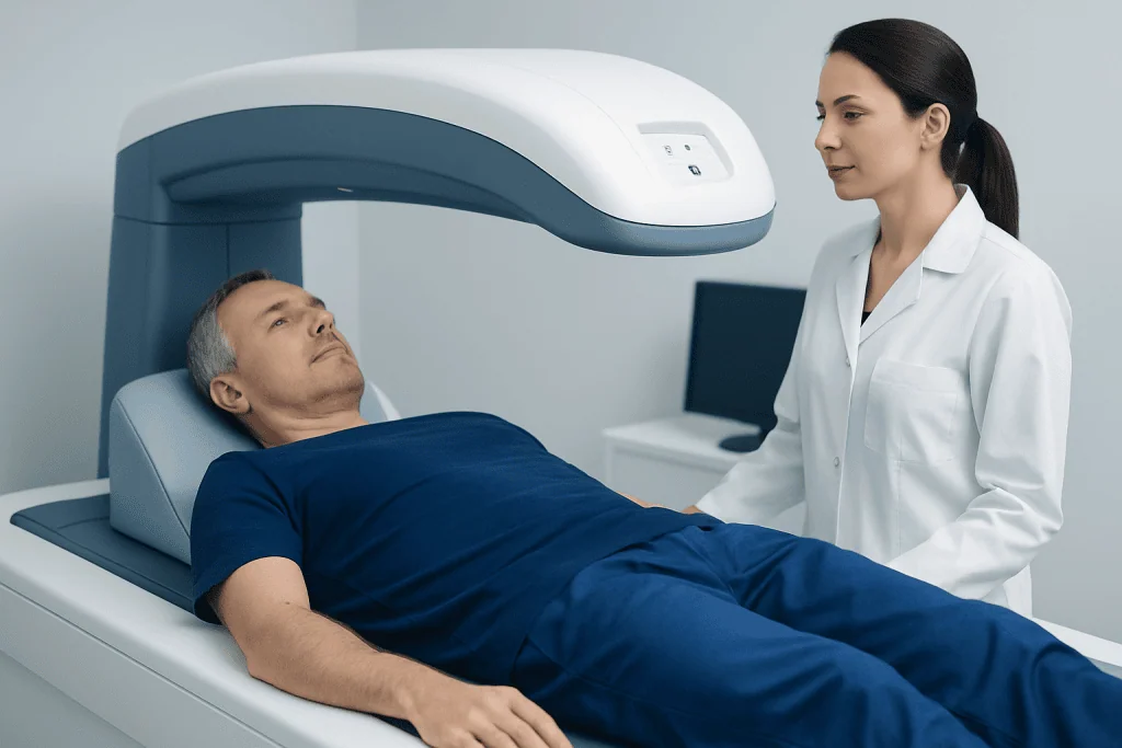
A doctor may suggest a bone scan when a person has symptoms affecting the joints or spine that need further evaluation. This may include persistent pain in the hip, knee, shoulder, sacroiliac joint, or spine, especially if the cause is not clear. It can also be useful if there is concern about a tiny fracture that is difficult to see on an X-ray, such as a stress fracture or an early compression fracture in the spine.
Bone scans can help in the evaluation of a range of conditions. These include inflammatory joint disease, osteoarthritis, infection in the bone or joint, reduced blood supply to bone, and some benign or malignant bone lesions. When pain comes from wear-and-tear changes, a scan may support the clinical picture of osteoarthritis or help distinguish one painful area from another in people who have changes in several joints.
In the spine, a bone scan may be used to investigate unexplained pain, identify active vertebral fractures, or look for inflammation around joints and spinal structures. It may also help doctors assess whether symptoms could be related to conditions such as scoliosis when structural changes are present but pain patterns are unclear. In some cases, additional imaging such as MRI or CT scanning may still be needed to clarify the diagnosis.
What the Scan Can Show
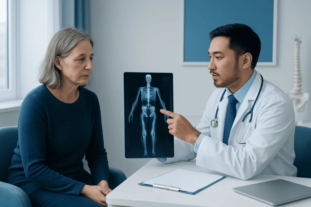
A bone scan does not simply take a picture of the bones. Instead, it shows where the tracer collects, which reflects blood flow and bone turnover. Areas that are healing, inflamed, infected, or under unusual stress often absorb more tracer and appear brighter on the images. This is why the scan can detect problems before structural changes become obvious on ordinary X-rays.
For joint problems, the scan may show patterns suggesting arthritis, inflammation, or stress around a prosthetic joint. After joint replacement surgery, a bone scan may sometimes help assess ongoing pain, although interpretation can be complex because healing itself can increase tracer uptake. In the spine, increased activity may be seen with compression fractures, degeneration, inflammation, or infection.
It is important to know that a bone scan is sensitive but not always specific. In simple terms, it is good at showing that something is happening in the bone, but it may not always show exactly what that problem is. Because of this, doctors often combine bone scan findings with a person’s medical history and may request further tests such as PET/CT in selected cases where more detailed assessment is needed.
What to Expect Before, During, and After the Test
Before the scan, a healthcare professional injects a small amount of tracer into a vein, usually in the arm. The injection itself is similar to a routine blood test or IV placement. After that, there is a waiting period, often a few hours, to allow the tracer to circulate and collect in the bones. During this time, patients are usually encouraged to drink water and empty the bladder as advised.
When it is time for imaging, the patient lies still on a padded table while a special gamma camera moves around the body or over the area of concern. The scan itself is painless, although staying still may be uncomfortable for people with significant back or joint pain. The test usually does not require anesthesia, but patients should tell the team if they feel anxious, claustrophobic, or unable to remain still.
After the scan, most people can return to their usual daily activities right away. Drinking fluids for the rest of the day helps the body clear the tracer naturally. The radioactive material used for a bone scan loses activity quickly and is generally considered safe in the small amounts used for medical imaging. The care team may give special instructions for pregnant or breastfeeding patients, so it is important to mention this in advance.
- Wear comfortable clothing and remove metal items if asked.
- Bring a list of medications and previous imaging reports if available.
- Inform the team about pregnancy, breastfeeding, allergies, or recent nuclear medicine tests.
- Ask about timing, since the waiting period can make the appointment longer than expected.
Benefits, Limitations, and Safety
One of the main benefits of a bone scan is that it can detect abnormal bone activity early. This can be helpful when symptoms are present but X-rays are normal, or when pain may be coming from more than one area. A whole-body bone scan can also look at many bones at once, which may be useful when symptoms are widespread or when the doctor wants a broader overview.
At the same time, the test has limitations. A bone scan may show an abnormal area without clearly identifying the exact cause. Increased tracer uptake can be seen in several different conditions, including arthritis, healing fractures, infection, and tumors. This means the result often needs to be interpreted together with other tests and sometimes followed by targeted imaging or laboratory studies.
For most adults, bone scans are considered safe. The radiation exposure is generally low and comparable to other medical imaging tests that use radiation. Allergic reactions to the tracer are uncommon. Still, every imaging decision should be individualized, especially for children, pregnant patients, and people who may need repeated scans over time.
How Results Are Interpreted
A specialist in nuclear medicine or radiology reviews the images and prepares a report for the referring doctor. The report describes where tracer uptake is increased or decreased and whether the pattern looks more consistent with arthritis, fracture, infection, degeneration, or another process. However, these descriptions are part of a larger clinical picture and are rarely interpreted in isolation.
For example, a bright area in the spine may suggest an active compression fracture, but the doctor may still need MRI to see nearby nerves, discs, and soft tissues more clearly. A hot spot near a joint could be related to inflammation, but blood tests or joint evaluation may be needed if infection is a concern. In this way, the scan acts as one tool among several.
If results are unclear, the doctor may recommend follow-up imaging, repeat scanning after a period of time, or referral to a specialist such as an orthopedist, rheumatologist, spine specialist, or oncologist. In complex cases, a multidisciplinary team approach can be helpful. Acibadem International’s multidisciplinary specialists in JCI-accredited hospitals diagnose and treat bone, joint, and spine conditions for international patients when advanced evaluation is needed.
When to Speak With a Doctor
People should speak with a doctor if they have joint or back pain that persists, worsens, or limits daily activities. Medical advice is also important for pain after an injury, repeated stress-related pain from exercise, or pain associated with swelling, fever, numbness, weakness, or unexplained weight loss. These symptoms do not always mean a serious problem, but they should be assessed properly.
A bone scan may be part of the evaluation when symptoms are difficult to explain or when there is a need to detect active bone changes. It is especially useful when the goal is to find out whether pain is linked to a hidden fracture, inflammation, infection, or another process affecting the skeleton. Early assessment can help guide appropriate treatment and avoid unnecessary delay.
Anyone preparing for a bone scan should feel comfortable asking questions about why the test is recommended, what the results may show, and whether any other imaging might also be needed. Understanding the purpose of the scan often makes the process feel more manageable. A qualified doctor can explain how the findings relate to the person’s symptoms and the next best steps for care.
Frequently asked questions
Is a bone scan painful?
The scan itself is not painful. Most people only feel a brief pinch during the tracer injection, and then they lie still while images are taken. If a person already has significant joint or back pain, staying in one position may be the most uncomfortable part.
How long does a bone scan take?
The imaging part is often completed within about 30 to 60 minutes, but the full appointment is usually longer because there is a waiting period after the injection. In many cases, patients wait a few hours for the tracer to collect in the bones. The exact schedule can vary between hospitals.
Can a bone scan detect arthritis in the spine or joints?
A bone scan can show increased bone activity that may be related to arthritis or inflammation in joints and the spine. It is helpful for identifying active areas, especially when more than one region may be involved. However, the scan does not always tell exactly which type of arthritis is present, so other tests may still be needed.
What is the difference between a bone scan and an MRI?
A bone scan shows bone metabolism and highlights areas where bone is unusually active. MRI provides detailed images of bones, discs, nerves, joints, and soft tissues. Doctors may choose one or both tests depending on the symptoms and the question they are trying to answer.
Is the radiation from a bone scan dangerous?
The amount of radiation used in a bone scan is generally low and is considered safe for most people when medically appropriate. The tracer is used in a small quantity and leaves the body over time. Patients who are pregnant or breastfeeding should discuss this with their doctor before the test.
Do patients need to prepare for a bone scan?
Preparation is usually simple. Patients may be advised to drink fluids, avoid certain metal items during imaging, and tell the team about pregnancy, breastfeeding, medicines, or recent imaging tests. The hospital or imaging center will provide specific instructions if anything extra is needed.
References
- World Health Organization
- Radiological Society of North America
- Society of Nuclear Medicine and Molecular Imaging
- American College of Radiology
- National Institute of Arthritis and Musculoskeletal and Skin Diseases
This article is for general information only and is not a substitute for professional medical advice. Please consult a qualified doctor about your individual situation.
Medical weight-loss options in Turkey — costs & eligibility
JCI-accredited · board-certified surgeons · reply within 24h
Add us as a Preferred Source to see more of our trusted health content across Google Search, AI Overviews and Discover.