Brain Anatomy: How the Human Brain Is Organized
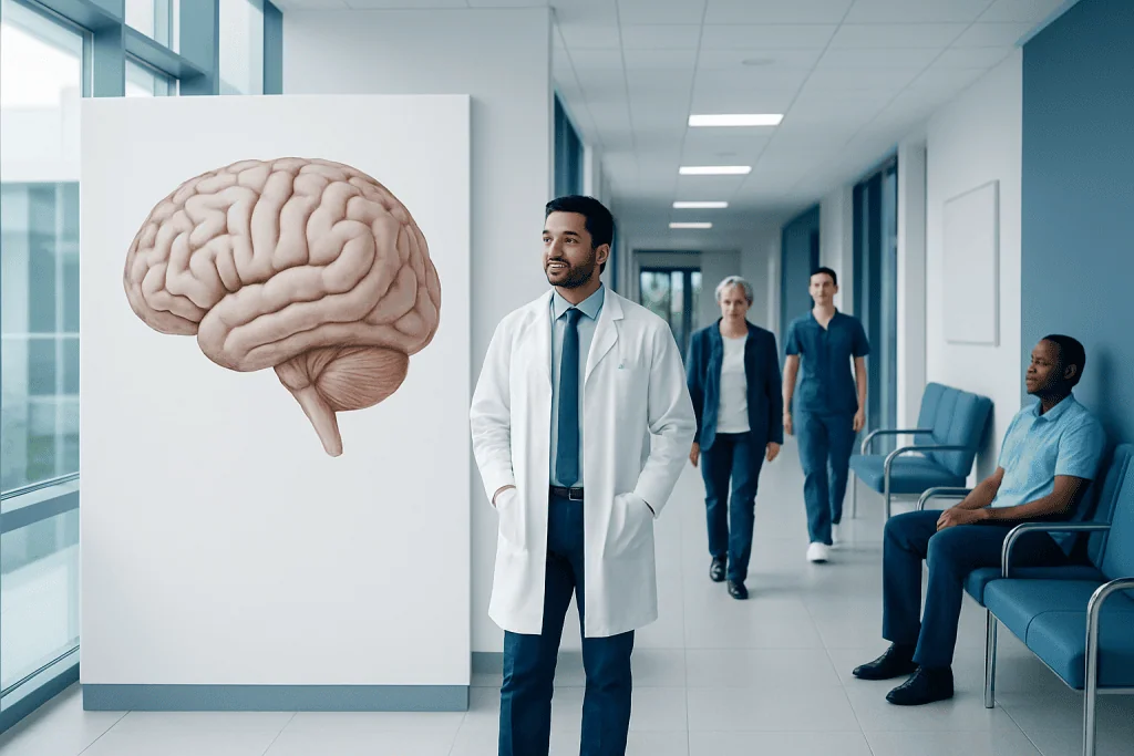
The brain is divided into major regions, including the cerebrum, cerebellum, brainstem, and deeper internal structures. Each area has specific roles, but normal brain function depends on constant communication between regions.
Key Takeaways
- The brain is divided into major regions, including the cerebrum, cerebellum, brainstem, and deeper internal structures.
- Each area has specific roles, but normal brain function depends on constant communication between regions.
- Symptoms from brain disorders depend on which structures are affected and may include weakness, speech changes, balance problems, or memory concerns.
- Doctors may use neurological examination, imaging, and other tests to assess brain structure and function.
- Healthy habits that support blood flow, sleep, and overall wellness also help support brain health.
Brain anatomy explains how the human brain is organized into specialized regions that work together to control movement, sensation, memory, language, and vital body functions. Understanding these structures can make common neurological symptoms and tests easier to understand.
Overview
Brain anatomy describes how the human brain is arranged and how its parts work together. The brain is the body’s central control organ: it processes information, coordinates movement, supports memory and language, and helps regulate breathing, heart rate, temperature, and many other functions.
Although the brain is often discussed as one organ, it is organized into several major regions. The largest part, the cerebrum, handles thinking, voluntary movement, and sensory processing. The cerebellum helps with balance and coordination, while the brainstem connects the brain to the spinal cord and supports essential life functions.
Understanding basic brain anatomy can help patients make sense of medical terms such as lobes, nerves, cortex, and white matter. It can also help explain why different neurological conditions cause different symptoms. For example, a problem affecting the speech centers may look very different from one affecting balance or vision.
Main Parts of the Brain
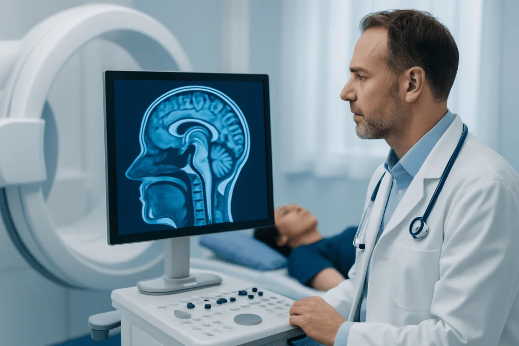
The brain is commonly described in several layers and regions. The outer portion, called the cerebral cortex, is the wrinkled surface of the cerebrum and is responsible for many higher functions such as reasoning, perception, and voluntary movement. Beneath it are deeper structures that help relay signals, regulate emotion, and support memory.
The cerebrum is divided into right and left hemispheres, which communicate through a thick bundle of nerve fibers called the corpus callosum. Each hemisphere has four main lobes: frontal, parietal, temporal, and occipital. These lobes are not isolated; they continuously work together to support everyday activities such as speaking, reading, recognizing faces, and planning tasks.
The cerebellum sits at the back of the brain, under the cerebrum. The brainstem lies at the base of the brain and includes the midbrain, pons, and medulla. The brain also contains structures such as the thalamus, hypothalamus, hippocampus, and basal ganglia, which are important for sensory relay, hormone control, memory, and movement. These structures are part of the broader network sometimes discussed in brain conditions such as stroke and other neurological disorders.
What Each Brain Lobe Does
Each lobe of the cerebrum has characteristic responsibilities, although most brain tasks involve more than one lobe at a time. The frontal lobe is involved in planning, decision-making, personality, problem-solving, and voluntary movement. It also contains areas that help produce speech.
The parietal lobe processes touch, pressure, temperature, and spatial awareness. It helps a person judge where the body is in relation to the surroundings. The temporal lobe is important for hearing, language understanding, memory, and emotion. The occipital lobe primarily processes visual information from the eyes.
Because brain functions overlap, damage to one area may produce several symptoms. For example, a problem in the temporal lobe may affect both memory and language comprehension, while a problem in the parietal lobe may affect sensation and navigation. This networked design is one reason brain anatomy is so closely linked to clinical neurology, including evaluation for seizure disorders such as epilepsy.
How Brain Tissue Is Organized
Brain tissue is often described as gray matter and white matter. Gray matter contains many nerve cell bodies and is heavily involved in processing information. White matter contains nerve fibers, also called axons, that carry signals between brain regions and to the rest of the nervous system.
The brain is covered by protective membranes called meninges: the dura mater, arachnoid mater, and pia mater. The brain also floats in cerebrospinal fluid, which cushions it and helps protect it from injury. Blood vessels supply the brain with oxygen and nutrients; because the brain has a high energy demand, even brief interruptions in blood flow can affect function.
Inside the brain are fluid-filled spaces called ventricles. These spaces produce and circulate cerebrospinal fluid. Together, these structures help keep the brain protected, nourished, and working in a stable internal environment.
Symptoms That Reflect Different Brain Regions
Symptoms related to the brain vary widely because different regions control different functions. A person may notice weakness, numbness, speech difficulty, vision changes, dizziness, memory problems, tremor, or difficulty with balance. Mood changes, confusion, and personality changes can also occur depending on the area involved.
Some symptoms suggest a specific location. For example, trouble with coordination may point toward the cerebellum, while difficulty forming words may involve language centers in the dominant hemisphere. Problems with swallowing, breathing, or consciousness can involve the brainstem and require prompt medical assessment.
- Weakness or loss of coordination
- Speech or language problems
- Changes in memory or attention
- Vision or hearing changes
- Seizures or episodes of confusion
These symptoms do not always mean a serious brain disorder, but they do deserve evaluation when new, persistent, or worsening.
Common Causes and Risk Factors for Brain Problems
Many conditions can affect brain anatomy and function. These include stroke, trauma, infections, tumors, neurodegenerative diseases, autoimmune conditions, and congenital abnormalities. Some problems mainly affect the brain’s structure, while others affect how brain circuits communicate.
Risk factors vary by condition. High blood pressure, diabetes, smoking, high cholesterol, and atrial fibrillation increase the risk of some vascular brain problems. Head injuries may occur during falls, sports, or accidents. Certain infections, genetic conditions, and age-related changes can also influence the risk of neurological disease.
Lifestyle factors do not cause every brain disorder, but they can influence brain health over time. Good sleep, regular physical activity, balanced nutrition, and treatment of chronic medical conditions support healthy brain function and may lower the risk of some problems.
How Doctors Evaluate the Brain
Doctors usually begin with a detailed medical history and neurological examination. The exam may assess strength, reflexes, coordination, sensation, eye movements, speech, and memory. These findings help determine which brain regions may be involved.
If more information is needed, doctors may order imaging tests such as CT or MRI scans to look at brain structure. In some situations, additional tests such as electroencephalography (EEG), blood tests, lumbar puncture, or specialized neuropsychological testing may be useful. The choice of test depends on the symptoms and the suspected cause.
Brain anatomy is often reviewed alongside test results because location matters. A scan may show where an abnormality is, but the doctor also considers how that location matches the patient’s symptoms and examination findings. This approach helps guide appropriate care.
Treatment, Prevention, and Brain Health
Treatment depends entirely on the underlying cause. Some conditions may require urgent care, such as stroke, severe head injury, or infection. Others are managed with medication, rehabilitation, monitoring, or surgery. When needed, specialized care may involve neurology, neurosurgery, rehabilitation medicine, speech therapy, or occupational therapy.
Not every brain problem can be prevented, but healthy habits can support long-term brain function. Regular exercise, blood pressure control, diabetes management, not smoking, adequate sleep, and hearing and vision care all contribute to overall neurological wellness. Using seat belts, helmets, and fall-prevention strategies also helps protect the brain from injury.
For patients who need advanced evaluation or treatment, multidisciplinary teams can be helpful. Acibadem International’s multidisciplinary specialists and JCI-accredited hospitals diagnose and treat many neurological conditions for international patients in a coordinated setting.
When to See a Doctor
A doctor should be consulted for new or persistent neurological symptoms, especially if they interfere with daily life. Warning signs may include unexplained weakness, sudden speech changes, new seizures, repeated falls, significant memory loss, or ongoing headaches with other neurological symptoms.
Emergency care is important for sudden facial droop, one-sided weakness, severe confusion, loss of consciousness, or sudden vision loss, because these can indicate a serious problem such as stroke. Early evaluation can make a major difference in outcome for time-sensitive conditions.
Even when symptoms are mild, a medical assessment can provide reassurance and help identify whether the cause is temporary, treatable, or needs follow-up. Patients should not try to self-diagnose brain symptoms, since many different conditions can overlap.
Frequently asked questions
What are the main parts of the brain?
The main parts are the cerebrum, cerebellum, and brainstem. The cerebrum handles thinking, sensation, and voluntary movement, the cerebellum supports balance and coordination, and the brainstem helps control vital functions such as breathing and heart rate.
What does the cerebral cortex do?
The cerebral cortex is the outer layer of the cerebrum and is responsible for many higher brain functions. It helps with perception, language, memory, decision-making, and voluntary movement.
Why do different brain problems cause different symptoms?
Different brain regions have different jobs, so the symptoms depend on which area is affected. A problem in the occipital lobe may affect vision, while a problem in the frontal lobe may affect planning, movement, or behavior.
Can brain anatomy be seen on imaging tests?
Yes. MRI and CT scans can show the brain’s structure and may reveal injury, bleeding, swelling, tumors, or other abnormalities. These tests are interpreted together with the patient’s symptoms and examination findings.
What is the difference between gray matter and white matter?
Gray matter contains many nerve cell bodies and is involved in processing information. White matter contains nerve fibers that connect different brain regions and help signals travel efficiently.
How can someone support brain health?
Brain health is supported by good control of blood pressure and blood sugar, regular physical activity, healthy sleep, and not smoking. Protecting the head from injury and seeking care for new neurological symptoms are also important.
References
- National Institute of Neurological Disorders and Stroke
- National Institutes of Health
- Mayo Clinic
- Cleveland Clinic
- World Health Organization
This article is for general information only and is not a substitute for professional medical advice. Please consult a qualified doctor about your individual situation.
Heart care in Turkey — expert evaluation and treatment
JCI-accredited · board-certified surgeons · reply within 24h
Add us as a Preferred Source to see more of our trusted health content across Google Search, AI Overviews and Discover.
Check your numbers in seconds
BMI, calories, due date, blood pressure and 30+ more clinical calculators — free, instant, doctor-reviewed ranges.