Breast Ca Mammogram: What Is Normal, What Is Not, and When to Seek Care
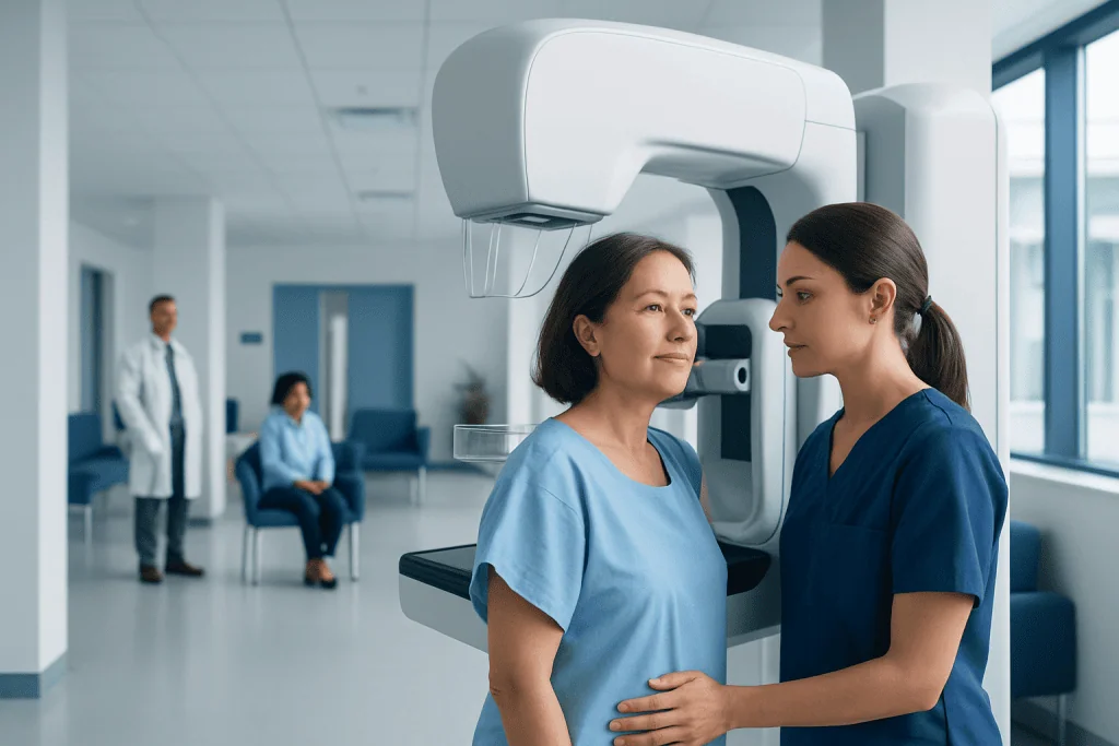
A normal mammogram means no suspicious findings were seen, but routine screening is still important. An abnormal mammogram does not automatically mean breast cancer; many findings are benign.
Key Takeaways
- A normal mammogram means no suspicious findings were seen, but routine screening is still important.
- An abnormal mammogram does not automatically mean breast cancer; many findings are benign.
- BI-RADS categories help explain whether a result is negative, probably benign, suspicious, or needs urgent evaluation.
- Further tests may include diagnostic mammography, breast ultrasound, MRI, or biopsy.
- New breast symptoms such as a lump, nipple discharge, or skin changes should be assessed even if a recent mammogram was normal.
A breast ca mammogram looks for changes in breast tissue that may be benign, need short-term follow-up, or require more testing. Most abnormal results do not mean cancer, but understanding what is normal, what is not, and when to seek care can make next steps clearer and less stressful.
Overview: what a breast ca mammogram can and cannot show
A breast ca mammogram is an X-ray examination of the breast used to look for signs of breast cancer and other breast changes. A result may be normal, probably benign, or suspicious enough to need additional imaging or a biopsy. In many cases, a callback after screening is a precaution and does not mean cancer has been found.
Mammograms are used both for routine screening in people without symptoms and for diagnostic evaluation when there is a concern such as a lump, nipple discharge, focal pain, or skin changes. Screening mammograms aim to detect problems early, sometimes before they can be felt. Diagnostic mammograms take extra views and are tailored to a specific area of concern.
Mammography is one of the main tools for early detection, but it is not perfect. Some cancers may not be visible, especially in dense breasts, and some visible changes may turn out to be noncancerous. This is why mammogram findings are interpreted together with age, symptoms, physical examination, family history, and sometimes other tests.
What is considered normal on a mammogram
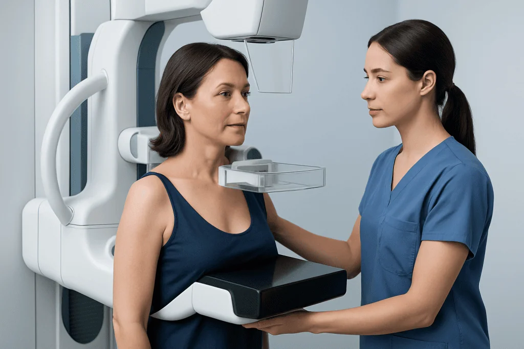
A normal mammogram generally means the radiologist did not see a suspicious mass, suspicious calcifications, or architectural distortion that suggests cancer. Many reports describe this as a negative examination or assign a BI-RADS category 1. A BI-RADS category 2 means benign findings were seen, such as simple cysts, stable calcifications, or a harmless lymph node, but there is no sign of cancer.
Normal does not always mean the breasts have no changes at all. It is common for a mammogram to show benign findings that are simply documented for future comparison. Breasts also vary naturally in density and structure, so a report may note dense breast tissue without calling the study abnormal.
Breast density matters because dense tissue can make mammograms harder to interpret and can sometimes hide small cancers. A person with dense breasts may still have a normal mammogram, but the report may recommend discussing whether additional imaging is appropriate based on overall risk. Routine screening should continue at intervals recommended by a qualified clinician.
What is not normal: common abnormal findings and what they may mean
An abnormal mammogram means something was seen that needs a closer look, not that breast cancer is confirmed. Common findings include a mass, asymmetry, clustered calcifications, architectural distortion, or a new change compared with prior images. Many of these findings are caused by benign conditions such as cysts, fibroadenomas, hormonal changes, scar tissue, or overlapping normal tissue.
Radiologists use the BI-RADS system to guide follow-up. BI-RADS 0 means the screening exam is incomplete and more images are needed. BI-RADS 3 means the finding is probably benign and is usually followed with repeat imaging after a short interval. BI-RADS 4 and 5 mean the finding is suspicious or highly suggestive of malignancy and usually requires biopsy. BI-RADS 6 is used when cancer has already been confirmed by biopsy.
Microcalcifications can be a useful example. Many calcifications are harmless and related to aging or prior inflammation, while some patterns may suggest precancerous change or cancer and need tissue sampling. A mass may be a simple fluid-filled cyst on ultrasound, which is often benign, or a solid lesion that needs biopsy for certainty. If cancer is diagnosed, the next steps may involve evaluation for breast cancer and a tailored treatment plan.
Why mammogram results vary: risk factors, breast density, and symptoms
Mammogram interpretation depends on several factors beyond the image itself. Age, menopausal status, hormone use, prior breast surgery, family history of breast or ovarian cancer, known genetic variants, and personal history of breast cancer can all influence how findings are assessed. Prior mammograms are especially valuable because stability over time often supports a benign explanation.
Breast density is one of the most important factors. Dense tissue appears white on a mammogram, and so do many abnormalities, which can make small cancers less obvious. Dense breasts are common and not abnormal by themselves, but they may affect the sensitivity of screening and prompt discussion of supplemental imaging in selected patients.
Symptoms also matter. A mammogram done for screening is interpreted differently from a mammogram done because of a new lump or nipple discharge. Even if a screening mammogram is normal, persistent symptoms deserve medical attention. In some cases, clinicians add targeted ultrasound or breast MRI when the clinical picture suggests more evaluation is needed.
How doctors confirm whether a finding is benign or cancerous
If a screening mammogram is unclear or abnormal, the next step is often a diagnostic mammogram with additional views. These images can help determine whether the original finding was caused by overlapping tissue or whether it remains concerning. Targeted breast ultrasound is commonly used to evaluate a specific area, especially a mass or focal symptom.
When imaging still does not provide a definite answer, a biopsy may be recommended. This may involve a needle procedure guided by mammography, ultrasound, or MRI, depending on where the abnormality is best seen. A biopsy is the only way to confirm whether cells are benign, precancerous, or malignant.
If cancer is diagnosed, additional testing may help define the stage and guide treatment. This can include imaging, pathology review, and discussions with specialists in radiology, breast surgery, oncology, and pathology. A coordinated approach helps match treatment to the type and extent of disease rather than to the mammogram result alone.
Treatment options when a mammogram leads to a cancer diagnosis
Treatment depends on the biopsy result, the size and location of the lesion, whether lymph nodes are involved, hormone receptor status, HER2 status, and the patient’s overall health and preferences. Some findings are noninvasive or very early-stage, while others require broader treatment planning. The goal is to treat effectively while preserving quality of life and avoiding unnecessary procedures when possible.
Common treatment options may include surgery, radiation therapy, systemic therapies such as hormone therapy, targeted therapy, or chemotherapy, or a combination of these. Surgical care may involve breast cancer surgery to remove the tumor and assess nearby lymph nodes. In selected cases, treatment is given before surgery to shrink the cancer and improve options.
Not every abnormal mammogram leads to cancer treatment. Many patients only need short-term imaging follow-up or reassurance after a benign biopsy. For those who do need treatment, decisions are individualized and are usually made with a multidisciplinary team. Acibadem International’s multidisciplinary specialists and JCI-accredited hospitals diagnose and treat this condition for international patients.
Prevention, screening, and self-care between mammograms
The best way to reduce the chance of a delayed diagnosis is to follow a screening plan recommended by a qualified doctor. The right age to begin and the right interval for mammography vary depending on personal risk, family history, and local guideline recommendations. People at higher risk may need earlier screening or additional tests.
It can help to know what the breasts normally look and feel like, although formal self-exams are not a substitute for screening. Any new persistent change should be reported, especially a lump, skin dimpling, nipple inversion, one-sided discharge, or swelling under the arm. Keeping prior mammograms available for comparison can also improve accuracy and reduce unnecessary callbacks.
General health habits support breast health overall, even though they cannot prevent every case of cancer. These include limiting alcohol, maintaining a healthy weight, staying physically active, avoiding tobacco, and discussing hormone therapy risks and benefits with a doctor. Patients with strong family histories may benefit from risk assessment and genetic counseling.
When to seek medical care
Medical care should be sought promptly if a mammogram report recommends additional imaging, short-interval follow-up, or biopsy. It is also important to seek evaluation for new breast symptoms even after a recent normal screening mammogram. Symptoms that deserve attention include a new lump, bloody or spontaneous nipple discharge, skin thickening or dimpling, redness that does not improve, nipple inversion, or swelling in the armpit.
Urgency does not always mean emergency, but it does mean the finding should not be ignored. Most callbacks are resolved with more imaging, and many biopsies are benign. Still, timely follow-up matters because it helps clarify whether a change is harmless or needs treatment.
Anyone who feels uncertain about a report can ask for the BI-RADS category, the exact finding, and the recommended next step. A second radiology review or specialist consultation may be appropriate in selected cases, particularly for dense breasts, persistent symptoms, or a strong family history. Early clarification often reduces anxiety and supports better decision-making.
Frequently asked questions
Does an abnormal breast ca mammogram mean cancer?
No. An abnormal mammogram means the radiologist saw something that needs more evaluation, but many findings are benign. Extra imaging or a biopsy may be recommended to find out exactly what the change represents.
What is the difference between BI-RADS 1, 2, 3, and 4?
BI-RADS 1 means negative and BI-RADS 2 means benign findings with no sign of cancer. BI-RADS 3 means probably benign and usually needs short-term follow-up imaging, while BI-RADS 4 means suspicious and often leads to biopsy.
Can a mammogram be normal if symptoms are present?
Yes. A mammogram can be normal even when a person has a lump or other breast symptom, especially in dense breasts or when a change is subtle. Persistent symptoms should still be assessed by a doctor, often with targeted ultrasound or other testing.
What happens after a callback from a screening mammogram?
A callback usually means more pictures are needed to clarify an area seen on the screening study. The next step may be a diagnostic mammogram, ultrasound, or both. Many callbacks end with reassurance and routine follow-up.
Do dense breasts mean a person has breast cancer?
No. Dense breasts are common and are not cancer by themselves. They can, however, make mammograms harder to read, so a doctor may discuss whether additional screening is appropriate based on overall risk.
When should someone seek care after a normal mammogram?
Care should be sought if there is a new lump, persistent focal pain, nipple discharge, nipple inversion, skin dimpling, redness, or swelling under the arm. New symptoms should be evaluated even if a recent mammogram was reported as normal.
References
- American Cancer Society
- Breast Imaging Reporting and Data System (BI-RADS), American College of Radiology
- National Cancer Institute
- U.S. Preventive Services Task Force
- World Health Organization
This article is for general information only and is not a substitute for professional medical advice. Please consult a qualified doctor about your individual situation.
Oncology care in Turkey — second opinion and treatment plan
JCI-accredited · board-certified surgeons · reply within 24h
Add us as a Preferred Source to see more of our trusted health content across Google Search, AI Overviews and Discover.