

Broken Forearm Xray: Preparation, Procedure and Results

Forearm X-rays are quick, painless imaging tests commonly used after falls, sports injuries and direct blows. Images usually include at least two views of the forearm and may include the wrist or elbow.
Key Takeaways
- Forearm X-rays are quick, painless imaging tests commonly used after falls, sports injuries and direct blows.
- Images usually include at least two views of the forearm and may include the wrist or elbow.
- A fracture may still allow some movement, so being able to move the arm does not rule out a broken bone.
- Treatment depends on fracture location, alignment, stability, age and associated injuries.
- Follow-up X-rays may be needed to check that bones remain aligned and are healing as expected.
A broken forearm xray is the standard first imaging test when a fracture of the radius, ulna, or both bones is suspected. It can confirm whether a bone is broken, show the location and alignment of the injury, and help clinicians plan treatment and monitor healing.
Broken Forearm Xray: What It Shows
A broken forearm xray is an imaging examination used to look for fractures in the two long bones between the elbow and wrist: the radius and ulna. It is usually requested after an injury causing pain, swelling, bruising, deformity or difficulty using the arm. The test helps the clinical team determine whether one or both bones are affected and whether the broken pieces have moved out of their normal position.
X-rays use a small amount of radiation to create images of bone. They are fast, widely available and particularly useful in urgent injury assessment. A radiologist or treating clinician reviews the images alongside the person’s symptoms and physical examination. If the X-ray does not fully explain ongoing symptoms, further imaging such as CT or MRI may occasionally be recommended.
In addition to identifying a fracture, the images can show angulation, shortening, rotation, joint involvement or signs of a dislocation. These details are important because they influence whether the arm can be treated with a splint or cast, needs the bone to be realigned, or may require orthopedic surgery.
How the X-Ray Procedure Works
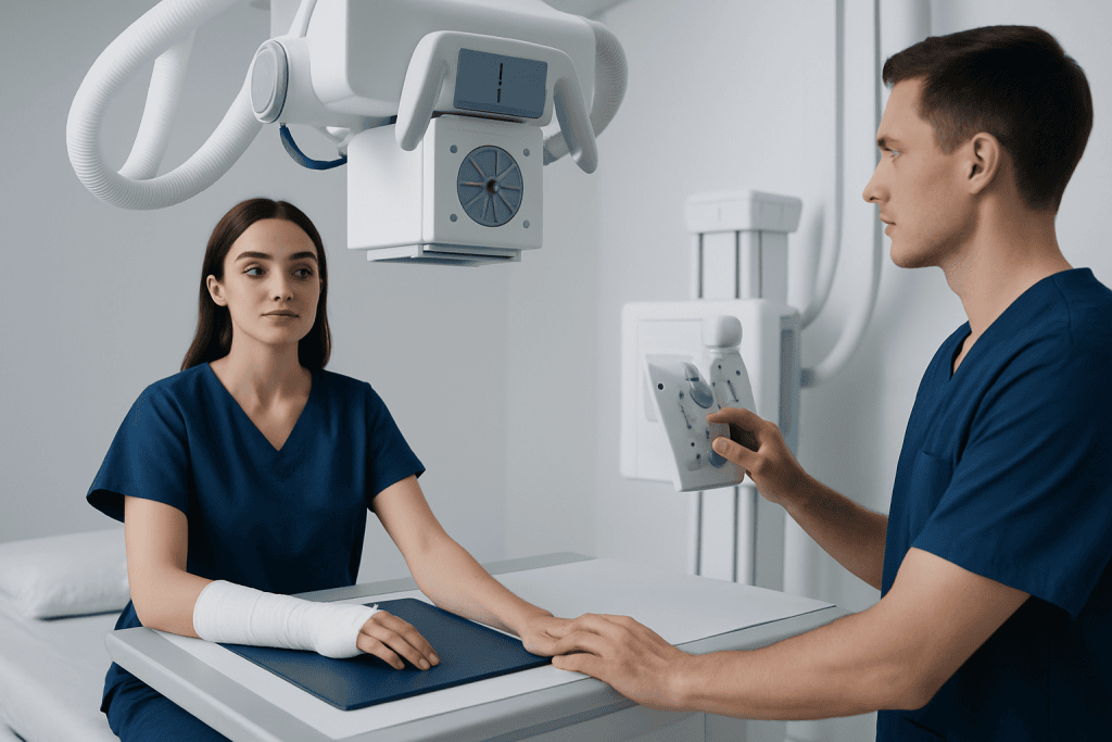
During an X-ray, the forearm is positioned between an X-ray machine and a digital detector. The machine sends a carefully controlled beam through the arm. Dense tissues such as bone absorb more of the beam and appear lighter on the image, while soft tissues appear in shades of gray.
For suspected forearm fractures, clinicians commonly request at least two views taken from different angles. This is important because a fracture line or displacement can be easier to see in one view than another. The elbow and wrist may also be imaged, especially if pain extends to either joint or the injury mechanism raises concern for a related injury.
There is usually no special preparation. The person may be asked to remove jewelry, watches or other metal objects near the arm. Pregnancy should be mentioned before imaging so the radiology team can take appropriate precautions, although an arm X-ray exposes the abdomen and pelvis only minimally.
Who Needs a Broken Forearm Xray and What Happens Step by Step?
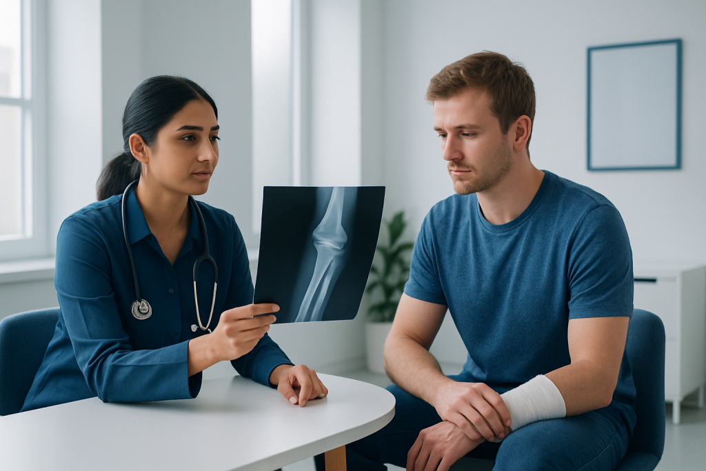
A broken forearm xray is appropriate when a clinician suspects a fracture after a fall onto an outstretched hand, a collision, a twisting injury, a sports accident or a direct impact. Children commonly need imaging after playground or sports injuries, while adults may sustain forearm fractures after falls, road traffic incidents or higher-energy trauma.
In a broken forearm x ray child assessment, the same basic approach is used, with positioning adapted gently to reduce discomfort. Children’s bones are still growing and may fracture differently from adult bones, including incomplete patterns sometimes called greenstick fractures. Clinicians also assess whether a fracture is close to a growth plate, since this can affect follow-up planning.
The procedure generally follows a simple sequence:
- The injured arm is supported in the most comfortable safe position possible.
- A radiographer explains the required positions and takes the requested images.
- The person is asked to stay still for a few seconds for each image.
- The images are checked for quality and reviewed by the treating team, often on the same day.
- If a fracture is found, the arm may be splinted while definitive treatment is arranged.
Moving a newly injured arm can be painful. The radiographer should be told if any position causes significant discomfort, numbness or worsening pain. Images can usually be obtained without forcing the arm into an unsafe position.
How Serious Is a Broken Forearm?
A broken forearm can range from a stable, uncomplicated crack in one bone to a more complex injury involving both bones, the wrist or elbow joint, nearby nerves or blood vessels, or an open wound. Many forearm fractures heal well with prompt assessment and appropriate immobilization, but they should always be evaluated by a qualified clinician.
Fractures are more concerning when the arm looks bent or shortened, bone is visible through the skin, fingers become pale, blue, cold, numb or weak, or pain and swelling increase rapidly. These signs can indicate impaired circulation, nerve involvement, severe swelling or an open fracture, all of which require urgent medical attention.
The position of the bones matters in the forearm because the radius and ulna work together to allow the palm to turn upward and downward. A displaced fracture that heals in poor alignment can limit this motion. For this reason, clinicians may arrange repeat imaging after a cast is applied or after the bone has been realigned.
How Painful Is a Forearm Fracture?
A forearm fracture is often painful, particularly immediately after the injury and with movement, gripping or rotation of the hand. Pain may be accompanied by swelling, tenderness, bruising, a crackling sensation, or an obvious change in the arm’s shape. However, pain levels vary widely depending on the injury, the person and whether other structures were injured.
First aid should focus on keeping the arm still and supported. A sling or improvised support can help if it does not force the arm into a painful position. Ice wrapped in cloth may reduce swelling, but it should not be placed directly on the skin. Food and drink may need to be avoided if urgent sedation or surgery could be required, so it is sensible to follow local emergency-service advice.
Do not try to straighten a visibly deformed arm or push protruding bone back under the skin. Pain relief may be recommended by a clinician based on the person’s age, health conditions and other medicines. A splint and appropriate treatment of the fracture usually improve comfort substantially.
Can You Still Move Your Forearm If It Is Fractured?
Yes. Some people can still move their fingers, wrist, elbow or even part of the forearm after a fracture. A stable or incomplete break may allow more movement than a displaced fracture, and adrenaline immediately after an accident can temporarily mask pain. Therefore, movement does not exclude a broken bone.
Trying to test the arm repeatedly can worsen pain and may move an unstable fracture. After an injury, it is safer to stop activity, support the arm and seek medical assessment if there is persistent pain, swelling, tenderness over a bone, reduced function or concern about deformity.
An X-ray provides clearer information than symptoms alone. In some cases, early X-rays can appear normal despite a small fracture or injury near a joint. If symptoms remain significant, the clinician may recommend a temporary splint, repeat X-rays after a period of time, or different imaging.
Broken Arm X Ray Before and After: Treatment and Recovery
A broken arm x ray before and after treatment can look quite different when a displaced fracture has been realigned. Before treatment, images may show a gap, angulation or overlap between bone fragments. After reduction, splinting, casting or surgical fixation, X-rays are used to confirm that alignment is acceptable and to monitor it during healing.
Stable fractures may be treated with a splint or cast and follow-up appointments. When bones are significantly displaced, unstable, open, involve a joint, or cannot be held in a safe position in a cast, a specialist may recommend reduction and fixation with pins, plates, screws or other devices. Rehabilitation may be advised once the clinician confirms it is safe to restore movement and strength.
How long does it take to recover from a broken bone in the forearm? Bone healing often takes several weeks, but the full timeline depends on age, fracture type, whether surgery was needed, smoking status, overall health and adherence to follow-up advice. Children often heal faster than adults, while return to heavy lifting, contact sport or physically demanding work may take longer than initial bone healing. Follow-up X-rays help the clinical team guide a safe return to usual activities.
Acibadem International’s multidisciplinary specialists and JCI-accredited hospitals assess and treat forearm fractures for international patients, including imaging, orthopedic care and rehabilitation planning when needed.
When to Seek Medical Care
Urgent medical care is needed after a significant arm injury when there is severe pain, a visible deformity, an open wound, uncontrolled bleeding, rapidly increasing swelling, or suspected fracture. Emergency assessment is also important if the fingers are numb, weak, unusually pale or blue, cold, or difficult to move.
A same-day clinical assessment is appropriate for ongoing forearm pain after a fall or impact, especially when there is focal tenderness, bruising, swelling, pain with rotation of the palm, or difficulty using the hand and arm. A clinician can decide whether a broken forearm xray or other imaging is needed.
After a fracture has been treated, seek prompt advice if pain becomes markedly worse, a cast feels excessively tight or loose, there is a new fever or drainage from a wound, or the fingers develop tingling, numbness, color change or reduced movement. These symptoms should not be managed by simply waiting for the next routine appointment.
Frequently asked questions
How long does a broken forearm X-ray take?
The image-taking part usually takes only a few minutes. Positioning the arm carefully and reviewing image quality can make the overall appointment somewhat longer, particularly when the injury is painful or the person is a child.
Do I need to prepare for a broken forearm xray?
Usually, no special preparation is required. The person may need to remove jewelry, a watch or other metal objects from the affected arm, and should tell the radiology team about possible pregnancy.
Will an X-ray always show a forearm fracture?
Most forearm fractures are visible on standard X-rays, especially when images are taken from more than one angle. Very small, early or subtle fractures may not be obvious at first, so persistent symptoms may require follow-up imaging or reassessment.
Can a child have an X-ray for a suspected broken forearm?
Yes. X-rays are commonly used to assess suspected fractures in children, with radiation exposure kept as low as reasonably achievable. The results are interpreted with attention to developing bones and growth plates.
Can I drive home after a forearm X-ray?
The X-ray itself does not affect alertness, so it does not normally prevent driving. However, driving may be unsafe or inappropriate because of pain, a splint or cast, reduced arm control, medication, or the nature of the injury.
Why are follow-up X-rays needed after a forearm fracture?
Follow-up images help confirm that the fracture remains in an acceptable position while it heals. They can also help the clinician decide when immobilization can be changed or removed and when activity can be gradually resumed.
References
- American Academy of Orthopaedic Surgeons
- RadiologyInfo.org
- National Institute for Health and Care Excellence
- American College of Radiology
- Royal College of Radiologists
This article is for general information only and is not a substitute for professional medical advice. Please consult a qualified doctor about your individual situation.
Joint and spine care in Turkey — expert assessment & treatment
JCI-accredited · board-certified surgeons · reply within 24h
Add us as a Preferred Source to see more of our trusted health content across Google Search, AI Overviews and Discover.

More from the Health Library



Related Specialists

Prof. Dr. Dilaver Kaya
Neurology
Dr. Mehmet Yakupçebioğlu
Physical Medicine & Rehabilitation
Dr. Ayşen Yücetürk
Vitro Fertilization and Reproductive Medicine Center
Dr. Şaban Selçuk Dinçer
Anesthesiology