Can a CT Scan Detect an Aneurysm: Preparation, Procedure and Results
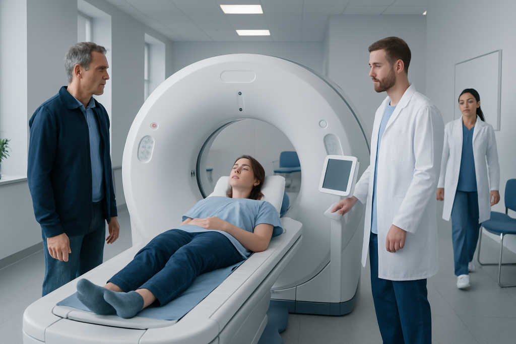
CT angiography is commonly used to assess suspected aneurysms in the brain, aorta and other arteries. A contrast dye may be needed to make blood vessels clearer on the scan.
Key Takeaways
- CT angiography is commonly used to assess suspected aneurysms in the brain, aorta and other arteries.
- A contrast dye may be needed to make blood vessels clearer on the scan.
- CT is fast and particularly valuable when symptoms could indicate a ruptured or leaking aneurysm.
- Not every aneurysm requires immediate treatment; decisions depend on location, size, growth and individual health factors.
- Sudden severe headache, chest or abdominal pain, fainting, weakness or speech difficulty require urgent medical assessment.
Yes, a CT scan can detect many aneurysms, especially when CT angiography (CTA) is used to create detailed images of blood vessels. It can show an aneurysm’s location, size, shape and possible complications, helping clinicians decide whether monitoring or treatment is needed.
Can a CT Scan Detect an Aneurysm?
Yes. A CT scan can detect many aneurysms, which are weakened, bulging areas in the wall of an artery. When clinicians need a close view of blood vessels, they commonly use computed tomography angiography (CTA), a specialized CT examination performed with contrast dye. CTA can identify an aneurysm and provide detailed information about its size, location, shape and relationship to nearby structures.
CT is often chosen because it is quick, widely available and highly useful in urgent situations. It can help detect bleeding caused by a ruptured aneurysm, including bleeding around the brain or within the chest or abdomen. A normal CT result does not always exclude every possible vascular problem, however, so further tests may occasionally be needed depending on symptoms and clinical concern.
An aneurysm may develop in different arteries. Examples include a brain aneurysm, an abdominal aortic aneurysm and an aneurysm in the chest portion of the aorta. The most appropriate scan and next steps depend on which blood vessel is being assessed and whether symptoms are present.
How CT and CT Angiography Work
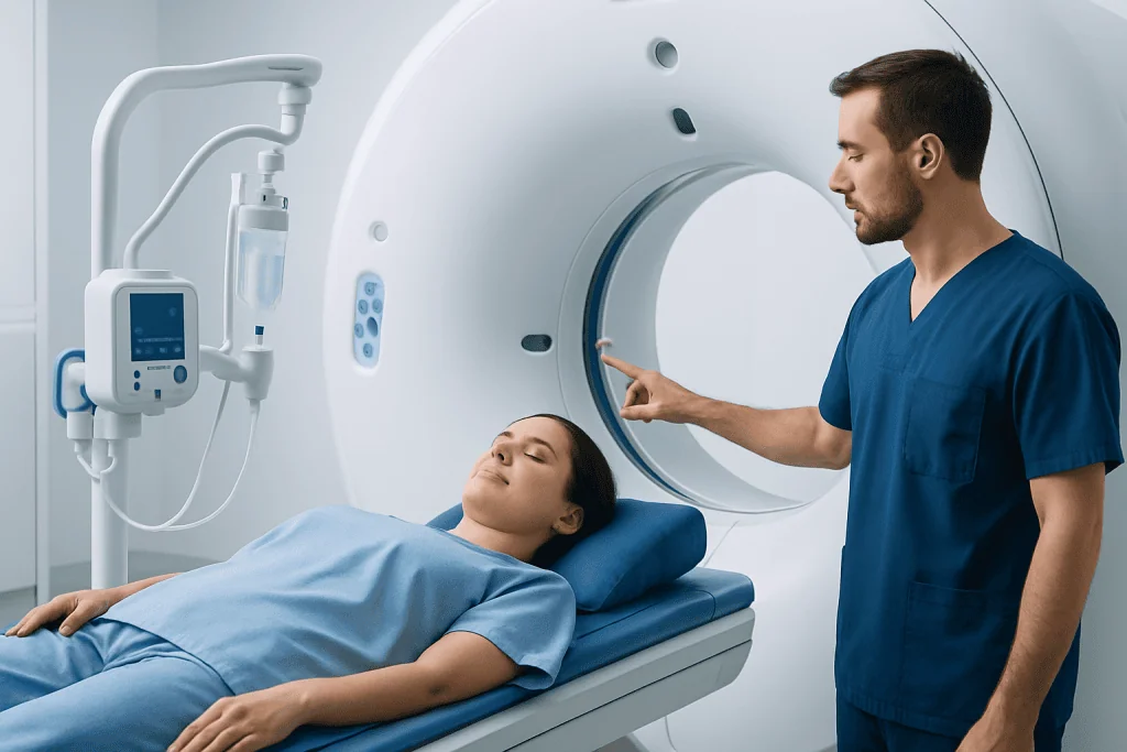
A CT scanner uses a series of X-ray images taken from multiple angles to create cross-sectional pictures of the body. Computer processing turns these images into detailed views of tissues, bones and organs. For suspected aneurysms, a routine CT may show bleeding, calcium in an artery or a large vessel enlargement.
CTA adds an iodine-containing contrast material, usually injected into a vein in the arm. The dye travels through the bloodstream and makes arteries appear more clearly on the images. This allows the radiologist to examine the vessel wall and blood flow pathway, measure an aneurysm accurately and look for features such as clot within the aneurysm or signs of a leak.
CTA is not the only imaging option. Ultrasound is often used to screen for or monitor abdominal aortic aneurysms because it does not use radiation. Magnetic resonance angiography may be appropriate for some people, particularly when repeated imaging is needed or iodinated contrast is unsuitable. Catheter angiography can provide very detailed vascular images and may be used when treatment is being planned.
Who May Need a CT Scan for an Aneurysm

A clinician may recommend CT or CTA when symptoms, examination findings or another test raise concern about an aneurysm. In urgent care, a head CT is commonly performed for a sudden, unusually severe headache to look for bleeding around the brain. If bleeding is found or strongly suspected, CTA may help identify a possible source.
CTA of the chest, abdomen or pelvis may be requested for sudden severe chest, back or abdominal pain, particularly when an aortic aneurysm or aortic dissection is a concern. It may also be used after an aneurysm has been found on ultrasound, X-ray or another scan, to obtain measurements that support treatment planning.
People with a family history of certain aneurysms, connective tissue disorders, smoking history, high blood pressure or established vascular disease may need individualized assessment. Imaging is not routinely needed for everyone with these risk factors. A doctor can determine whether screening or surveillance is appropriate based on the person’s age, medical history and family history.
- CT may be used to investigate possible rupture, leakage or dissection urgently.
- CTA can clarify the anatomy before surgery or minimally invasive vascular treatment.
- Follow-up scans can monitor a known aneurysm for growth or changes over time.
Preparing for the Scan and What Happens During It
Preparation depends on whether contrast is planned. The imaging team may ask about allergies, previous reactions to contrast dye, kidney disease, diabetes medicines, thyroid conditions and pregnancy. Some patients may be asked not to eat for a short period before a contrast-enhanced CT. Instructions vary, so it is important to follow the directions provided by the hospital or imaging center.
Before the scan, the patient usually removes metal objects and changes into a gown if needed. A radiographer places a small intravenous cannula in the arm when contrast will be used. The patient lies on a narrow table that moves through the circular CT scanner. The scanner is open at both ends and the examination is generally not painful.
During CTA, the contrast injection can briefly cause a warm sensation, a metallic taste or a feeling of needing to urinate. These sensations usually pass within seconds. The radiographer gives simple breathing instructions, especially for chest and abdominal scans, because staying still and holding the breath briefly helps produce clear images.
The image acquisition itself often takes only a few minutes, although the full appointment may take longer for registration, preparation and observation. The radiographer operates the scanner from an adjacent room but can see, hear and speak with the patient throughout the examination.
Results, Benefits and Possible Risks
A radiologist reviews the CT images and prepares a report for the referring clinician. The report may describe whether an aneurysm is present, where it is located, its measurements and whether there are signs of clot, bleeding, rupture or other changes. The clinician then explains what the findings mean in the context of the patient’s symptoms and overall health.
If an aneurysm is identified, the result does not automatically mean that surgery is required. Some small or stable aneurysms can be monitored with scheduled imaging and risk-factor management. Others may need prompt assessment by vascular, cardiac, neurosurgical or neurointerventional specialists. Treatment may include open surgery or minimally invasive procedures such as endovascular aneurysm repair, depending on the affected vessel and anatomy.
The benefits of CT include speed, detailed images and the ability to detect internal bleeding and assess several body structures at once. CT does involve radiation, but imaging teams use the lowest practical dose for the clinical question. Contrast dye can rarely cause an allergic reaction and may pose additional considerations for people with significantly reduced kidney function; the care team reviews these risks beforehand.
Most people can return to usual activities immediately after a CT scan. Those who receive contrast are generally encouraged to drink fluids unless they have been told to limit intake for another medical reason. The intravenous site may be mildly tender or bruised for a short time.
What Happens After an Aneurysm Is Found
Follow-up is tailored to the aneurysm’s location, size, rate of growth, symptoms and the person’s health. For example, a known abdominal aortic aneurysm may be monitored with ultrasound or CT at intervals recommended by the treating team. Aneurysms that are larger, growing, symptomatic or considered at higher risk may require specialist treatment planning.
Risk-factor management is an important part of care. This may include stopping smoking, controlling blood pressure, managing cholesterol and diabetes where relevant, staying physically active within medical guidance and attending scheduled imaging appointments. Medicines should only be adjusted with advice from a qualified clinician.
Depending on the type of aneurysm, patients may be referred to vascular surgery, cardiology, cardiac surgery, neurology, neurosurgery or interventional radiology. For aneurysms involving the brain, teams may discuss options such as observation, microsurgical clipping or endovascular coiling for a brain aneurysm. A coordinated review helps match treatment to the individual anatomy and risks.
Acibadem International’s multidisciplinary specialists and JCI-accredited hospitals assess and treat aneurysm conditions for international patients, with imaging findings reviewed alongside each patient’s broader medical needs.
When to Seek Medical Care
Emergency medical care is needed for symptoms that could indicate a ruptured aneurysm or another serious vascular emergency. These include a sudden, severe headache unlike previous headaches; loss of consciousness; new weakness or numbness; trouble speaking; sudden vision changes; a seizure; or severe neck stiffness. Do not drive oneself to hospital when these symptoms occur.
Urgent assessment is also important for sudden severe chest, upper back, abdominal or flank pain, especially if it is accompanied by sweating, shortness of breath, dizziness, fainting, weakness or a rapid heartbeat. These symptoms can have several causes, but prompt evaluation is essential because some require immediate treatment.
People with a known aneurysm should contact their treating team if they develop new or worsening symptoms, or if they have questions about activity, travel, medicines or follow-up imaging. Regular surveillance is designed to detect meaningful changes before complications occur, and attending scheduled appointments is an important part of safe care.
Frequently asked questions
Can a regular CT scan detect an aneurysm?
A regular CT scan can sometimes show an aneurysm, particularly if it is large or associated with bleeding. However, CT angiography is usually more informative because contrast dye makes the blood vessels easier to see and measure. The clinician selects the test based on the suspected location and urgency.
Is CT angiography accurate for aneurysms?
CT angiography provides detailed images and is widely used to evaluate aneurysms in the brain, aorta and other arteries. It can show size, shape and surrounding anatomy, which are important for monitoring and treatment planning. In selected situations, further imaging may still be necessary.
How long does a CT scan for an aneurysm take?
The scanning portion is often completed within minutes. The total visit may take longer because of check-in, preparation, placement of an intravenous line and any necessary observation after contrast. Timing varies according to the body area being scanned and local procedures.
Does a CT scan for an aneurysm hurt?
The CT scan itself is painless. If contrast is injected, there may be a brief warm feeling or metallic taste, and the intravenous line may cause a short pinch when placed. Most people tolerate the examination well.
Can CT contrast dye be used if a person has kidney disease?
Kidney function is considered before iodinated contrast is given, particularly in people with known kidney disease or other relevant health conditions. The care team may order blood tests, adjust the plan or choose another imaging method when appropriate. Patients should tell the imaging team about kidney problems and all regular medicines.
What should a person do after a CT angiogram?
Most people can resume normal daily activities immediately unless their care team gives different instructions. Drinking fluids may be advised after contrast administration if medically appropriate. New rash, swelling, breathing difficulty or worsening symptoms should be reported urgently, as these may need prompt assessment.
References
- American Heart Association
- American College of Radiology
- National Institute of Neurological Disorders and Stroke
- Society for Vascular Surgery
- National Health Service
This article is for general information only and is not a substitute for professional medical advice. Please consult a qualified doctor about your individual situation.
Explore treatments in Turkey — costs, top hospitals & a free quote
JCI-accredited · board-certified surgeons · reply within 24h
Add us as a Preferred Source to see more of our trusted health content across Google Search, AI Overviews and Discover.
Check your numbers in seconds
BMI, calories, due date, blood pressure and 30+ more clinical calculators — free, instant, doctor-reviewed ranges.