

Cardiac CT Before Coronary Bypass Surgery: Why Imaging Matters

Cardiac CT can show the coronary arteries, aorta, calcium, prior bypass grafts, heart structures and nearby chest anatomy in one noninvasive scan. Before coronary artery bypass grafting, CT may help surgeons choose safe access routes, plan graft placement and identify aortic calcification or other findings that could change the surgical approach.
Key Takeaways
- Cardiac CT can show the coronary arteries, aorta, calcium, prior bypass grafts, heart structures and nearby chest anatomy in one noninvasive scan.
- Before coronary artery bypass grafting, CT may help surgeons choose safe access routes, plan graft placement and identify aortic calcification or other findings that could change the surgical approach.
- The scan usually involves an intravenous contrast dye and ECG-synchronized imaging, and the team checks kidney function, allergies and heart rhythm beforehand.
- Cardiac CT does not replace all other tests; it is interpreted together with coronary angiography, echocardiography, blood tests and the patient’s overall health.
- Patients should ask their cardiologist or cardiac surgeon why CT is recommended, how to prepare and how the results may influence the surgical plan.
Cardiac CT before coronary bypass surgery gives the heart team detailed images of the coronary arteries, aorta, bypass targets and chest anatomy. This information can support careful surgical planning, especially when anatomy is complex or previous procedures may affect the operation.
Overview
Coronary artery bypass grafting, often called CABG or coronary bypass surgery, is an operation used to improve blood flow to the heart muscle when coronary arteries are significantly narrowed or blocked. Before surgery, the heart team needs a clear understanding of the patient’s coronary artery disease, heart function, blood vessels and chest anatomy. Cardiac CT before coronary bypass surgery is one imaging method that can add valuable detail to this planning.
Cardiac CT uses computed tomography and specialized heart imaging techniques to create cross-sectional and three-dimensional pictures. In many cases, contrast dye is injected into a vein so that blood vessels can be seen more clearly. The scan can show the coronary arteries, the aorta, areas of calcification, previous stents or bypass grafts, and structures around the heart.
Not every patient needs the same imaging tests before bypass surgery. The choice depends on the clinical situation, previous test results, kidney function, heart rhythm, urgency of surgery and the surgeon’s specific questions. When used appropriately, cardiac CT can help the team plan a more individualized and well-prepared operation.
Why Imaging Matters Before Coronary Bypass Surgery
Bypass surgery is planned around both the diseased coronary arteries and the available blood vessels that can be used as grafts. Surgeons need to know which coronary arteries are suitable targets, where blockages are located, and whether there are technical factors that may affect access to the heart. Traditional coronary angiography remains central for assessing the inside of the coronary arteries, but CT can provide complementary information about the vessel wall, calcification and surrounding anatomy.
Cardiac CT can also evaluate the aorta, the large artery that carries blood from the heart to the body. If the aorta has heavy calcification or plaque, the surgeon may consider techniques that reduce manipulation of that area. This type of planning can be especially useful in people with advanced atherosclerosis, older age, prior chest surgery or other conditions that may make surgery more complex.
Another important role is mapping anatomy when previous procedures are present. For patients who have had earlier bypass surgery, cardiac CT can show the course of old grafts behind the breastbone and around the heart. This helps the surgical team understand what lies beneath the chest wall before reopening the chest and can guide a safer operative strategy.
What Cardiac CT Can Show

A cardiac CT scan can answer several practical questions before coronary bypass surgery. It may help identify the location and severity of coronary artery calcification, the path of coronary arteries, and whether prior stents or grafts are present. It can also show the size and shape of the heart chambers, the relationship of the heart to the sternum, and findings in the lungs or chest that may be relevant before surgery.
Depending on the protocol, the scan may include CT coronary angiography, a CT assessment of the aorta, or broader chest imaging. The radiologist and cardiology team tailor the examination to the clinical question. For example, one patient may need detailed graft mapping after prior surgery, while another may need evaluation of aortic calcification or unusual coronary anatomy.
- Coronary arteries: location of disease, calcium and vessel course.
- Aorta: calcification, plaque, enlargement or other structural findings.
- Prior bypass grafts: whether they appear open, where they run and how close they are to the sternum.
- Chest anatomy: relationship of the heart, blood vessels, lungs and chest wall.
- Incidental findings: selected lung, mediastinal or vascular findings that may need follow-up.
CT findings are not interpreted in isolation. They are combined with symptoms, physical examination, blood tests, electrocardiogram results, echocardiography and invasive angiography when performed. This combined view helps the heart team choose the most appropriate treatment plan.
Who May Benefit From Cardiac CT Before CABG
Cardiac CT may be considered when the expected benefit of additional anatomic detail is greater than the small risks of radiation and contrast dye. It can be particularly helpful when the surgical team needs more information about the aorta, previous grafts, chest anatomy or complex coronary anatomy. It may also be used when earlier imaging is incomplete or when there is a need to confirm the course of important blood vessels before surgery.
Patients with previous bypass surgery are a common group in whom CT may be useful. In repeat operations, old grafts can run close to the sternum and may be at risk during re-entry. CT can help locate these grafts and show whether they are open, narrowed or occluded, giving the surgeon important information before the first incision.
Cardiac CT may not be suitable for everyone. People with significantly reduced kidney function, a history of severe contrast allergy, certain heart rhythm problems, or inability to lie flat may need alternative imaging or special precautions. Pregnant patients or those who may be pregnant should inform their care team so that the need for imaging and radiation exposure can be assessed carefully.
Preparing for the Scan
Preparation depends on the CT protocol and the patient’s health. The care team usually reviews kidney function, medications, allergies, heart rhythm and previous reactions to contrast dye. Patients should tell the team about kidney disease, thyroid disease, asthma, diabetes, implanted devices, pregnancy, breastfeeding and any previous contrast reaction.
Many cardiac CT scans require an intravenous contrast agent to highlight the blood vessels. Some patients may be asked to avoid food for a short period before the scan, while drinking water may be encouraged unless a doctor advises otherwise. The imaging team may also give instructions about caffeine or certain medications, particularly if heart-rate control is needed for clearer images.
It is helpful for patients to bring or share previous imaging results, such as coronary angiography, echocardiography, stress tests, earlier CT scans or operation reports. These records allow the radiologist, cardiologist and surgeon to compare findings and focus the scan on the most important questions. Patients should ask in advance whether they need to adjust any medication; changes should only be made with medical guidance.
What Happens During Cardiac CT
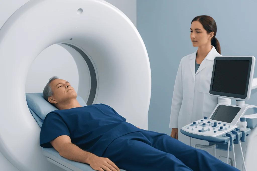
During the scan, the patient lies on a CT table that moves through a ring-shaped scanner. Small ECG electrodes are placed on the chest so the scan can be timed with the heartbeat. This ECG synchronization helps reduce motion blur and improves the quality of heart images.
An intravenous line is usually placed in the arm for contrast injection. When contrast is given, patients may feel a brief warm sensation or a metallic taste; this typically passes quickly. The imaging team remains in contact throughout the examination and may ask the patient to hold their breath for short periods so that images are as clear as possible.
The scanning itself is usually brief, although preparation and monitoring take additional time. Afterward, most patients can return to normal activities unless they have received specific instructions. Drinking fluids may be recommended to help clear contrast from the body, but patients with heart failure or kidney disease should follow their doctor’s personalized advice on fluid intake.
How Results Guide the Heart Team
After the scan, a radiologist with experience in cardiovascular imaging reviews the images and prepares a report. The report may describe coronary calcification, graft location, aortic findings, heart and chest anatomy, and any other relevant observations. The cardiac surgeon and cardiologist then consider these findings alongside the patient’s symptoms, angiogram, echocardiogram and overall surgical risk.
CT results may influence several parts of the surgical plan. For example, the surgeon may choose a different site for connecting a graft to the aorta, adjust the approach for a repeat sternotomy, select specific bypass targets, or request additional testing before surgery. In some cases, CT may confirm that the planned operation can proceed as expected.
It is important for patients to understand that imaging supports decision-making, but it does not make decisions by itself. The best plan depends on the full clinical picture, including coronary anatomy, heart function, other medical conditions, patient preferences and the urgency of treatment. A clear discussion with the heart team can help patients understand what the scan showed and what it means for their operation.
Safety, Limitations and When to Speak With a Doctor
Cardiac CT is a widely used diagnostic imaging test, but like all medical tests it has benefits and limitations. It involves exposure to ionizing radiation, and contrast dye may not be appropriate for some people with kidney problems or previous severe allergic reactions. Modern CT protocols aim to use the lowest radiation dose that can still provide useful diagnostic images.
Limitations can include image blurring from irregular heart rhythms, very fast heart rates, heavy calcium, movement during the scan or inability to use contrast. Sometimes CT shows findings that require follow-up but are not directly related to bypass surgery. In other cases, the scan may not answer every surgical question, and the team may rely on additional imaging or invasive angiography.
Patients should contact their doctor promptly if they develop symptoms such as worsening chest pain, shortness of breath, fainting, new weakness or signs of an allergic reaction after contrast. Before the scan, they should also ask what the test is expected to clarify, whether contrast will be used, and how the results may affect timing or planning of surgery. For international patients, Acibadem International’s multidisciplinary specialists and JCI-accredited hospitals provide diagnostic evaluation and treatment planning for coronary artery disease, including advanced cardiac imaging and bypass surgery when appropriate.
Frequently asked questions
Is cardiac CT required for everyone before coronary bypass surgery?
No. Cardiac CT is not required for every patient before coronary bypass surgery. It is usually considered when the heart team needs extra anatomic detail, such as aortic calcification, prior graft location, complex anatomy or unclear findings from other tests.
Does cardiac CT replace coronary angiography?
In many patients being considered for bypass surgery, invasive coronary angiography remains an important test for defining coronary artery blockages. Cardiac CT may add information about the vessel wall, calcium, the aorta, previous grafts and chest anatomy. The two tests can be complementary rather than interchangeable.
Will the CT scan show whether bypass surgery is the right treatment?
Cardiac CT can provide useful information for planning, but it does not decide treatment on its own. The decision for bypass surgery depends on symptoms, coronary anatomy, heart function, medical conditions, test results and patient goals. The cardiologist and cardiac surgeon review all of these factors together.
Is contrast dye always used for cardiac CT before bypass surgery?
Contrast dye is often used because it makes blood vessels and grafts easier to see. However, some CT assessments, such as calcium or certain chest anatomy evaluations, may be performed without contrast. The imaging team chooses the safest and most useful protocol for the clinical question.
What should patients tell the imaging team before the scan?
Patients should mention kidney disease, previous contrast reactions, asthma, thyroid disease, diabetes, pregnancy or possible pregnancy, breastfeeding, and all current medications. They should also share prior heart procedures, stents, bypass operations or implanted devices. This helps the team plan the scan safely.
How soon are cardiac CT results available?
Timing varies by hospital and urgency. The images must be processed and reviewed by a radiologist or cardiovascular imaging specialist, then discussed with the treating heart team. Patients should ask when and how they will receive results and who will explain the findings.
References
- American Heart Association
- European Society of Cardiology
- Society of Cardiovascular Computed Tomography
- American College of Radiology
- National Institute for Health and Care Excellence
This article is for general information only and is not a substitute for professional medical advice. Please consult a qualified doctor about your individual situation.
Heart care in Turkey — expert evaluation and treatment
JCI-accredited · board-certified surgeons · reply within 24h

More from the Health Library



Related Specialists

Prof. Dr. Ercan Karaarslan
Radiology
Prof. Dr. Bora Peynircioğlu
Interventional Radiology
Prof. Dr. Koray Güven
Interventional Radiology
Assoc. Prof. Dr. Deniz Can Aliş
Radiology