

Cerebral Angiography: Step-by-Step, Risks, and Recovery After Brain Vessel Imaging

Cerebral angiography uses a thin catheter, contrast dye, and X-ray imaging to show brain blood vessels clearly. It is often used when detailed vessel imaging is needed beyond CT or MRI scans.
Key Takeaways
- Cerebral angiography uses a thin catheter, contrast dye, and X-ray imaging to show brain blood vessels clearly.
- It is often used when detailed vessel imaging is needed beyond CT or MRI scans.
- Most people recover well, but temporary soreness, bruising, and short observation after the procedure are common.
- Although generally safe, cerebral angiography carries small risks such as bleeding, allergic reaction, kidney strain, or stroke.
- Careful preparation and following aftercare instructions can help reduce complications.
Cerebral angiography is a specialized imaging procedure used to examine blood vessels in the brain and neck in great detail. It can help doctors diagnose aneurysms, narrowing, bleeding, stroke-related problems, and other vascular conditions, while also guiding treatment when needed.
Overview
Cerebral angiography, also called a cerebral angiogram, is a diagnostic imaging procedure that allows doctors to see the blood vessels of the brain, head, and neck in very fine detail. It is considered one of the most precise ways to evaluate how blood flows through these vessels and whether there are abnormalities such as narrowing, blockages, aneurysms, or vessel malformations.
During the procedure, a doctor inserts a thin tube called a catheter into a blood vessel, usually in the groin or wrist, and gently guides it toward the arteries that supply the brain. A contrast dye is then injected, and a series of X-ray images are taken. These images help the medical team assess the size, shape, and pathway of the vessels.
Cerebral angiography may be performed as a purely diagnostic test, or it may be combined with a treatment in the same session. For example, if a doctor identifies a vascular problem that can be treated through the catheter, an interventional procedure may follow. In some cases, cerebral angiography is used alongside other evaluations for brain aneurysm or to guide minimally invasive interventional neuroradiology treatment.
Why Cerebral Angiography Is Done
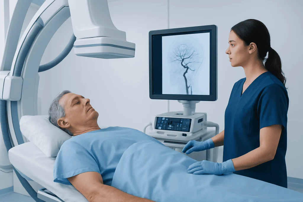
Doctors recommend cerebral angiography when they need more detailed information than noninvasive scans such as CT angiography or MR angiography can provide. It is especially useful when a diagnosis remains uncertain, when treatment planning requires precise vessel mapping, or when a problem in the brain’s circulation is suspected.
This test may help evaluate several conditions affecting the brain’s blood vessels. These include aneurysms, arteriovenous malformations, narrowing of the carotid or intracranial arteries, blood vessel inflammation, unexplained bleeding, and some stroke-related conditions. It can also be used before surgery or endovascular treatment to clarify anatomy.
Common reasons for cerebral angiography include:
- Suspected aneurysm or ruptured aneurysm
- Unexplained bleeding in or around the brain
- Blood vessel narrowing or blockage
- Evaluation after an abnormal CT or MRI result
- Planning for endovascular treatment or neurosurgery
- Assessment of certain tumors with unusual blood supply
In selected cases, the same catheter-based approach may also support treatments related to stroke treatment or other vascular procedures in the brain.
Step-by-Step: What Happens During the Procedure

Before the procedure, the patient is usually asked not to eat or drink for several hours. The medical team reviews medical history, allergies, medications, and kidney function, and may order blood tests. A small intravenous line is placed for fluids or medicines, and the patient changes into a hospital gown. The skin at the catheter entry site is cleaned carefully.
In the angiography suite, the patient lies on an X-ray table. Local anesthetic is used to numb the skin, and sedation may be given to help relaxation. The doctor inserts a catheter into an artery, most often through the groin or the wrist, and guides it under imaging toward the vessels leading to the brain. Contrast dye is injected, and the patient may briefly feel warmth or flushing while multiple images are captured.
Once the imaging is complete, the catheter is removed. Pressure or a closure device is used to stop bleeding at the puncture site. The patient is then moved to a recovery area for monitoring. Depending on the reason for the test and the patient’s condition, the procedure may take from about one hour to several hours, particularly if treatment is performed at the same time.
If the images reveal a condition that may benefit from a minimally invasive approach, the team may discuss additional procedures such as aneurysm treatment during the same admission or at a later time.
Preparation and How to Get Ready
Good preparation helps the procedure go smoothly and reduces avoidable risks. Patients should tell their doctor about all medicines they take, including blood thinners, diabetes medicines, over-the-counter products, and supplements. It is also important to mention any history of contrast dye allergy, asthma, kidney disease, or previous bleeding problems.
Doctors may advise stopping or adjusting certain medicines before the test, but this should only be done under medical guidance. Patients should also report pregnancy or the possibility of pregnancy. In many cases, they will be asked to arrange transportation home, since sedation or post-procedure instructions may make driving unsafe.
Helpful preparation steps often include:
- Following fasting instructions exactly
- Bringing a list of medicines and allergies
- Drinking fluids as advised before and after the test, unless instructed otherwise
- Wearing comfortable clothing and leaving valuables at home
- Asking in advance when usual medicines can be restarted
For people with known vascular or neurological conditions, preparation may be coordinated with specialists in neurology, neurosurgery, or interventional neuroradiology to ensure the safest plan.
Risks and Possible Complications
Cerebral angiography is generally safe when performed by experienced teams, but like all invasive procedures, it carries some risks. Most complications are uncommon, and the care team takes several steps to reduce them. Patients are monitored closely before, during, and after the procedure for any early signs of problems.
The most common minor issues are temporary bruising, tenderness, or swelling at the catheter insertion site. Some patients also notice brief flushing, nausea, or a metallic taste when contrast dye is injected. These effects usually pass quickly.
Less common but more serious risks can include:
- Bleeding or hematoma at the puncture site
- Allergic reaction to contrast dye
- Kidney strain related to contrast, especially in people with kidney disease
- Blood vessel injury or spasm
- Blood clot formation
- Transient neurological symptoms or stroke
- Infection, though this is uncommon
The doctor will weigh these risks against the expected benefit of accurate diagnosis or treatment planning. In many situations, the detailed information from cerebral angiography can be essential for managing serious conditions, including evaluation of stroke or other abnormalities affecting brain circulation.
Recovery After Brain Vessel Imaging
After cerebral angiography, the patient remains under observation while the puncture site and vital signs are monitored. If the catheter was placed through the groin, lying flat for a period may be necessary to reduce bleeding risk. If wrist access was used, recovery may be more comfortable, though activity restrictions still apply. Many people go home the same day, while others stay longer depending on their overall condition and the reason for the procedure.
Mild soreness, bruising, and tiredness can occur for a day or two. Drinking fluids is often encouraged to help clear the contrast dye, unless the doctor recommends fluid restriction. Patients are typically advised to avoid heavy lifting, intense exercise, or straining for a short period.
It is important to follow discharge instructions carefully, including guidance on bathing, wound care, and restarting medicines. The puncture site should be checked for increased swelling, ongoing bleeding, worsening pain, or signs of infection. Most people return to normal routines soon, but recovery can vary if a treatment was done during the same session.
Near the end of care planning, some patients may continue follow-up with multidisciplinary specialists. Acibadem International’s JCI-accredited hospitals provide diagnosis and treatment for international patients with cerebrovascular conditions through coordinated neurology, neurosurgery, and interventional teams.
When to Seek Medical Advice
Patients should contact a doctor promptly if they develop unusual symptoms after returning home. Early medical attention can help address complications quickly and safely. Most concerns do not turn out to be serious, but it is always better to ask if something feels wrong.
Warning signs that need urgent medical review include bleeding that does not stop with pressure, rapidly enlarging swelling at the puncture site, severe pain, fever, shortness of breath, rash, or reduced urine output. Any new neurological symptom should also be treated as urgent.
Seek immediate medical care if there is:
- Sudden weakness or numbness
- Difficulty speaking or understanding speech
- Vision changes
- Severe headache unlike usual headaches
- Confusion, loss of balance, or fainting
- Coldness, color change, or severe pain in the accessed arm or leg
Follow-up appointments are also important, even when recovery seems smooth. They allow the doctor to review the angiography findings, explain next steps, and decide whether monitoring, medication, surgery, or endovascular therapy is the best option.
Frequently asked questions
Is cerebral angiography the same as a CT or MRI scan?
No. Cerebral angiography is an invasive catheter-based test that provides highly detailed images of brain blood vessels. CT and MRI vessel scans are noninvasive and may be used first, but cerebral angiography is often chosen when more precise information is needed.
Is cerebral angiography painful?
Most people do not describe the procedure as painful, although there can be brief discomfort. The skin is numbed with local anesthetic, and some patients receive sedation to help them relax. Mild soreness at the catheter site afterward is common.
How long does recovery take after cerebral angiography?
Initial recovery usually takes a few hours of observation in the hospital or clinic. Many people feel ready for light daily activities within a day or two, although heavy lifting and strenuous activity are often limited for a short time. The exact timeline depends on the access site, overall health, and whether any treatment was performed.
What are the main risks of cerebral angiography?
The main risks include bruising or bleeding at the puncture site, allergic reaction to contrast dye, and kidney strain in susceptible patients. Rare but serious complications can include blood vessel injury, blood clot, or stroke. Doctors take precautions to keep these risks as low as possible.
Why would a doctor order cerebral angiography instead of another imaging test?
A doctor may order it when detailed vessel anatomy is needed to confirm a diagnosis or plan treatment. It is especially useful for evaluating aneurysms, vascular malformations, narrowing, and some causes of bleeding or stroke. It can also allow treatment through the same catheter in selected cases.
Can a person go home the same day after cerebral angiography?
Yes, many patients go home the same day after a period of monitoring. However, some people stay longer if they have other medical conditions, develop symptoms that need observation, or undergo an additional treatment during the same session.
References
- American Heart Association
- National Institute of Neurological Disorders and Stroke
- Radiological Society of North America
- National Health Service
- Society of NeuroInterventional Surgery
This article is for general information only and is not a substitute for professional medical advice. Please consult a qualified doctor about your individual situation.
Explore treatments in Turkey — costs, top hospitals & a free quote
JCI-accredited · board-certified surgeons · reply within 24h
Add us as a Preferred Source to see more of our trusted health content across Google Search, AI Overviews and Discover.

More from the Health Library



Related Specialists

Assoc. Prof. Dr. Recep Kurnaz
Orthopedic Surgery & Traumatology
Dr. Cemile Ertan
Dermatology
Dr. Canan Savaş İyigün
Dermatology
Prof. Dr. Hakan Ağır
Aesthetic Plastic & Reconstructive Surgery