Chondroma Treatment: How It Works, Results and What to Expect

A chondroma is usually a benign tumor made of cartilage cells. Observation with repeat imaging may be appropriate for a stable, symptom-free lesion.
Key Takeaways
- A chondroma is usually a benign tumor made of cartilage cells.
- Observation with repeat imaging may be appropriate for a stable, symptom-free lesion.
- Surgery is considered for pain, fracture risk, growth, functional problems or concern about a malignant tumor.
- Curettage, sometimes combined with bone grafting or fixation, is a common surgical approach for chondromas inside bone.
- Most people have an excellent outlook after appropriate assessment and treatment, but follow-up is important.
Chondroma treatment is tailored to the tumor’s location, symptoms and imaging features. Many small, typical chondromas can be monitored, while painful, growing or uncertain lesions may need surgery and laboratory examination to confirm the diagnosis.
Chondroma treatment: how it works
Chondroma treatment aims to confirm that a cartilage tumor is benign, relieve symptoms when present and protect the affected bone or nearby joint. In many people, a chondroma is found incidentally on an X-ray or scan performed for another reason. If the lesion has typical benign features and causes no symptoms, the safest approach may be observation rather than immediate surgery.
When treatment is needed, surgery is usually designed to remove or thoroughly clear the abnormal cartilage while preserving as much normal bone and function as possible. The orthopedic team considers the lesion’s size, location, appearance on imaging, effect on bone strength and whether pain is likely to be caused by the tumor. A tissue sample may be examined by a pathologist, particularly when imaging cannot reliably distinguish a benign chondroma from a low-grade cartilage cancer.
Chondromas may occur within the bone, where they are often called enchondromas, or on the bone surface. They are most commonly identified in the small bones of the hands and feet, although they can occur in longer bones and other sites. Management should be individualized because cartilage tumors can look different depending on where they arise.
Is chondroma benign or malignant?
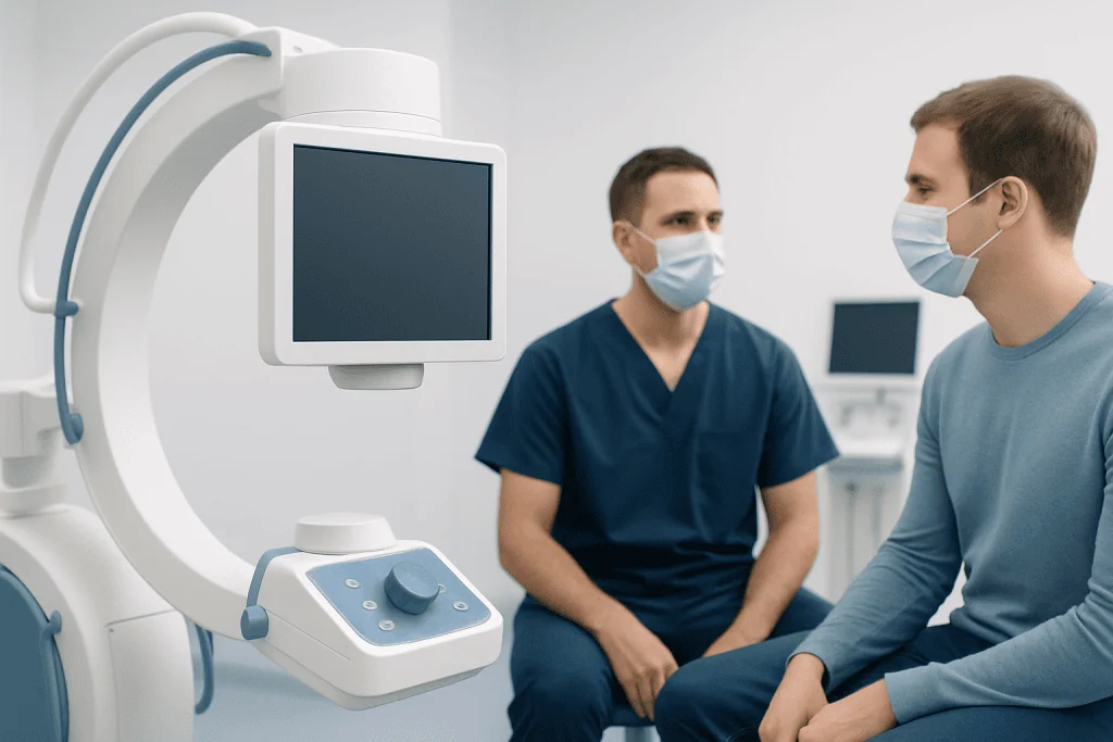
A chondroma is, by definition, a benign cartilage tumor. It does not have the invasive behavior of cancer and does not spread to distant organs. However, a cartilage lesion can sometimes be difficult to classify from symptoms and scans alone, especially in the pelvis, shoulder region or long bones, where benign lesions and low-grade chondrosarcoma may overlap in appearance.
For this reason, doctors assess the complete clinical picture rather than relying on one test. Persistent or increasing pain, growth after skeletal maturity, destruction of surrounding bone, a soft-tissue mass or certain MRI features may lead to referral to an orthopedic oncology team. If surgery is performed, pathology review helps establish the most accurate diagnosis.
A diagnosis of chondroma should not be assumed to explain every ache near the affected area. Joint, tendon, nerve and arthritis-related conditions can also cause pain. A specialist can determine whether the lesion is likely to be responsible and whether monitoring or further evaluation is appropriate.
Who may need treatment and how diagnosis guides planning
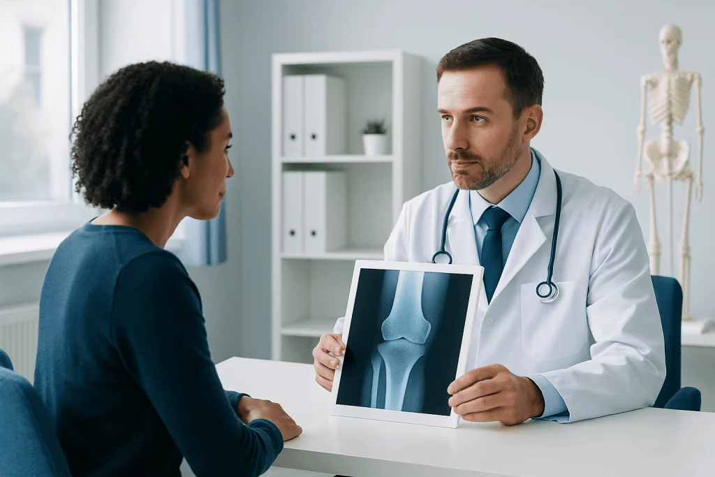
People may be candidates for chondroma treatment when the lesion causes pain that is clearly linked to the affected bone, weakens the bone, contributes to a fracture, limits hand or joint function, or has imaging findings that are not clearly benign. Surgery may also be discussed if a lesion changes during follow-up or if a definite diagnosis is needed to guide care.
Assessment usually begins with a medical history and physical examination, followed by plain X-rays. X-rays can show the lesion’s position, its effect on the bone and features such as calcification. MRI may be used to assess cartilage tissue, the bone marrow and surrounding soft tissues. CT can be useful for evaluating mineralization and bone structure, particularly in complex anatomical areas.
A biopsy is not necessary for every suspected chondroma. In selected cases, however, it can be important before definitive treatment. Biopsy planning should be coordinated by the surgical team that would treat the lesion, because the biopsy pathway and placement can affect later surgery. Review by musculoskeletal radiologists, orthopedic surgeons and pathologists supports careful decision-making.
- Stable, painless lesions with reassuring imaging may be monitored.
- Symptomatic or structurally significant lesions may need surgical management.
- Indeterminate lesions may require advanced imaging, biopsy or specialist review.
Chondroma surgery: step by step
The exact procedure depends on the tumor’s site and size. For many enchondromas, the surgeon performs curettage: an opening is made in the bone and the lesion is carefully scraped out. The remaining cavity may be treated with surgical tools or other local methods chosen by the surgeon to reduce the chance that abnormal cartilage remains.
After curettage, the bone cavity may be left to heal naturally or filled with bone graft, bone graft substitute or cement, depending on the bone involved and the size of the defect. If the bone is at risk of breaking, the surgeon may stabilize it with plates, screws, pins or another fixation method. Surface chondromas may be removed with a margin of surrounding tissue when appropriate.
The operation is performed with anesthesia, and the procedure may be outpatient or require a short hospital stay depending on its complexity and the patient’s general health. Removed tissue is sent for pathology. The final pathology result, together with imaging and operative findings, informs the follow-up plan.
For lesions requiring specialist orthopedic tumor care, treatment may include orthopedic oncology assessment and treatment as part of a coordinated plan. The goal is always to balance complete and safe lesion management with preservation of movement, strength and day-to-day function.
Benefits, risks and recovery timeline
The potential benefits of surgery include diagnosis confirmation, relief of tumor-related pain, improved function and reduced risk of fracture in a weakened bone. For a benign lesion that is completely treated, surgery can be definitive. Not every symptom improves immediately, however, because pain may also reflect nearby joint or soft-tissue conditions.
Recovery varies with the surgical site, size of the bone cavity, whether fixation was needed and a person’s overall health. After surgery in a finger or hand bone, swelling and stiffness may be noticeable early on, and prescribed exercises or hand therapy can be important. Procedures involving weight-bearing bones often require temporary restrictions on walking, lifting or sport while the bone heals.
Many patients return gradually to light daily activities over days to weeks, while full bone healing and return to impact activities can take several months. Follow-up appointments and X-rays help the team confirm healing and identify uncommon recurrence or complications. The treating surgeon provides individualized guidance on work, driving, activity and rehabilitation.
Possible surgical risks include infection, bleeding, nerve or blood-vessel injury, stiffness, persistent pain, fracture, delayed bone healing, recurrence and risks related to anesthesia. These complications are not expected for most patients, but they should be discussed before treatment so that informed decisions can be made.
Can a chondroma turn cancerous?
Most solitary chondromas do not turn into cancer. A benign cartilage tumor can, however, resemble a low-grade chondrosarcoma, and distinguishing between the two may require expert review of imaging, symptoms and pathology. This is why new or changing symptoms should not be ignored, even after a previous benign diagnosis.
The likelihood of malignant change is higher in uncommon conditions involving multiple enchondromas, such as Ollier disease and Maffucci syndrome, than it is with a single isolated chondroma. These conditions need ongoing specialist surveillance. A doctor may recommend repeat imaging if pain becomes persistent or worse, if a lump enlarges, or if prior scans showed features requiring observation.
It is helpful to remember that pain alone does not prove cancer. Still, pain at rest or at night, progressive symptoms, swelling, a growing mass or a change in function warrants medical assessment. Early specialist evaluation can clarify the cause and avoid unnecessary delay.
What is the prognosis for chondroma?
The prognosis for a solitary chondroma is generally excellent. These tumors are benign, and many remain stable without treatment. When surgery is recommended and the lesion is appropriately managed, people commonly regain good function, although recovery depends on the location of the tumor, the amount of bone involved and whether a fracture or reconstruction was present.
Some lesions can recur, particularly if removal was incomplete or when multiple cartilage tumors are present. Scheduled clinical reviews and imaging allow the care team to check bone healing and compare the area with earlier scans. Follow-up plans are individualized; a stable, typical lesion may need less monitoring than a lesion with uncertain features.
If examination or pathology identifies chondrosarcoma rather than chondroma, the outlook and treatment plan are different. Chondrosarcoma care is guided by tumor grade, location and extent, often with surgery as the main treatment. Clear diagnosis is therefore a central part of achieving the best possible outcome.
How quickly does chondrosarcoma spread? When to seek medical care
Chondrosarcoma does not have one fixed rate of growth or spread. Low-grade tumors often grow slowly and may remain localized for a long time, whereas higher-grade tumors can behave more aggressively and have a greater chance of spreading, commonly to the lungs. Tumor grade, size, location and whether it can be completely removed all influence the outlook.
A person should seek medical care promptly for persistent or progressively worsening bone pain, pain that occurs at rest or wakes them from sleep, an enlarging lump, unexplained swelling, reduced use of a limb, or pain after a minor injury in an area known to contain a bone lesion. Urgent assessment is also appropriate after a suspected fracture, particularly if the bone was previously weakened by a tumor.
People already being monitored for a chondroma should keep scheduled appointments and report new symptoms rather than waiting for the next scan. Acibadem International’s multidisciplinary specialists and JCI-accredited hospitals can assess cartilage tumors and coordinate treatment for international patients when needed.
Frequently asked questions
Do all chondromas need surgery?
No. A small, painless lesion with reassuring imaging findings may be monitored with clinical review and repeat imaging. Surgery is more often considered when there is pain, bone weakening, fracture risk, growth, functional impairment or uncertainty about the diagnosis.
What type of doctor treats a chondroma?
An orthopedic surgeon with experience in bone tumors or orthopedic oncology commonly leads care for a chondroma. Depending on the location and findings, radiologists, pathologists, rehabilitation specialists and other surgeons may also contribute to the treatment plan.
Is a biopsy always needed for chondroma?
No. Some lesions have a typical appearance on X-ray and MRI and can be monitored without biopsy. If imaging is unclear or there are concerning features, a carefully planned biopsy may be needed to distinguish a benign lesion from chondrosarcoma.
Can a chondroma come back after surgery?
Recurrence is possible but is not common after appropriate treatment of a solitary benign chondroma. The chance depends on the lesion’s site, whether it was fully cleared and whether the person has a condition associated with multiple cartilage tumors. Follow-up imaging helps detect changes early.
How long does recovery from chondroma surgery take?
Recovery can range from several weeks for lighter activities to several months for full bone healing and return to demanding activity. The timeline depends on the bone treated, the size of the surgical cavity, the need for fixation and whether weight-bearing must be restricted.
Can exercise make a chondroma worse?
A stable, painless chondroma does not always require activity restriction, but this should be confirmed by the treating clinician. High-impact activity or heavy loading may be limited when the lesion weakens a bone or after surgery, until imaging shows sufficient healing.
References
- American Academy of Orthopaedic Surgeons
- National Cancer Institute
- American Cancer Society
- European Society for Medical Oncology
- Orphanet
This article is for general information only and is not a substitute for professional medical advice. Please consult a qualified doctor about your individual situation.
Joint and spine care in Turkey — expert assessment & treatment
JCI-accredited · board-certified surgeons · reply within 24h
Add us as a Preferred Source to see more of our trusted health content across Google Search, AI Overviews and Discover.

More from the Health Library



Related Specialists

Prof. Dr. İlgin Özden
General Surgery
Dr. Kadir Özyılmaz
Anesthesiology
Dr. Sezgi Güllü Erciyestepe
Gynecology & Obstetrics
Prof. Dr. İbrahim Yıldız
Medical Oncology