

Chondrosarcoma on Xray: Preparation, Procedure and Results

X-rays are often the first imaging test used when persistent bone pain, swelling or an unexplained bone lesion is present. Chondrosarcoma commonly affects the pelvis, hip region, shoulder region, ribs and long bones such as the femur.
Key Takeaways
- X-rays are often the first imaging test used when persistent bone pain, swelling or an unexplained bone lesion is present.
- Chondrosarcoma commonly affects the pelvis, hip region, shoulder region, ribs and long bones such as the femur.
- Typical X-ray patterns may include ring-and-arc calcifications, bone remodeling and cortical changes, but appearance alone cannot always confirm the diagnosis.
- Most chondrosarcomas grow slowly, although higher-grade tumors may behave more aggressively and can spread.
- Specialist assessment with MRI, CT and image-guided biopsy helps distinguish chondrosarcoma from benign cartilage lesions and other bone tumors.
- Surgery is the main treatment for many localized chondrosarcomas, with care tailored to tumor grade, location and stage.
Chondrosarcoma on Xray can show changes in bone caused by a cartilage-producing tumor, including areas of calcification, bone expansion, or damage to the surrounding cortex. An X-ray is usually an important first test, but CT, MRI and sometimes biopsy are needed to determine whether a lesion is chondrosarcoma and to plan treatment safely.
Chondrosarcoma on Xray: what the image can show
Chondrosarcoma on Xray may appear as an abnormal area within a bone where cartilage-producing tumor tissue has changed the bone’s internal structure. Doctors often identify patterns of mineralized cartilage, changes in bone shape, thinning or disruption of the outer bone layer, and sometimes a soft-tissue mass. However, an X-ray cannot reliably establish the tumor grade or confirm every diagnosis on its own.
Chondrosarcoma is a malignant tumor that produces cartilage matrix. It is uncommon and is most often diagnosed in adults, although the age range and clinical features vary by subtype. Many lesions are found after an X-ray is arranged for persistent pain, a lump, reduced movement, or an injury that leads to imaging for another reason.
When an X-ray raises concern, the next step is usually referral to an orthopedic oncology or sarcoma team. Chondrosarcoma is assessed using the imaging appearance, symptoms, examination findings and, when appropriate, carefully planned biopsy results. This combined approach is important because benign cartilage lesions can sometimes resemble low-grade chondrosarcoma on initial images.
How X-ray assessment works and who may need it
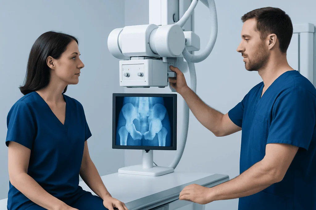
A plain X-ray uses a small amount of ionizing radiation to create images of bone. It is quick, widely available and particularly useful for showing mineralization, fractures, bone contour and changes to the hard outer shell of bone, called the cortex. For suspected bone tumors, images are commonly taken from at least two directions to give a clearer view of the lesion.
An X-ray may be considered for a person with unexplained bone pain that persists or worsens, a growing swelling near a bone, reduced function of a nearby joint, or an abnormality detected during another examination. It may also be used to investigate a fracture that occurred with little force, especially when there are features suggesting an underlying bone lesion.
Most people can have an X-ray. Pregnant people or those who may be pregnant should tell the radiology team beforehand so imaging can be adapted or postponed where appropriate. X-rays are not a screening test for people without symptoms, and they do not replace specialist evaluation when a bone abnormality is identified.
Preparation and step-by-step X-ray procedure

Preparation for a bone X-ray is usually simple. The person may be asked to remove jewelry, belts, clothing with metal fasteners, or other objects that could obscure the area being examined. There is generally no need to fast, stop regular medicines, receive contrast dye or arrange recovery time after the scan.
During the procedure, a radiographer positions the body part on or near the detector and may use supports to help keep it still. Several positions can be needed, particularly for the pelvis, shoulder, chest wall, hand or long bones. The person may be asked to briefly hold a position while each image is taken; the X-ray itself is painless and usually takes only a few minutes.
The images are reviewed by a radiologist and then considered alongside the clinical history. If findings are suspicious or uncertain, MRI is often used to define the tumor’s extent in bone and soft tissues, while CT can show mineralized cartilage and cortical bone in greater detail. A chest CT may also be part of staging when a diagnosis is confirmed or strongly suspected.
What are the typical radiological findings of chondrosarcoma?
Typical radiological findings of chondrosarcoma reflect the tumor’s cartilage-producing nature. On chondrosarcoma radiographs, calcified cartilage may create ring-and-arc, stippled, or popcorn-like areas of density within a bone lesion. The lesion may be lytic, meaning it appears darker than normal bone, with calcifications distributed within it.
Other findings can include widening of the affected bone, endosteal scalloping where the lesion erodes the inner cortex, cortical thinning or breakthrough, and a soft-tissue component. Features such as deep endosteal scalloping, progressive growth, cortical destruction, pain attributable to the lesion, or a soft-tissue mass can make a more aggressive cartilage tumor more likely. Still, no single feature is definitive in every case.
Imaging is interpreted in context. Low-grade chondrosarcoma and enchondroma, a benign cartilage lesion, can overlap in appearance, especially in certain locations. MRI helps show marrow and soft-tissue involvement, while a biopsy may be necessary when imaging and symptoms do not provide a sufficiently clear answer. Biopsy planning should be performed by the team that would undertake definitive treatment, as the biopsy pathway may affect surgical planning.
Which body part is most commonly affected by chondrosarcoma?
Chondrosarcoma most commonly arises in the pelvis, particularly around the hip region. It also frequently affects the upper femur, shoulder girdle, ribs and other bones of the trunk. These locations contain cartilage-rich areas and are common sites for conventional chondrosarcoma in adults.
The tumor can occur in other bones, including the hands and feet, but this is less common. A chondrosarcoma hand Xray may show a cartilage-type lesion within a small bone of the finger or hand, often with expansion or thinning of the bone. Because benign cartilage lesions are also relatively common in the hand, specialist interpretation is essential.
Location influences symptoms, imaging choices and treatment planning. A pelvic lesion may cause deep hip, buttock or groin pain, while a lesion near the shoulder can produce discomfort and limited arm movement. Tumors in the ribs may be noticed as a firm chest-wall swelling or localized pain.
What are the early signs of chondrosarcoma?
Early signs of chondrosarcoma can be subtle. The most common symptom is persistent, localized bone pain that gradually becomes more noticeable over time. Pain may occur at rest or at night, although these symptoms can also have many non-cancerous causes and do not by themselves diagnose a bone tumor.
Some people develop a slowly enlarging lump, swelling, stiffness, reduced range of motion near a joint, or tenderness over the affected area. If the tumor weakens bone, a fracture can occasionally occur after a minor injury. In some cases, there are no clear symptoms and an abnormality is first seen on imaging performed for a different reason.
Symptoms should be assessed according to their pattern rather than assumed to be caused by chondrosarcoma. Persistent or unexplained symptoms deserve medical review, especially when they are worsening, interfere with sleep or daily activities, or are accompanied by a noticeable mass. Early assessment allows appropriate imaging and referral without unnecessary delay.
How fast does chondrosarcoma spread?
How fast chondrosarcoma spreads depends mainly on its subtype and grade. Many conventional, low-grade chondrosarcomas grow slowly and may remain localized for a long time. Higher-grade tumors, dedifferentiated chondrosarcoma and some rarer subtypes tend to grow more quickly and have a greater risk of spreading to other parts of the body.
When chondrosarcoma metastasizes, the lungs are a common site. The treatment team uses imaging, tumor pathology and staging tests to estimate risk and decide on follow-up. It is not possible to determine the pace of an individual tumor from symptoms or a single X-ray alone.
Regular follow-up is important after treatment because recurrence can occur, sometimes years later depending on tumor type and location. Follow-up schedules vary, but often include examinations and imaging of the treatment area and chest. A sarcoma specialist can explain what the grade and stage mean for the individual situation.
Results, benefits, limitations and next steps
The benefit of an X-ray is that it can quickly identify a structural bone abnormality and help guide the next investigation. There is no recovery period, and normal activities can usually continue immediately. The radiation dose is generally low, though it is kept as low as reasonably achievable and used only when clinically justified.
The main limitation is that X-rays provide limited detail about bone marrow, cartilage activity and soft tissues. A normal or non-specific X-ray does not always rule out an early or subtle problem, while an abnormal image does not automatically mean cancer. MRI, CT, bone scanning in selected circumstances, chest imaging and biopsy can each contribute different information.
If chondrosarcoma is diagnosed, surgery is the main treatment for many localized tumors because conventional chondrosarcoma often does not respond strongly to standard chemotherapy or radiotherapy. The operation aims to remove the tumor with an appropriate margin while preserving function where possible. Bone tumor surgery may involve reconstruction, joint-preserving techniques or other procedures depending on the location and extent of disease.
Care is usually coordinated by orthopedic oncologists, radiologists, pathologists, medical oncologists, radiation oncologists and rehabilitation professionals. Acibadem International’s multidisciplinary specialists and JCI-accredited hospitals assess and treat chondrosarcoma for international patients, with plans based on tumor type, anatomy and individual health needs.
When to seek medical care
A person should arrange medical assessment for bone pain that persists for several weeks, becomes progressively worse, occurs at rest or at night, or cannot be explained by an injury or known condition. A new or enlarging lump near a bone, unexplained swelling, declining joint movement, or a fracture after minor force should also be evaluated.
Urgent assessment is appropriate for severe pain after an injury, inability to use a limb, sudden marked swelling, or symptoms suggesting nerve or blood-vessel compression, such as weakness, numbness, a cold limb or color change. These symptoms are not specific to chondrosarcoma but should not be ignored.
People who have previously had a cartilage lesion, bone tumor or cancer should discuss new persistent symptoms with their clinician. A primary care doctor can arrange initial evaluation and imaging, then refer to a specialist sarcoma service when needed. Self-diagnosis based on online images or chondrosarcoma xrays is not reliable.
Frequently asked questions
Can an X-ray diagnose chondrosarcoma?
An X-ray can identify features that suggest a cartilage-forming bone tumor, but it usually cannot confirm chondrosarcoma by itself. MRI and CT provide more detail, and a carefully planned biopsy may be needed to establish the diagnosis and tumor grade.
What does chondrosarcoma look like on an X-ray?
It may appear as a bone lesion with characteristic calcified cartilage patterns, often described as rings and arcs, stippling or popcorn-like calcification. The bone may also show expansion, inner cortical erosion, thinning, destruction or an associated soft-tissue mass.
Is chondrosarcoma painful in the early stages?
Persistent localized pain is a common early symptom, but some tumors cause little or no discomfort initially. Pain may gradually worsen and can occur at rest or during the night, although these symptoms have many possible causes.
How is chondrosarcoma different from an enchondroma on imaging?
Both are cartilage-forming lesions and may look similar, especially when the tumor is low grade. Features such as pain attributable to the lesion, marked endosteal scalloping, cortical breakthrough, growth over time and a soft-tissue mass may raise concern for chondrosarcoma, but expert review is needed.
Does having an X-ray for suspected chondrosarcoma require special preparation?
Usually, no special preparation is required. The person may need to remove metal objects or clothing items from the area being scanned, and should inform the radiology team about possible pregnancy.
What happens after a suspicious bone X-ray?
The clinician may arrange MRI or CT and refer the person to an orthopedic oncology or sarcoma team. If biopsy is needed, it should be planned by the specialist team so that any future surgery can be performed safely and effectively.
References
- National Cancer Institute
- American Cancer Society
- European Society for Medical Oncology
- Radiopaedia
- American College of Radiology
This article is for general information only and is not a substitute for professional medical advice. Please consult a qualified doctor about your individual situation.
Oncology care in Turkey — second opinion and treatment plan
JCI-accredited · board-certified surgeons · reply within 24h
Add us as a Preferred Source to see more of our trusted health content across Google Search, AI Overviews and Discover.

More from the Health Library



Related Specialists

Dr. Nuh Hakan Temiz
Nuclear Medicine
Ferît Pekgöz
Physical Medicine & Rehabilitation
Dr. Aylin Tatlıadım
Gynecology & Obstetrics
Prof. Dr. Canan Ayabakan
Pediatric Cardiology