Cisternogram Procedure: An Evidence-Based Patient Guide

Cisternography is most often used when a CSF leak is suspected but has not been clearly located with standard imaging. CT cisternography combines a lumbar puncture, intrathecal contrast material, and targeted CT scans.
Key Takeaways
- Cisternography is most often used when a CSF leak is suspected but has not been clearly located with standard imaging.
- CT cisternography combines a lumbar puncture, intrathecal contrast material, and targeted CT scans.
- The procedure is planned individually because timing, positioning, and scan areas depend on the suspected leak or CSF-flow question.
- Temporary headache, back soreness, nausea, and contrast reactions are possible, while serious complications are uncommon.
- Patients should follow their imaging team's instructions on medicines, hydration, activity, and symptoms to report after the test.
A cisternogram procedure is a specialized imaging test that helps clinicians assess the flow of cerebrospinal fluid (CSF) and, in selected cases, locate a suspected CSF leak. It usually involves placing a small amount of contrast material into the fluid around the spinal cord through a lumbar puncture, followed by CT or nuclear medicine imaging.
Overview: what is a cisternogram procedure?
A cisternogram procedure, also called cisternography, is an imaging examination used to follow cerebrospinal fluid (CSF) as it circulates around the brain and spinal cord. CSF cushions the brain and spinal cord and supports normal nervous-system function. The test may help identify an abnormal pathway of CSF, particularly a leak from the skull base that can cause clear drainage from the nose or ear.
There are different forms of cisternography. CT cisternography uses CT images after contrast material is introduced into the CSF through a lumbar puncture. Radionuclide cisternography uses a small amount of radioactive tracer and special nuclear medicine imaging. The choice depends on the clinical question, the likely location of a leak, and the results of earlier tests.
This is not usually a first-line investigation. Clinicians commonly begin with a history, examination, laboratory testing of suspicious drainage, and standard CT or MRI. A cisternogram is considered when those assessments do not provide enough information for diagnosis or treatment planning, including possible care for a cerebrospinal fluid leak.
Why it may be recommended and who is a candidate
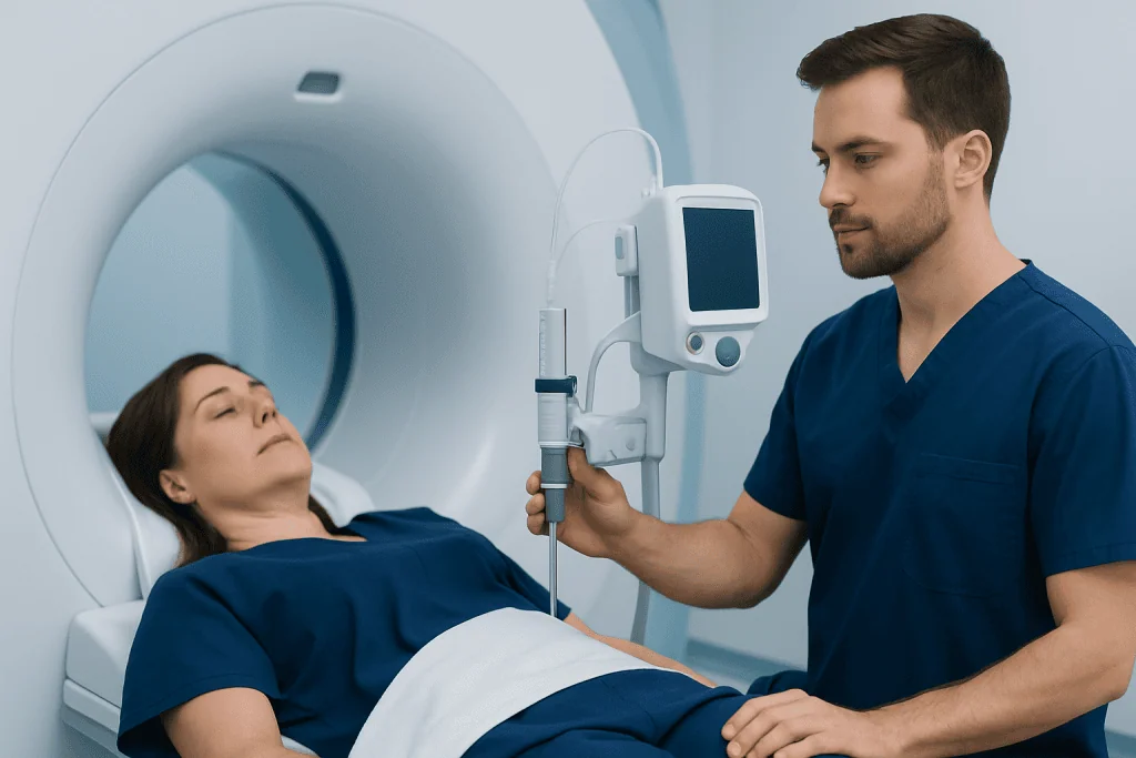
A clinician may recommend a cisternogram when there is persistent or intermittent clear, watery drainage from one nostril or ear and testing suggests it may be CSF. It can also be considered after head trauma, sinus or ear surgery, neurosurgery, or when a person has symptoms and imaging findings that raise concern for a spontaneous skull-base leak. In carefully selected situations, radionuclide cisternography may help evaluate CSF circulation.
Not every person with a suspected leak needs this test. CT and MRI techniques can often show a skull-base defect or related changes without intrathecal contrast. A multidisciplinary assessment involving ENT, neuroradiology, neurology, and neurosurgery may help determine whether the potential benefit of cisternography outweighs the invasiveness of a lumbar puncture.
Before scheduling, the team reviews pregnancy status, kidney problems, bleeding disorders, allergies to contrast material or medicines, infections, and current medications. Blood-thinning medicines may need special planning, but they should never be stopped without instructions from the prescribing clinician. The test may be postponed if there is an infection near the puncture site or another reason that makes lumbar puncture unsafe.
What is the protocol for a CT cisternogram?

A CT cisternogram protocol is individualized rather than identical for every patient. In broad terms, it includes pre-procedure review, a lumbar puncture performed under sterile conditions, injection of a clinician-selected amount of water-soluble iodinated contrast into the CSF, positioning to help contrast reach the region of interest, and thin-section CT imaging of the skull base or another suspected site.
The patient may lie on their side or stomach for the lumbar puncture, sometimes with imaging guidance. Local anesthetic is used to numb the skin. After contrast is introduced, the radiology team may ask the patient to remain in a particular position for a limited period. CT images are then obtained, often with focused views of the sinuses, temporal bones, or other areas where leakage is suspected.
Timing is important because a leak may be intermittent. Some protocols include immediate images and, when clinically appropriate, delayed imaging. The interpreting radiologist looks for contrast outside the usual CSF spaces, which can indicate the pathway of a leak. The examination should be performed by an experienced team using established safety procedures for intrathecal contrast.
Administrative labels such as a cisternography CPT code, cisternogram CPT code, or cisternogram ICD-10 code are used for billing and documentation and can vary by setting, technique, and reason for testing. They do not describe an individual’s diagnosis or replace a clinician’s explanation of the planned examination.
How long does a cisternogram take?
The active lumbar puncture and contrast administration may take a relatively short time, but the total visit is usually longer because it includes check-in, preparation, positioning, imaging, observation, and discharge instructions. For CT cisternography, patients should plan for several hours at the imaging facility. Additional or delayed scans may extend the visit.
Radionuclide cisternography may take longer overall because images can be obtained at more than one time point, sometimes over one or more days. The exact schedule depends on the tracer used and the question being investigated. The imaging department should provide clear instructions about arrival time, food and drink, medicines, transportation, and expected duration.
In some cases, a person may need to remain under observation after the procedure, particularly if sedation has been used, symptoms develop, or additional imaging is planned. It is sensible to arrange for a responsible adult to accompany the patient or be available to take them home if the care team advises this.
What should I expect after a cisternogram?
After a cisternogram, patients are monitored for a period determined by the type of examination and their individual circumstances. Mild tenderness or bruising at the lower-back puncture site can occur. Some people develop a headache after lumbar puncture, often made worse by being upright and improved by lying down. The care team may recommend rest, fluids if appropriate, and simple pain relief that is safe for the individual.
Most people can return to gentle activities after the period advised by their clinician, while avoiding strenuous exercise, heavy lifting, or activities that could worsen symptoms for a short time. Instructions vary, so the imaging team’s written guidance should take priority. Patients should not drive if they have received sedating medication or do not feel well enough to drive safely.
The radiologist reviews the images and sends a report to the referring clinician, who explains what the findings mean in the context of symptoms and prior tests. If a leak is confirmed, treatment may involve observation, targeted management of contributing factors, or repair. For certain skull-base leaks, CSF leak repair may be discussed with an appropriate specialist.
What are the risks of a CT cisternogram?
A CT cisternogram has risks related to both lumbar puncture and intrathecal contrast. The most common effects are temporary headache, lower-back discomfort, nausea, dizziness, or fatigue. A post-lumbar-puncture headache usually improves with conservative care, but occasionally requires further medical treatment.
Less common risks include bleeding, infection, nerve irritation, and an allergic or other adverse reaction to contrast material. Serious neurologic complications are rare, but intrathecal contrast must be used carefully and only when appropriate. CT also uses ionizing radiation; the team aims to use the lowest radiation exposure needed to answer the clinical question.
The main benefit is that the examination may identify a leak or clarify CSF flow when other tests have not answered the question. Locating the site can support safer, more precise planning for treatment. However, a normal result does not always exclude an intermittent leak, and the clinician may recommend additional testing if symptoms and prior evidence remain convincing.
Patients should tell the team promptly about past contrast reactions, seizures, kidney disease, pregnancy, blood-thinner use, or new symptoms before the examination. These details allow the team to select the safest approach and discuss alternatives when needed.
Preparing safely and when to seek medical care
Preparation instructions may include guidance about eating, drinking, regular medicines, diabetes medicines, and blood thinners. Patients should bring a current medication list and details of prior imaging and surgeries. They should not make medication changes independently. If there is any chance of pregnancy, this should be discussed with the care team before CT imaging.
After the procedure, urgent medical advice is appropriate for a severe or worsening headache, fever, increasing redness or drainage at the puncture site, new weakness or numbness, confusion, a seizure, persistent vomiting, trouble breathing, or symptoms of a significant allergic reaction such as facial swelling or widespread hives. These symptoms do not necessarily mean a serious complication, but they need timely assessment.
Clear nasal or ear drainage after a significant head injury, especially with severe headache, confusion, neck stiffness, vision changes, or worsening drowsiness, also requires prompt medical evaluation. A confirmed CSF leak should be assessed because persistent leaks can increase the risk of infection around the brain and spinal cord.
Acibadem International’s multidisciplinary specialists and JCI-accredited hospitals can assess suspected CSF leaks and coordinate diagnostic imaging and treatment for international patients when appropriate.
Frequently asked questions
Is a cisternogram painful?
The lumbar puncture can cause pressure or brief discomfort, despite the use of local anesthetic to numb the skin. Some people have back soreness or a headache afterward. The team can explain comfort measures and what symptoms should be reported.
Is CT cisternography the same as a regular CT scan?
No. A regular CT scan creates images without placing contrast into the cerebrospinal fluid. CT cisternography includes a lumbar puncture and intrathecal contrast so that clinicians can look for abnormal escape or movement of CSF.
Why might a cisternogram be needed if MRI or CT has already been done?
Standard CT or MRI may show structural changes but may not demonstrate an active or intermittent CSF leak. A cisternogram can sometimes provide additional functional information by tracking contrast or tracer through the CSF spaces. It is generally reserved for selected cases because it is more invasive.
Can a cisternogram detect an intermittent CSF leak?
It may detect an intermittent leak if the leak is active while the contrast reaches the relevant area. However, a negative study cannot always rule out a leak that occurs only occasionally. The clinician interprets the result alongside symptoms, laboratory findings, and other imaging.
How soon are cisternogram results available?
Preliminary imaging observations may be available soon after the scan, but the formal report is usually prepared by a radiologist and sent to the referring clinician. Timing can vary according to the imaging method and whether delayed images are needed. The referring team will discuss the results and next steps.
Can I go home after a cisternogram procedure?
Many patients can go home after the recommended observation period, provided they are stable and have no concerning symptoms. The team may advise having someone available to take them home, especially if sedation was used. Individual instructions depend on the technique and the patient's health status.
References
- American College of Radiology
- Radiological Society of North America
- National Institute of Neurological Disorders and Stroke
- U.S. Food and Drug Administration
- Merck Manual Professional Edition
This article is for general information only and is not a substitute for professional medical advice. Please consult a qualified doctor about your individual situation.
Joint and spine care in Turkey — expert assessment & treatment
JCI-accredited · board-certified surgeons · reply within 24h
Add us as a Preferred Source to see more of our trusted health content across Google Search, AI Overviews and Discover.