Cone Beam CT: How This Low-Dose 3D Scan Is Used in Dental and Facial Care
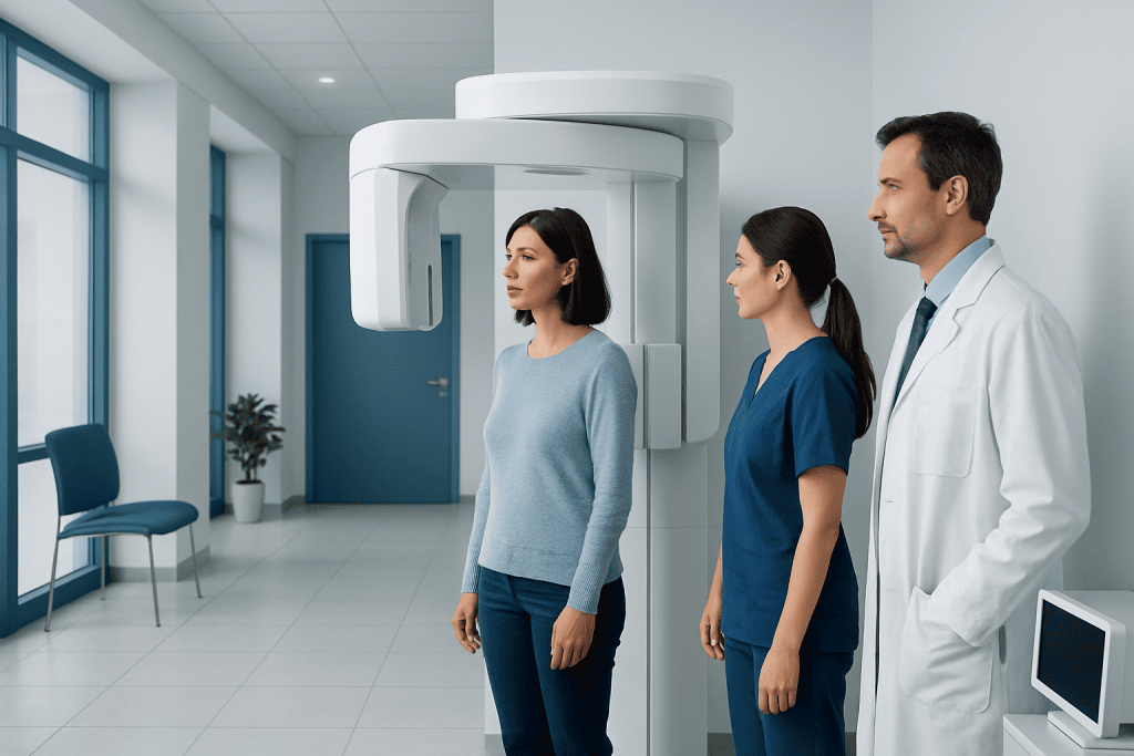
Cone Beam CT creates detailed 3D images of teeth, jawbones, facial bones, and sinuses. It is commonly used for dental implants, orthodontics, oral surgery, and complex root canal assessment.
Key Takeaways
- Cone Beam CT creates detailed 3D images of teeth, jawbones, facial bones, and sinuses.
- It is commonly used for dental implants, orthodontics, oral surgery, and complex root canal assessment.
- For many dental indications, it uses less radiation than a standard medical CT, though exposure is still kept as low as reasonably achievable.
- The scan is quick, noninvasive, and usually does not require special preparation.
- Cone Beam CT is ordered when 3D information is likely to improve diagnosis or treatment planning.
Cone Beam CT is a specialized 3D imaging scan that gives detailed views of teeth, jaws, facial bones, and nearby structures using a relatively low radiation dose compared with conventional medical CT for many dental uses. It helps dentists and specialists diagnose problems more precisely and plan treatments such as implants, orthodontics, and jaw procedures with greater accuracy.
Overview: What Cone Beam CT Is
Cone Beam CT, often called CBCT, is an imaging technology that produces three-dimensional views of the teeth, jaws, facial bones, airways, and nearby structures. Unlike standard dental X-rays, which show flat two-dimensional images, Cone Beam CT captures detailed 3D data that can be viewed from different angles. This allows clinicians to better understand anatomy that may be difficult to assess on routine imaging.
The scan uses a cone-shaped X-ray beam that rotates around the head and collects many images in a single sweep. A computer then reconstructs these images into a 3D model. Because it is designed specifically for the head and face, Cone Beam CT is especially useful in dental and maxillofacial care.
For many dental applications, Cone Beam CT provides more detail than conventional dental X-rays while using a lower radiation dose than a typical medical CT of the same region. The exact dose depends on the machine, scan area, and settings used. For this reason, the scan is generally recommended only when the extra 3D information is expected to help with diagnosis or treatment planning.
How Cone Beam CT Is Used in Dental and Facial Care
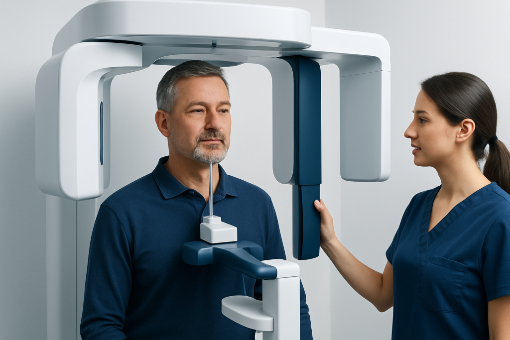
Cone Beam CT has become an important tool across several dental and facial specialties. One of its most common uses is planning dental implants. The scan helps measure bone height, width, and density, and shows the position of important structures such as nerves and the maxillary sinus. This can help the care team choose the safest and most suitable implant position, often as part of dental implant planning.
It is also widely used in orthodontics, oral and maxillofacial surgery, periodontics, and endodontics. In orthodontic care, CBCT may help evaluate impacted teeth, jaw relationships, and facial asymmetry. In root canal treatment, it can be useful when standard imaging does not clearly show complex root anatomy, hidden canals, or persistent infection around the tooth, sometimes supporting root canal treatment decisions.
Other common uses include assessing wisdom teeth, jaw joint problems, facial trauma, sinus anatomy, cleft-related planning, and certain airway evaluations. Specialists may also use it to investigate cysts, tumors, or bone abnormalities and to better understand conditions affecting the jaw or facial skeleton. In selected cases, CBCT can support the assessment of temporomandibular joint disorders or chronic dental infections.
- Dental implant assessment and surgical planning
- Evaluation of impacted teeth, especially wisdom teeth and canines
- Orthodontic and jaw alignment planning
- Complex root canal and tooth root evaluation
- Assessment of facial bones, trauma, and sinus relationships
What the Scan Can Show

Cone Beam CT provides highly detailed images of hard tissues, especially bone and teeth. It can show the shape and position of tooth roots, the thickness and contour of jawbone, the location of the mandibular nerve, sinus floor anatomy, and the relationship between teeth and surrounding bone. This level of detail can be valuable when treatment needs to be precise.
It is particularly helpful when anatomy is crowded, overlapping, or unusual. For example, a routine dental X-ray may not fully reveal whether an impacted tooth is touching a nearby root, or how close a planned implant site is to a nerve canal. A 3D image can often answer these questions more clearly and help reduce uncertainty before treatment.
Although Cone Beam CT is excellent for bone and dental structures, it is not the best test for every problem. Soft tissues such as muscles, glands, and some internal structures are usually better assessed with other imaging methods, depending on the situation. The most appropriate scan depends on the symptoms, the body area being evaluated, and the clinical question the doctor is trying to answer.
What to Expect Before, During, and After the Scan
In most cases, Cone Beam CT requires very little preparation. Patients are usually asked to remove metal items from the head and neck area, such as earrings, necklaces, removable dentures, hearing aids, or glasses, because metal can interfere with image quality. It is important to tell the imaging team about pregnancy or the possibility of pregnancy before any X-ray-based scan.
During the scan, the patient may sit or stand depending on the machine. The head is positioned carefully to keep it still while the scanner rotates around it. The scan itself is quick, often lasting less than a minute, though positioning and setup may take a little longer. The procedure is painless, noninvasive, and does not usually require injections, sedation, or recovery time.
After the scan, normal activities can be resumed right away. A dentist, oral surgeon, orthodontist, or radiologist reviews the images and prepares an interpretation based on the reason the scan was ordered. The results are then used to guide the next steps, whether that means reassurance, monitoring, or planning treatment such as orthodontic treatment or oral surgery.
Benefits and Limitations of Cone Beam CT
The main advantage of Cone Beam CT is that it provides a detailed 3D view in a relatively fast and accessible way. This can improve diagnostic confidence and support more precise treatment planning. In many situations, it helps clinicians identify anatomical details that are not visible on standard 2D dental X-rays, which may reduce surprises during treatment.
Another benefit is radiation efficiency for the specific tasks it is designed to perform. For many head and dental uses, CBCT exposes patients to less radiation than a conventional medical CT. Even so, it still involves ionizing radiation, so it should be used thoughtfully. Clinicians generally follow the principle of using the smallest scan area and lowest settings needed to answer the clinical question.
Cone Beam CT also has limitations. It is less effective than some other imaging methods for evaluating soft tissues, and image quality can be affected by patient movement or metal restorations. Not every dental issue requires 3D imaging, and a standard dental X-ray may often be enough. The decision to order CBCT should be individualized based on likely benefit.
Safety, Radiation Dose, and Who May Need Extra Caution
Many patients understandably ask whether Cone Beam CT is safe. In general, it is considered safe when used appropriately and when the expected diagnostic value outweighs the small radiation risk. The goal is not to avoid imaging when it is needed, but to use it carefully and only for justified clinical reasons.
Radiation dose from CBCT varies widely depending on the device, field of view, scan resolution, and the body area examined. A small, focused scan of one part of the jaw can involve much less radiation than a larger scan of the whole facial region. Children and teenagers may need extra caution because growing tissues are more sensitive to radiation, so providers are especially careful to use CBCT only when it is likely to change management.
Pregnant patients should always inform the care team before the scan. If imaging is not urgent, it may sometimes be postponed or replaced with another method. If it is necessary, the dental and radiology team will consider the safest approach. Good imaging practice includes choosing the right test, limiting the scan area, and avoiding repeat scans unless they are truly needed.
When Cone Beam CT May Be Recommended
A dentist or specialist may recommend Cone Beam CT when standard examination and routine X-rays do not provide enough information. This often happens in more complex cases where treatment accuracy matters, such as implant placement, impacted teeth, jaw surgery, or unexplained facial pain. The scan may also be helpful when symptoms persist and the cause remains unclear on basic imaging.
CBCT can be especially useful before surgical procedures because it helps map anatomy in three dimensions. This may be important when a tooth lies close to a nerve, when bone appears limited, or when sinus anatomy affects treatment options. In some situations, it also helps identify hidden sources of infection, bone loss, or structural changes related to sinus disease or previous dental problems.
Near the end of the care pathway, patients may also want to know where comprehensive evaluation is available. Acibadem International’s multidisciplinary specialists and JCI-accredited hospitals diagnose and treat dental and facial conditions for international patients, using advanced imaging where clinically appropriate, including support for procedures such as oral and maxillofacial surgery.
Questions to Ask the Dentist or Specialist
When Cone Beam CT is suggested, patients may feel more comfortable if they understand why it is being ordered and how it will affect care. A useful first question is whether the scan is expected to change the diagnosis or treatment plan. If the answer is yes, the added imaging may offer clear value.
It can also help to ask what area will be scanned, whether a smaller field of view can be used, and whether another imaging option could answer the same question. Patients may wish to ask about expected benefits, possible limitations, and what the doctor hopes to learn from the scan. These are reasonable and important questions.
In general, informed imaging decisions are a partnership between patient and clinician. The aim is to gather enough information to provide safe, effective care while avoiding unnecessary radiation or testing. If anything about the recommendation is unclear, asking for an explanation is appropriate and encouraged.
Frequently asked questions
What is the difference between Cone Beam CT and a regular dental X-ray?
A regular dental X-ray produces a two-dimensional image, while Cone Beam CT creates a three-dimensional view of the teeth, jaw, and surrounding structures. This extra detail can be helpful when anatomy is complex or when treatment needs very precise planning.
Is Cone Beam CT the same as a medical CT scan?
No. Cone Beam CT is designed mainly for dental and maxillofacial imaging and uses a cone-shaped beam to capture the head and face. For many dental purposes, it typically involves less radiation than a standard medical CT, but the tests are used for different clinical questions.
Is a Cone Beam CT scan painful?
No, the scan is painless and noninvasive. The patient simply stays still while the machine rotates around the head for a short time.
How long does a CBCT scan take?
The actual image capture is usually very quick, often under a minute. The full appointment may take a little longer because careful positioning is needed to get clear images.
Does everyone need Cone Beam CT before dental treatment?
No. Many dental problems can be assessed well with examination and standard dental X-rays. Cone Beam CT is usually reserved for situations where 3D information is likely to improve diagnosis, safety, or treatment planning.
Is Cone Beam CT safe for children?
It can be safe when it is clearly needed and performed with child-appropriate settings. Because children are more sensitive to radiation, clinicians use CBCT selectively and try to keep exposure as low as reasonably achievable.
Can Cone Beam CT show infections or jaw problems?
It can often help detect signs of dental infection, bone loss, impacted teeth, and some jaw abnormalities. However, it is not the best test for every condition, especially if the concern involves mainly soft tissues rather than bone or teeth.
References
- American Dental Association
- International Atomic Energy Agency
- Radiological Society of North America
- American Association of Oral and Maxillofacial Surgeons
- European Academy of Dentomaxillofacial Radiology
This article is for general information only and is not a substitute for professional medical advice. Please consult a qualified doctor about your individual situation.
Explore treatments in Turkey — costs, top hospitals & a free quote
JCI-accredited · board-certified surgeons · reply within 24h
Add us as a Preferred Source to see more of our trusted health content across Google Search, AI Overviews and Discover.