Contrast Enhanced Ultrasound: Preparation, Procedure and Results

Contrast enhanced ultrasound uses microbubble contrast to make blood flow and tissue perfusion easier to see. Preparation depends on the body area being examined; some abdominal scans require fasting, while others do not.
Key Takeaways
- Contrast enhanced ultrasound uses microbubble contrast to make blood flow and tissue perfusion easier to see.
- Preparation depends on the body area being examined; some abdominal scans require fasting, while others do not.
- The scan commonly lasts about 20 to 45 minutes, including preparation and observation when needed.
- Microbubble contrast is generally well tolerated, but allergic-like reactions and heart or lung symptoms are possible and uncommon.
- Results are interpreted alongside symptoms, medical history and other tests; further imaging may sometimes still be needed.
Contrast enhanced ultrasound is an imaging examination that combines standard ultrasound with tiny gas-filled microbubbles delivered through a vein. It helps clinicians observe blood flow and assess certain findings in real time, usually without ionising radiation and with little recovery time.
Overview: what is contrast enhanced ultrasound?
Contrast enhanced ultrasound, also called CEUS, is an imaging test that uses sound waves together with an ultrasound contrast agent. The agent contains very small gas-filled microbubbles that are injected into a vein. As the microbubbles travel through the bloodstream, they reflect ultrasound waves strongly, helping the radiologist see blood flow and how a tissue or organ receives blood in real time.
It may be used to evaluate findings in the liver, kidneys, spleen, pancreas, bowel, heart, blood vessels or other soft tissues, depending on local expertise and the clinical question. For example, it can help clarify the appearance of a lesion detected on a standard ultrasound or another imaging examination. It does not use X-rays, and it is different from the iodine-based contrast commonly used for CT scans or the gadolinium-based contrast sometimes used for MRI.
The examination is usually performed by a trained sonographer, radiologist or another imaging specialist. It is a diagnostic tool rather than a treatment, and its findings are considered together with a person’s symptoms, laboratory tests and previous scans.
How does contrast enhanced ultrasound work?
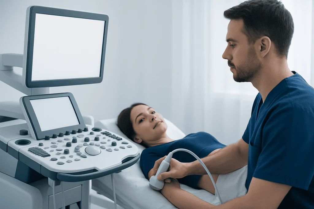
A standard ultrasound probe sends high-frequency sound waves into the body and converts returning echoes into images. Microbubbles circulating in the blood produce a distinctive echo pattern. Special ultrasound settings can separate the microbubble signal from surrounding tissue, allowing clinicians to watch contrast enter and leave an area over seconds to minutes.
This real-time information can show patterns of enhancement, meaning how quickly and in what way an area receives blood. These patterns may help distinguish between different types of tissue changes, assess organ perfusion, or examine blood flow after certain procedures. The exact interpretation depends on the organ being studied and the person’s wider medical context.
Unlike CT contrast, ultrasound microbubbles remain within blood vessels rather than passing into surrounding tissue. They are broken down naturally, with the gas commonly exhaled through the lungs and the remaining components processed by the body. This characteristic can make CEUS useful for some people in whom avoiding iodinated CT contrast is desirable, although suitability must always be assessed individually.
Candidacy, preparation and safety checks
A clinician may recommend contrast enhanced ultrasound when standard ultrasound has not fully answered a question, when an abnormality needs further characterisation, or when a dynamic view of blood flow is useful. It may also be considered when reducing radiation exposure is important. However, it is not the best test for every concern, and CT or MRI may provide more complete information in some situations.
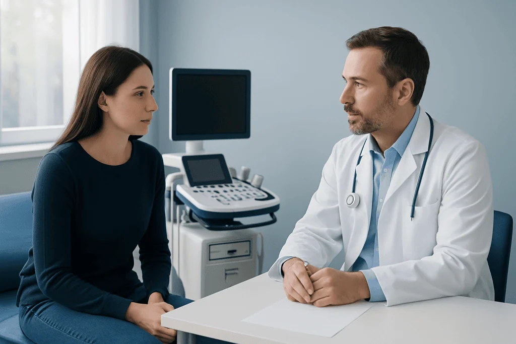
Before the examination, patients should tell the imaging team about previous reactions to contrast agents, allergies, heart or lung disease, pregnancy, breastfeeding, current medicines and any significant recent illness. The team will review whether microbubble contrast is appropriate and explain the planned test. Certain contrast products may have specific precautions, particularly for people with unstable heart or lung conditions.
Is there anything you shouldn’t do before an ultrasound? Preparation depends on the area being scanned. For some abdominal examinations, patients may be asked not to eat for several hours beforehand because food and bowel gas can reduce image quality. For pelvic or urinary tract scans, the instructions may instead include drinking water and arriving with a comfortably full bladder. Patients should not stop prescribed medicines unless their clinician specifically advises it, and should follow the instructions provided by the imaging department.
- Bring previous imaging reports or images if they are available.
- Wear comfortable clothing that allows access to the area being examined.
- Ask in advance about fasting, fluids and usual medication if the instructions are unclear.
- Inform the team if there is any chance of pregnancy or if a serious medical condition has changed recently.
What happens during the procedure?
The patient usually lies on an examination couch while the specialist applies gel to the skin and places an ultrasound probe over the area of interest. Initial images are often obtained without contrast. A small intravenous cannula is then placed in a hand or arm vein if one is not already present.
The microbubble contrast agent is injected through the cannula, typically followed by a saline flush. The imaging team records ultrasound images continuously or at planned intervals as the contrast circulates. The patient may be asked to change position, hold their breath briefly, or remain still so that clear images can be obtained. The scan itself is generally painless, although the cannula insertion and probe pressure may cause mild temporary discomfort.
How long does a contrast ultrasound take? The imaging portion often takes around 20 to 45 minutes, although the timing varies by body area, clinical question and whether additional standard ultrasound images are needed. The contrast is usually observed in the area of interest for only several minutes, but preparation, review of safety information and short observation afterward can add time. The imaging department can provide the most accurate estimate for an individual appointment.
Recovery timeline, benefits and possible disadvantages
Most people can return to normal activities immediately after contrast enhanced ultrasound. The cannula is removed, and pressure may be applied briefly to the injection site. Patients are commonly asked to remain nearby for a short period if the team considers observation appropriate, especially after a first exposure or when there are relevant health concerns.
Potential benefits include no ionising radiation, real-time imaging, no need for sedation in most cases, and a contrast agent with different handling by the body than CT or MRI contrast. CEUS can also be repeated during the same visit when clinically appropriate. Its value is strongest when it is used for a clearly defined question and interpreted by an experienced team.
What are the disadvantages of contrast-enhanced ultrasound? The test is operator-dependent and image quality can be reduced by bowel gas, body habitus, deep location of a structure, movement or limited acoustic windows. It may not assess the entire body or provide the broad anatomical detail available with CT or MRI. Microbubble contrast reactions are uncommon but can occur, including headache, nausea, flushing, rash, breathing difficulty or changes in blood pressure; severe reactions are rare but require prompt medical attention. In some cases, an additional test or biopsy may still be needed.
Results and choosing between ultrasound and CT
After the examination, a radiologist reviews the recorded images and prepares a report for the clinician who requested the test. Results may be available quickly in urgent settings, but timing varies by hospital and by whether comparison with earlier scans is needed. The requesting clinician explains what the findings mean for the individual and whether follow-up imaging, blood tests, specialist review or treatment is appropriate.
Which is better, ultrasound or CT scan with contrast? Neither test is universally better; the most suitable choice depends on the clinical question. Contrast enhanced ultrasound can be particularly helpful for assessing blood flow in an accessible organ or lesion without radiation. CT with contrast can provide a wider view of the chest, abdomen or pelvis, show structures that ultrasound cannot reach well, and may be preferred in trauma, emergencies or when a comprehensive survey is needed.
CT uses X-rays and iodinated contrast, while CEUS uses sound waves and microbubbles. Factors such as kidney function, prior contrast reactions, pregnancy status, the body area being assessed and the availability of specialist expertise may influence the decision. A clinician may sometimes recommend both tests because they provide complementary information.
When to seek medical care
People should seek urgent medical help after contrast administration if they develop difficulty breathing, swelling of the face or throat, widespread hives, severe dizziness, fainting, chest pain or a sudden severe unwell feeling. Although serious reactions are unusual, they need immediate assessment. Patients should tell staff straight away if any new symptom occurs while they are still in the imaging department.
A person should also contact the clinician who arranged the examination if they have persistent pain, increasing redness, swelling or discharge at the injection site, or if they have concerns about the result or follow-up plan. Symptoms that led to the scan, such as ongoing abdominal pain, unexplained weight loss, jaundice, blood in urine or worsening shortness of breath, should not be ignored while waiting for results.
Acibadem International’s multidisciplinary specialists and JCI-accredited hospitals can assess imaging concerns and coordinate appropriate diagnostic care for international patients. The appropriate next step is based on the individual’s symptoms, examination findings and imaging results.
Frequently asked questions
Does contrast enhanced ultrasound use radiation?
No. Contrast enhanced ultrasound uses sound waves, not X-rays or other ionising radiation. The contrast agent contains microbubbles that improve the visibility of blood flow on ultrasound images.
Is contrast enhanced ultrasound painful?
The scan is usually not painful. A person may feel a brief needle pinch when the intravenous line is placed and mild pressure from the ultrasound probe. The contrast injection itself is often not felt or may cause only a brief sensation.
Can someone eat before a contrast enhanced ultrasound?
It depends on the type of examination. Abdominal scans may require fasting for several hours, while other scans may not require dietary changes. The imaging department's instructions should be followed carefully.
Is ultrasound contrast safe for people with kidney problems?
Microbubble ultrasound contrast is handled differently from iodinated CT contrast and gadolinium MRI contrast. It may be an option for some people with kidney disease, but the imaging clinician must review the person's overall health and the specific contrast product before the test.
What happens if the result is unclear?
An unclear result does not necessarily mean there is a serious condition. The clinician may compare the images with previous scans, arrange follow-up ultrasound, or recommend CT, MRI, laboratory tests or tissue sampling depending on the finding and symptoms.
Can contrast enhanced ultrasound diagnose cancer?
CEUS can provide useful information about the blood-flow pattern of a lesion and may help clinicians assess whether further investigation is needed. However, it does not always provide a final diagnosis on its own. A confirmed cancer diagnosis may require additional imaging, specialist assessment or a biopsy.
References
- World Health Organization
- European Federation of Societies for Ultrasound in Medicine and Biology
- American College of Radiology
- Radiological Society of North America
This article is for general information only and is not a substitute for professional medical advice. Please consult a qualified doctor about your individual situation.
Ultrasound in Turkey — costs, top hospitals & a free quote
JCI-accredited · board-certified surgeons · reply within 24h
Add us as a Preferred Source to see more of our trusted health content across Google Search, AI Overviews and Discover.

Check-up & Preventive Medicine
Comprehensive health screenings designed for early detection and prevention.
2 specialists in this unit