Cranial Fossa Surgery: Procedure, Recovery and Results

Cranial fossa surgery refers to approaches through areas of the skull base, not one single operation. A cranial fossa craniotomy may be performed for tumors, cerebrospinal fluid leaks, vascular conditions, trauma or congenital skull abnormalities.
Key Takeaways
- Cranial fossa surgery refers to approaches through areas of the skull base, not one single operation.
- A cranial fossa craniotomy may be performed for tumors, cerebrospinal fluid leaks, vascular conditions, trauma or congenital skull abnormalities.
- Planning commonly involves detailed MRI or CT imaging and collaboration between neurosurgery, ENT, plastic surgery and other specialties when needed.
- Initial hospital recovery is usually measured in days, while fuller cranial surgery recovery time often takes weeks to months.
- Every craniotomy has important risks, but the surgical team takes specific steps to protect brain function, nerves and blood vessels.
- New neurological symptoms, fever, wound concerns or clear fluid drainage after surgery need prompt medical advice.
Cranial fossa surgery is a specialized operation in which surgeons access the front, middle or back portion of the skull base to treat selected tumors, structural problems, fluid leaks, injuries or congenital conditions. Recovery varies with the reason for surgery and the approach used, but careful follow-up and rehabilitation can support a safe return to daily life.
Overview: what is cranial fossa surgery?
Cranial fossa surgery is a group of skull-base operations that allow surgeons to reach conditions located at the base of the skull. The cranial fossae are natural depressions inside the skull that support different parts of the brain: the anterior fossa lies toward the forehead, the middle fossa is near the temples and ears, and the posterior fossa is toward the back of the head.
The aim is to treat a specific problem while providing the safest possible route around important structures, including the brain, optic nerves, hearing and balance nerves, major blood vessels, sinuses and the spinal fluid spaces. Depending on the condition, a surgeon may perform a cranial fossa craniotomy, use an endoscopic route through the nose, or combine approaches with other specialists.
A cranial fossa approach can be used to remove or biopsy a tumor, repair a cerebrospinal fluid leak, relieve pressure, treat certain vascular abnormalities, correct trauma-related damage, or address congenital skull conditions. The exact procedure, expected results and recovery depend far more on the underlying diagnosis than on the name of the approach alone.
How it works and who may be a candidate
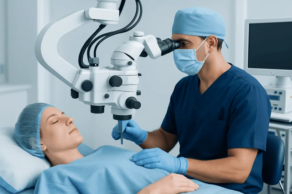
During cranial fossa surgery, the surgeon creates an access route to the skull base and then treats the identified problem. In an open craniotomy, a temporary bone opening is made in the skull, often behind the hairline or near the ear. The bone is usually replaced and secured at the end of the operation. In some cases, a minimally invasive endoscopic technique can be used through the nostrils or another smaller opening.
People may be considered for surgery when a structural problem is causing symptoms, has a risk of progression, cannot be managed adequately with observation or medicines, or requires a tissue diagnosis. Examples include selected skull-base tumors, persistent cerebrospinal fluid leakage, some congenital cranial abnormalities and certain injuries. A fossa repair may also be recommended when there is a defect between the skull base and nearby air spaces that needs closure.
Suitability is individualized. The team considers the location and size of the condition, symptoms, imaging findings, age, overall health, previous operations and a person’s priorities. Some small or slow-growing abnormalities can be monitored with scheduled scans, while others benefit from timely intervention. Surgery is recommended only when its likely benefits outweigh its potential risks.
- Detailed MRI and CT scans help map bone, blood vessels, nerves and the condition being treated.
- Hearing, vision, hormonal or neurological testing may be needed, depending on the surgical area.
- Preoperative review includes medicines, allergies, medical history and anesthesia assessment.
Step by step: what happens during the procedure?

Before surgery, the patient receives general anesthesia and is positioned carefully to protect the neck, skin and pressure points. The surgical team uses imaging and, when appropriate, navigation technology to confirm the planned route. Hair is often preserved as much as possible, although a small area may be trimmed if necessary for a safe incision.
For an open cranial fossa craniotomy, the surgeon makes an incision, temporarily lifts a section of bone and opens the protective covering of the brain, called the dura. Microsurgical instruments, an operating microscope and sometimes an endoscope help the team work precisely around delicate structures. The surgeon may remove a lesion, collect tissue, decompress an area, repair a leak or reconstruct a skull-base defect.
When a repair is required, the team may use the patient’s own tissue, surgical graft materials, sealants or small fixation devices to support healing. The dura and bone are closed or reconstructed as appropriate, and the incision is closed. Surgery length varies substantially according to the diagnosis, anatomy and whether reconstruction or monitoring is required.
Afterward, patients are transferred to a recovery area and may spend time in an intensive monitoring unit. Staff regularly assess alertness, strength, pupils, pain control, fluid balance and the surgical wound. The treating team can explain which aspects of the operation are expected to apply in an individual case.
Benefits, risks and expected results
The potential benefit of cranial fossa surgery is direct treatment of a problem that may otherwise affect neurological function, cause fluid leakage, produce pressure symptoms or continue to grow. For some conditions, surgery can provide a diagnosis as well as treatment. For others, the goal may be complete removal, partial removal, durable repair, symptom relief or preparation for additional treatment such as radiotherapy.
Results depend on the diagnosis and on whether nearby nerves, blood vessels or brain tissue are involved. Symptoms may improve gradually rather than immediately. In particular, recovery of hearing, balance, vision or facial nerve function can take time and is not always predictable. The surgeon should discuss the realistic goal of surgery and alternatives before consent is given.
Craniotomy is a major operation and carries risks. These can include bleeding, infection, blood clots, seizures, stroke, swelling, anesthesia complications, leakage of cerebrospinal fluid, wound-healing problems and the need for further treatment. Depending on the surgical site, there may also be risks involving memory, speech, vision, smell, hearing, balance, swallowing, facial movement or hormone function.
Risk reduction begins with careful planning and continues through surgery, monitoring and follow-up. Patients can support safety by sharing a full medication list, following instructions about fasting and blood-thinning medicines, avoiding smoking where possible, and attending all postoperative appointments.
Recovery timeline and daily life after surgery
Recovery begins in hospital, where the team manages pain, nausea, hydration, movement and neurological monitoring. Some patients go home after several days, while others need a longer stay because of the complexity of the operation, the condition treated or the need for rehabilitation. Tiredness, headaches, scalp tenderness, sleep changes and reduced concentration are common early experiences and generally improve over time.
During the first few weeks, activity is increased gradually. Walking and light daily tasks are often encouraged when the surgeon approves, but heavy lifting, strenuous exercise, driving and work duties may need to wait. Follow-up visits assess the incision, review pathology or imaging results when relevant, and guide a safe return to normal routines. Rehabilitation may include physiotherapy, occupational therapy, speech therapy, balance therapy or neuropsychological support.
Cranial surgery recovery time is highly individual. Many people feel noticeably stronger over four to eight weeks, but recovery of stamina, concentration and confidence may continue for several months. A return to work depends on the job, the person’s recovery and whether neurological symptoms remain. The care team can provide more individualized timing.
Family members can help by supporting rest, medication schedules, nutritious meals, short walks and follow-up care. Emotional adjustment is also important. Anxiety, low mood, irritability or changes in sleep can occur after a significant operation, and patients should feel comfortable raising these concerns with their healthcare team.
How long after a craniotomy do you feel normal?
There is no single point at which everyone feels normal after a craniotomy. Some people feel ready for much of their usual routine within several weeks, while others need several months for energy, sleep, concentration, mood and physical confidence to settle. The reason for surgery, the area of the brain involved, complications, previous health and rehabilitation needs all influence the timeline.
Improvement is often uneven. A patient may have a good day followed by a day with more fatigue or headache, especially during the first weeks. Gradual progress is usually more important than comparing recovery with another person’s experience. Keeping follow-up appointments helps the team identify whether symptoms are expected or need further assessment.
If fatigue, pain, cognitive changes or emotional symptoms are interfering with recovery, the clinician may recommend adjustments to activity, medication review, rehabilitation or additional support. New or worsening symptoms should not be assumed to be part of normal healing.
What is life like after craniosynostosis surgery?
Life after craniosynostosis surgery is often focused on healing, growth monitoring and supporting a child’s development. Craniosynostosis is a condition in which one or more skull sutures close too early. Surgery aims to create enough room for healthy brain growth and improve skull shape; the exact technique and follow-up plan depend on the child’s age and the sutures involved.
After the initial recovery period, many children return to feeding, sleep, play and developmental routines with guidance from their care team. Temporary swelling, changes in sleep and discomfort are common after surgery and are monitored closely. Families are given specific instructions about wound care, safe activity and symptoms that require urgent contact with the hospital.
Long-term care may involve neurosurgery, craniofacial or plastic surgery, pediatrics, ophthalmology, dentistry and developmental specialists. Some children need only routine observation, while others benefit from continued support for vision, breathing, learning or skull growth. Regular follow-up helps ensure that needs are recognized early.
What should you not do after a craniotomy? When to seek medical care
After a craniotomy, patients should not resume strenuous activity until their surgeon says it is safe. This commonly includes heavy lifting, contact sports, vigorous exercise, swimming before the wound has healed, and driving until medical clearance is given. Alcohol, smoking and non-prescribed medicines or supplements can interfere with recovery or interact with prescribed treatment, so these should be discussed with the care team.
Patients should also avoid changing or stopping prescribed medicines, including anti-seizure medicines, pain medicines or steroids, without medical advice. They should follow instructions about wound care and bathing, avoid scratching the incision, and ask their surgeon before travelling long distances or flying. Gradual activity, rest and reliable support at home are important in the early stage of recovery.
Medical advice should be sought promptly for fever, increasing redness or swelling around the wound, drainage from the incision, severe or worsening headache, repeated vomiting, confusion, fainting, a seizure, weakness, speech difficulty, new vision changes or clear watery drainage from the nose or ear. Emergency services should be contacted immediately for sudden signs of stroke, loss of consciousness, severe breathing difficulty or other rapidly worsening symptoms.
For patients travelling for complex care, Acibadem International’s multidisciplinary specialists and JCI-accredited hospitals can assess and treat appropriate cranial and skull-base conditions, with coordinated follow-up planning for international patients.
Frequently asked questions
Is a craniotomy a big surgery?
Yes. A craniotomy is considered major surgery because it involves opening the skull to access the brain or skull base. However, the size and complexity vary greatly, and the surgical team plans the approach carefully to address the condition while protecting surrounding structures.
How long is the hospital stay after cranial fossa surgery?
Hospital stay varies according to the reason for surgery, the approach used and the person’s recovery. Some patients may leave after a few days, while more complex procedures or recovery needs can require a longer stay and rehabilitation support.
Will hair grow back after a craniotomy?
In many operations, only a limited area of hair is trimmed or shaved. Hair commonly regrows over time, although the incision itself may leave a scar and hair growth around it can vary. The surgeon can explain the expected incision location and hair management before surgery.
Can a person exercise after a craniotomy?
Light walking is often introduced gradually after surgery when approved by the clinical team. Strenuous exercise, heavy lifting, swimming and contact sports should wait until the surgeon confirms that the wound and underlying healing are adequate.
Are headaches normal after cranial fossa surgery?
Headaches and scalp tenderness can be common during early recovery and often improve over time. A severe, sudden, worsening or unusual headache, especially with vomiting, fever, confusion or weakness, needs prompt medical assessment.
What follow-up is needed after cranial fossa surgery?
Follow-up may include wound checks, neurological examinations and repeat MRI or CT scans. Depending on the treated condition, hearing, vision, hormone, balance or rehabilitation assessments may also be part of longer-term care.
References
- National Institute of Neurological Disorders and Stroke
- American Association of Neurological Surgeons
- Mayo Clinic
- Johns Hopkins Medicine
- National Health Service
This article is for general information only and is not a substitute for professional medical advice. Please consult a qualified doctor about your individual situation.
Explore treatments in Turkey — costs, top hospitals & a free quote
JCI-accredited · board-certified surgeons · reply within 24h
Add us as a Preferred Source to see more of our trusted health content across Google Search, AI Overviews and Discover.