CT Scan Lymph Nodes: Preparation, Procedure and Results
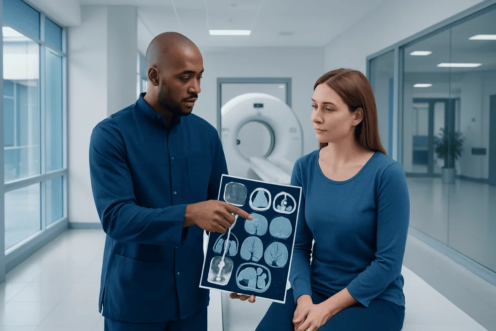
CT scans show the size, shape, location and distribution of lymph nodes throughout the body. Enlarged lymph nodes are common and may result from infection, inflammation, immune conditions or cancer.
Key Takeaways
- CT scans show the size, shape, location and distribution of lymph nodes throughout the body.
- Enlarged lymph nodes are common and may result from infection, inflammation, immune conditions or cancer.
- Contrast dye can improve image detail but may not be suitable for everyone, particularly people with certain kidney problems or previous contrast reactions.
- A suspicious lymph node on CT may require comparison with earlier scans, blood tests, PET imaging or a biopsy for diagnosis.
- Most CT examinations are quick, painless and allow patients to return to normal activities soon afterward.
A CT scan of lymph nodes is an imaging test that produces detailed cross-sectional pictures of the body. It can identify enlarged or unusual lymph nodes and help clinicians plan further assessment, but it cannot by itself confirm whether a node is cancerous.
Overview: What a CT Scan of Lymph Nodes Shows
A CT scan of lymph nodes uses X-rays and computer processing to create detailed images of areas inside the body. It may be requested to check lymph nodes in the neck, chest, abdomen, pelvis or several regions at once. The scan helps a doctor see whether nodes are enlarged, grouped together, pressing on nearby structures or changing over time.
Lymph nodes are small, bean-shaped structures that support the immune system by filtering lymph fluid and helping the body respond to infection. They can become temporarily enlarged during common infections, after inflammation or, less often, because of cancer. A CT scan of the lymphatic system does not image every lymphatic vessel in detail, but it provides valuable information about lymph node groups and nearby organs.
CT is often used when lymph nodes cannot be fully assessed during a physical examination, when symptoms persist, or when a known condition needs staging or follow-up. Results are interpreted alongside medical history, examination findings and other tests. An abnormal scan result is not, on its own, a diagnosis.
How the Scan Works and Who May Need It
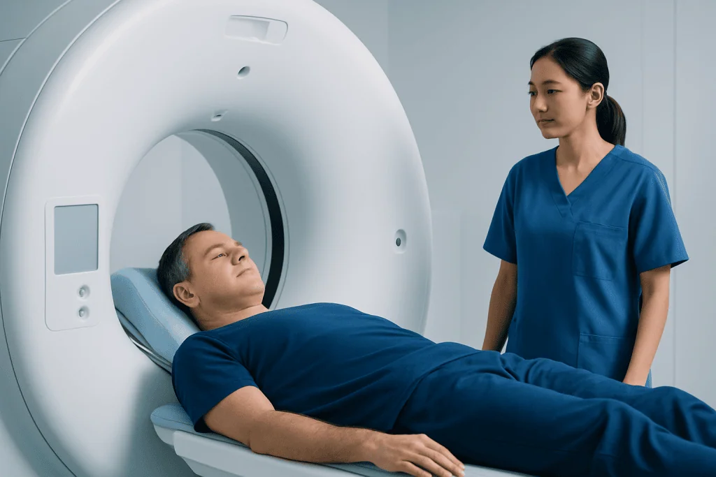
During CT, a rotating X-ray source takes multiple images from different angles. A computer combines these images into thin slices that allow the radiologist to evaluate structures in greater detail than a standard X-ray. A contrast material, usually given through a vein, may be used to make blood vessels, organs and some abnormal tissues easier to distinguish.
A clinician may arrange a CT scan to check lymph nodes after unexplained swelling, persistent fever, weight loss, night sweats, recurrent infections or abnormal findings on an ultrasound, chest X-ray or blood test. It may also be used to assess the extent of a diagnosed cancer, monitor treatment response or investigate symptoms involving the chest, abdomen or pelvis.
CT is not always the first imaging test. Ultrasound can be especially useful for superficial lymph nodes in the neck, armpit or groin, while MRI or PET/CT may be more appropriate in selected situations. The best test depends on the body area, the clinical question, prior imaging and the person’s health history.
- Tell the imaging team about pregnancy or a possible pregnancy.
- Report previous reactions to iodinated contrast material, asthma, kidney disease and thyroid conditions.
- Provide an up-to-date list of medicines and discuss diabetes medicines if contrast is planned.
Preparation and Step-by-Step Procedure
Preparation for a CT scan lymph node assessment depends on the area being examined and whether contrast is needed. Some people may be asked not to eat for several hours before a contrast-enhanced scan. Clear instructions should come from the imaging department, and regular medicines should only be changed if a clinician advises it.
Before the scan, the patient removes metal objects such as jewelry, belts or clothing with metal fasteners and changes into a gown if required. If intravenous contrast is used, a small cannula is placed in a vein, usually in the arm or hand. The radiographer reviews safety questions and explains how to communicate during the examination.
The patient lies on a padded table that moves slowly through the doughnut-shaped CT scanner. It is important to remain still, and brief breath-holds may be requested for scans of the chest or upper abdomen. The imaging itself commonly takes only a few minutes, although the full appointment can be longer because of registration, preparation and contrast administration.
When contrast enters the vein, some people notice a brief warm sensation, a metallic taste or the feeling of passing urine. These sensations usually pass quickly. The radiographer watches from an adjacent room and can speak with the patient throughout the procedure.
Benefits, Recovery and Possible Risks
The main benefit of CT is that it gives fast, detailed pictures of deep lymph node groups that cannot be felt from outside the body. It can help identify the most appropriate area for further testing, clarify whether nodes are changing and support treatment planning when cancer or another serious condition has already been diagnosed.
Most people can return to work, driving and usual daily activities immediately after the scan. If contrast was used, drinking fluids afterward may be advised unless a clinician has recommended fluid restriction. The imaging department may ask a patient to remain briefly for observation if there is a history of allergy or other individual concern.
CT uses ionizing radiation. The dose is carefully selected to obtain useful images while keeping exposure as low as reasonably achievable. Doctors balance this small potential risk against the expected benefit of obtaining information that may affect care. CT is generally avoided during pregnancy unless the expected benefit clearly outweighs the risk.
Contrast reactions are uncommon and are often mild, such as itching, nausea or a rash. Severe reactions are rare but need urgent treatment. Contrast can also require extra caution in people with significantly reduced kidney function. The care team may arrange blood testing or select a different imaging approach when appropriate.
Can a CT Scan Tell if Lymph Nodes Are Cancerous?
No. A CT scan cannot reliably prove that a lymph node is cancerous. It can identify features that raise or lower concern, but enlarged nodes may occur with viral or bacterial infections, autoimmune disease, inflammatory conditions and many other non-cancerous causes. Conversely, some cancer-containing lymph nodes may not be enlarged.
When CT findings are concerning, the next step may include repeat imaging, laboratory tests, PET/CT, endoscopic assessment or sampling tissue with a needle biopsy or surgical biopsy. A pathologist examines the tissue to determine whether cancer is present and, if so, its type. This is the most definitive way to diagnose lymph node cancer or cancer spread to lymph nodes.
For people who already have a cancer diagnosis, a CT scan for lymph nodes can help estimate whether nearby or distant node groups may be involved. It is an important part of assessment, but decisions are made by considering all available clinical and test information.
What Does a Cancerous Lymph Node Look Like on CT?
Radiologists assess several patterns rather than relying on one appearance. Features that may be more suspicious include a lymph node that is larger than expected for its location, rounded rather than oval, irregular in outline, clustered with other abnormal nodes, or showing areas of internal breakdown known as necrosis. The surrounding tissue and the pattern of lymph nodes across the body are also important.
However, none of these findings is specific to cancer. Infection, including certain bacterial infections, can also cause large, clustered or inflamed lymph nodes. The size considered notable varies by body region, and a small node may still deserve attention if it has an unusual appearance or is changing on serial imaging.
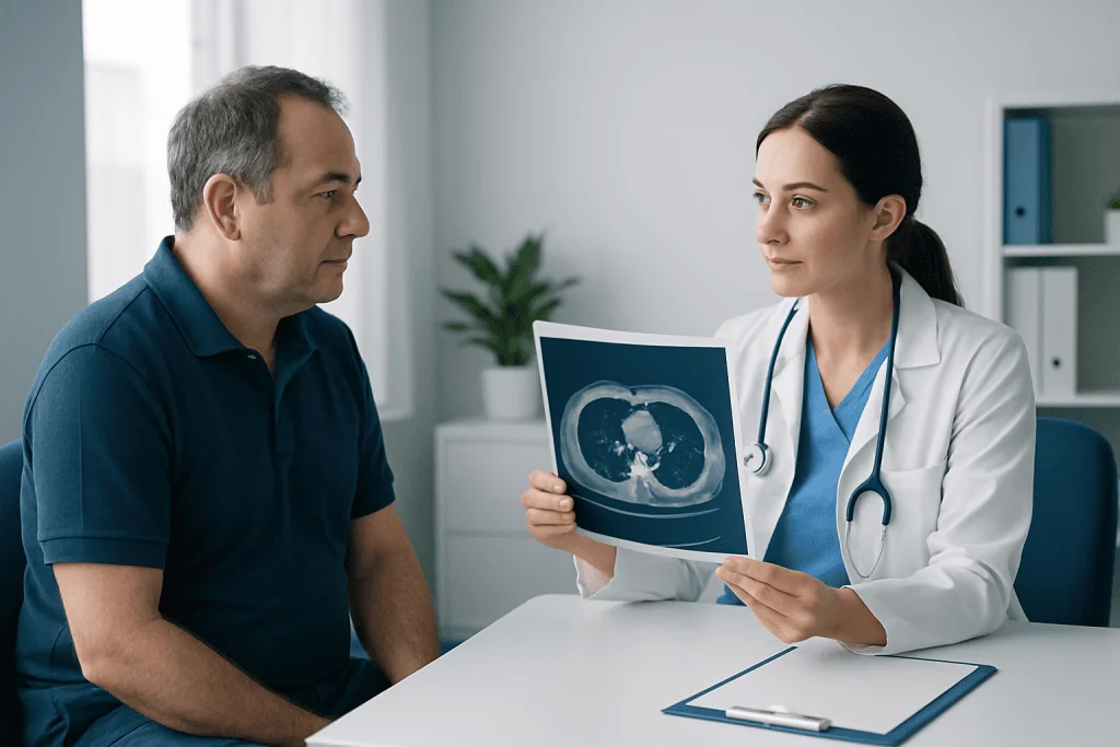
The radiology report may describe lymphadenopathy, which simply means enlarged or abnormal lymph nodes. It should be read in context by the referring doctor. If tissue confirmation is needed, a biopsy may be recommended to establish the cause safely and accurately.
What Are the First Signs of Lymph Node Cancer?
The first signs of lymphoma or another cancer affecting lymph nodes can vary. A painless lump in the neck, armpit or groin that persists or continues to enlarge is one possible sign. Some people notice no visible swelling when affected lymph nodes are deeper in the chest or abdomen.
Other symptoms can include ongoing unexplained fever, drenching night sweats, unintentional weight loss, persistent fatigue, itching, cough, shortness of breath, abdominal fullness or discomfort. These symptoms are not specific to cancer and are much more commonly related to other conditions. Their duration, severity and combination help guide medical evaluation.
A clinician may begin with a history, physical examination and blood tests, then choose ultrasound, CT, PET/CT or tissue sampling as needed. Conditions such as lymphoma require specialist assessment because the diagnosis and treatment plan depend on the precise subtype and extent of disease.
What Stage Is Cancer if It Is in the Lymph Nodes?
Cancer in lymph nodes does not automatically mean a single cancer stage. Staging depends on the original cancer type, the number and location of involved nodes, whether they are nearby or distant from the primary tumor, the tumor’s size or depth, and whether cancer has spread to other organs. Different cancers use different staging systems.
For many solid tumors, spread to regional lymph nodes may indicate a more advanced local or regional stage, but not necessarily distant metastatic disease. Cancer found in lymph nodes far from the original tumor may be classified differently. Lymphomas are staged using their own system, which considers the regions involved and whether disease is above or below the diaphragm, among other factors.
Accurate staging often combines pathology, imaging and clinical findings. A multidisciplinary team may include radiologists, pathologists, surgeons, medical oncologists and radiation oncologists. This approach helps ensure that staging is not based on a CT report alone.
When to Seek Medical Care
Medical advice is appropriate for a lymph node that remains enlarged for several weeks, is getting larger, feels hard or fixed, or occurs with persistent fever, night sweats, unexplained weight loss or marked fatigue. Prompt assessment is also sensible for swelling near the collarbone, breathing difficulty, trouble swallowing, severe pain or symptoms that are rapidly worsening.
People with a previous cancer diagnosis should contact their treating team if they notice a new lump or develop persistent new symptoms. A doctor can decide whether observation, examination, blood tests or imaging such as a CT scan is appropriate. It is best not to assume that a swollen node has a particular cause without assessment.
Acibadem International’s multidisciplinary specialists and JCI-accredited hospitals support international patients needing imaging assessment and care for lymph node-related conditions. The appropriate pathway may include imaging, pathology and oncology consultation based on individual findings.
Frequently asked questions
How long does a CT scan of lymph nodes take?
The scan itself often takes only a few minutes. The full visit may take longer because of registration, safety checks, positioning and intravenous contrast preparation if needed. The imaging center will provide appointment-specific instructions.
Do enlarged lymph nodes always mean cancer on a CT scan?
No. Enlarged lymph nodes are frequently caused by infections, inflammation or immune-system activity. CT can show the pattern and appearance of nodes, but further evaluation is sometimes needed to identify the cause.
Is contrast always needed for a CT scan to check lymph nodes?
No. Whether contrast is needed depends on the body area and the question the doctor needs answered. Contrast can improve assessment of blood vessels, organs and some tissue changes, but it may not be suitable for every patient.
When will CT scan lymph node results be available?
A radiologist reviews the images and sends a report to the referring clinician. Timing varies by facility and clinical urgency, so the patient should ask the imaging team or doctor when to expect results. The referring clinician explains what the findings mean in the individual situation.
Can a CT scan detect lymphoma?
CT can detect enlarged lymph nodes and other findings that may suggest lymphoma, including involvement of lymph nodes deep in the chest or abdomen. It cannot confirm lymphoma by itself. A biopsy is usually needed to diagnose the condition and determine its specific subtype.
What should someone do after a CT scan with contrast?
Most people can resume normal activities straight away. Drinking fluids may be recommended unless there is a medical reason to limit fluids. New rash, swelling, breathing difficulty or other concerning symptoms after contrast should be reported urgently.
References
- American Cancer Society
- National Cancer Institute
- Radiological Society of North America
- National Health Service
- World Health Organization
This article is for general information only and is not a substitute for professional medical advice. Please consult a qualified doctor about your individual situation.
Explore treatments in Turkey — costs, top hospitals & a free quote
JCI-accredited · board-certified surgeons · reply within 24h
Add us as a Preferred Source to see more of our trusted health content across Google Search, AI Overviews and Discover.